Acute pancreatitis
• Acutepancreatitis results from inappropriate release and activation of
pancreatic enzymes that, in turn, destroy pancreatic tissue and elicit
an acute inflammatory reaction.
Two factors havebeen proposed to contribute to alcohol indcuced
pancreatitis:
1. Duct obstruction
2. Acinar cell damage.
11.
Laboratory Investigations
• Elevationof serum amylase and lipase levels during the first 4 to 12
hours following the onset of pain.
• Serum amylase has a short half-life and may return to normal in 3 to 5
days, whereas lipase levels remain elevated for 8 to 14 days.
• Hypocalcemia may result from saponification of necrotic fat.
• Direct visualization of the enlarged inflamed pancreas by computed
tomography (CT) scanning is useful in the diagnosis of pancreatitis.
Chronic pancreatitis
• Prolongedinflammation of the pancreas associated with irreversible
destruction of exocrine parenchyma, fibrosis, and, in the late stages,
loss of endocrine parenchyma.
• The most common cause of chronic pancreatitis is long-term alcohol
use.
14.
Pathogenesis
• Chronic pancreatitisoften follows repeated episodes of acute
pancreatitis.
• It has been proposed that acute pancreatitis initiates a sequence of
perilobular fibrosis, duct distortion, and altered secretions that, as a
result of recurrent injury, leads to loss of exocrine parenchyma and
fibrosis.
• Chronic pancreatic injury of any cause leads to local production of
inflammatory mediators that promote fibrosis and acinar cell loss.
15.
• Chronic pancreatitisis characterized by parenchymal fibrosis, acinar atrophy and
dropout, and variable ductal dilatation.
• Grossly, the gland is hard, sometimes with visibly dilated ducts containing calcified
concretions.
• These changes are typically accompanied by a histologically evident chronic
inflammatory infiltrate that surrounds lobules and ducts.
• Acinar loss is a constant feature, but there is usually relative sparing of the islets of
Langerhans, which become embedded in the sclerotic tissue and may fuse and appear
enlarged.
• Ductal epithelium may be atrophic, hyperplastic, or metaplastic (squamous).
• Chronic pancreatitis caused by alcohol abuse is characterized by ductal dilation and
intraluminal protein plugs and calcifications
PANCREATIC CARCINOMA
• Infiltratingductal adenocarcinoma of the exocrine pancreas (commonly
known as “pancreatic cancer”) is the most frequent neoplasm of the pancreas
(about 85% of all neoplasms).
Risk Factors
• Cigarette smoking and alcohol use
• Consumption of a diet rich in fats
• Chronic pancreatitis
• Diabetes mellitus
• Inherited genetic defects: BRCA2 mutations
18.
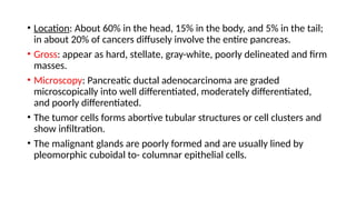
• Location: About60% in the head, 15% in the body, and 5% in the tail;
in about 20% of cancers diffusely involve the entire pancreas.
• Gross: appear as hard, stellate, gray-white, poorly delineated and firm
masses.
• Microscopy: Pancreatic ductal adenocarcinoma are graded
microscopically into well differentiated, moderately differentiated,
and poorly differentiated.
• The tumor cells forms abortive tubular structures or cell clusters and
show infiltration.
• The malignant glands are poorly formed and are usually lined by
pleomorphic cuboidal to- columnar epithelial cells.
Clinical Features
• Pancreaticcancers usually remain silent until they infiltrate into
adjacent structures.
• Pain: It is usually the first and the outstanding symptom.
• Obstructive jaundice: It is associated with carcinoma of the head of
the pancreas
• Weight loss, anorexia, and generalized malaise and weakness.
• Migratory thrombophlebitis (Trousseau sign): It is characterized by
spontaneously appearing and disappearing (migratory)
thrombosis.
• It is found in about 10% of patients.
• It is attributable to the production of platelet-aggregating factors
and procoagulants from the carcinoma or its necrotic products.
HEPATOCELLULARCARCINOMA
Arise from hepatocytes.
Etiology:
1.Hepatitis B
2. Hepatitis C
3. Alpha 1 antitrypsin def.
4. Hemochromatosis
5. Alcoholic liver ds
6. Cirrhosis
7. Toxin: Aflatoxin from fungus
8. Chemicals : vinyl chloride
9. Hormonal therapy: androgen , OCP.
27.
Gross
Expanding type: Mostfrequently,
it forms a single, yellow brown,
large mass, most often in the
right lobe of the liver with
central necrosis, haemorrhage
and occasional bile staining. It
may be deceptively
encapsulated.
Multifocal type: Less often,
multifocal, multiple masses,
3-5 cm in diameter, scattered
throughout the liver are seen.
Infiltrating type: Rarely, the
HCC forms diffusely
infiltrating tumour mass.
32.
Hepatoblastoma (Embryoma)
• Hepatoblastomais a rare malignant tumour arising from primitive
hepatic parenchymal cells.
• It presents before the age of 2 years as progressive abdominal distension
with anorexia, failure to thrive, fever and jaundice.
• It is more common in boys.
• The concentration of serum AFP is high.
• The tumour grows rapidly and causes death by haemorrhage, hepatic
failure or widespread metastases.
33.
MORPHOLOGIC FEATURES
• Grossly,the tumour is circumscribed and lobulated mass measuring 5-
25 cm in size, having areas of cystic degeneration, haemorrhage and
necrosis.
• Microscopically, hepatoblastoma consists of 2 components:
i) Epithelial component contains 2 types of cells— ‘embryonal’
hepatocytes are small with dark-staining, while ‘foetal’ hepatocytes are
larger with more cytoplasm.
ii) Mesenchymal component includes fibrous connective tissue,
cartilage and osteoid of variable degree of maturation.
Carcinoma of theGallbladder
• Primary carcinoma of the gallbladder is more prevalent.
• Like cholelithiasis and cholecystitis, it is more frequent in women than
in men with a peak incidence in 7th decade of life.
MORPHOLOGIC FEATURES
• Thecommonest site is the fundus.
• Grossly, cancer of the gallbladder is of 2 types—infiltrating and fungating
type
• Infiltrating type appears as an irregular area of diffuse thickening and
induration of the gallbladder wall.
• It may have deep ulceration causing direct invasion of the gallbladder wall
and liver bed.
• On section, the gallbladder wall is firm due to the growth.
• Fungating type grows like an irregular, friable, papillary or cauliflower-like
growth into the lumen as well as into the wall of the gallbladder and beyond.
39.
• Most gallbladdercancers are adenocarcinomas (90%).
• They may be papillary or infiltrative, well-differentiated or poorly-
differentiated.
• About 5% of gallbladder cancers are squamous cell carcinomas arising
from squamous metaplastic epithelium.
• A few cases show both squamous and adenocarcinoma pattern of
growth called adenosquamous carcinoma.
40.
Morphology
• GROSS:
• Appearas soft and hemorrhagic mass.
• Tumour mass may be solitary , diffuse infiltration lesion.
• Liver is enlarged.
• Yellowish white in colour.
• MICROSCOPIC:
• Tumour cells are polygons and large with granular cytoplasm.
• The tumour cells are arranged in clusters or cords.


![Ca pancreas [autosaved]](https://cdn.slidesharecdn.com/ss_thumbnails/capancreasautosaved-200627065511-thumbnail.jpg?width=640&height=640&fit=bounds)


