Downloaded 46 times

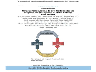
![Copyright © 2014, Canadian Cardiovascular Society
2014 CCS Guidelines on the Diagnosis and Management of Stable Ischemia Heart Disease
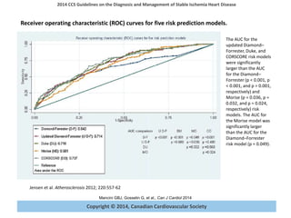
Mancini GBJ, Gosselin G, et al., Can J Cardiol 2014
Copyright © 2014, Canadian Cardiovascular Society
Meta regression analysis of the relationship of percentage of patients with
reperfusion therapy on the risk ratio of mortality with β-blockers.
• β-blockers reduced mortality in pre-
reperfusion[IRR=0.86, 95% CI=0.79-
0.94] but not in the reperfusion
era(IRR=0.98, 95% CI=0.92-1.05) where
there was reduction (short-term) in
myocardial infarction(IRR=0.72, 95%
CI=0.62-0.83) and angina(IRR=0.80,
95%CI=0.65-0.98) but increase in heart
failure(IRR=1.10, 95% CI=1.05-1.16),
cardiogenic shock(IRR=1.29, 95%
CI=1.18-1.41) and drug discontinuation.
• In contemporary treatment of MI, β-
blockers have no mortality benefit but
reduce myocardial infarction and
angina (short-term) with increase in
heart failure, cardiogenic shock and
drug discontinuation
Bangalore S, et al. The American Journal of Medicine, 2014 http://dx.doi.org/10.1016/j.amjmed.2014.05.032](https://image.slidesharecdn.com/sihdgui2014sden-170429140554/85/Stable-Ischemic-Heart-Disease-Guideline-27-320.jpg)

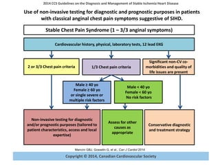
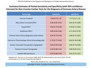
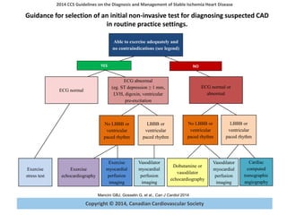
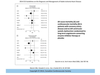
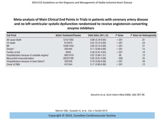
The document summarizes guidelines from the Canadian Cardiovascular Society (CCS) for the diagnosis and management of stable ischemic heart disease. It provides recommendations on establishing diagnosis and prognosis through history, physical exam, testing. It recommends non-invasive testing such as exercise ECG or imaging to diagnose patients with chest pain symptoms. The guidelines also discuss assessing prognosis based on factors like anatomical disease burden and left ventricular function. It provides guidance on selecting initial diagnostic tests and interpreting high risk features of test results.


![PERI-PROSTHETIC FRACTURE NAIL-PLATE CONSTRUCT [NPC].pptx](https://cdn.slidesharecdn.com/ss_thumbnails/drarunkumardrmohamedashrafperiprostheticfrasturenail-plateconstructnpc-260209164459-7e9d15a1-thumbnail.jpg?width=640&height=640&fit=bounds)


