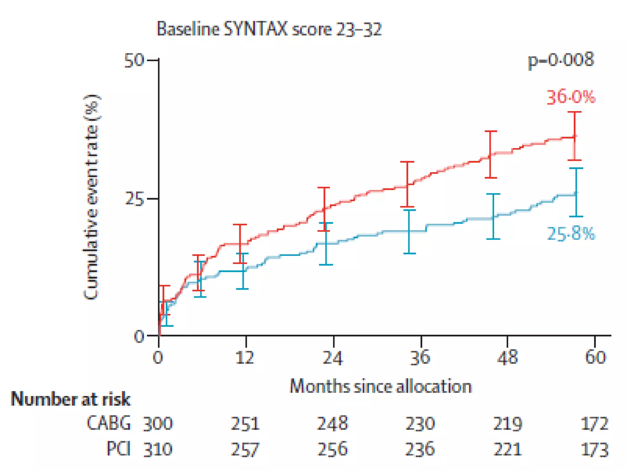
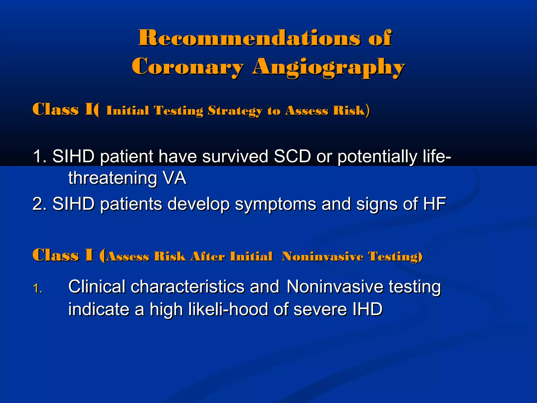
SlideShare a Scribd company logo
Stable IschemicStable Ischemic
Heart DiseaseHeart Disease
Patient ProfilingPatient Profiling
Local Heart team (surgeon &Local Heart team (surgeon &
interventional cardiologist)interventional cardiologist)
assessed each patient inassessed each patient in
regards toregards to ::
 Patient’s operative risk (EuroSCOREPatient’s operative risk (EuroSCORE
& Parsonnet score)& Parsonnet score)
 Coronary lesion complexity (NewlyCoronary lesion complexity (Newly
developed SYNTAX score)developed SYNTAX score)
 Goal: SYNTAX score to provideGoal: SYNTAX score to provide
guidance on optimalguidance on optimal
revascularization strategies forrevascularization strategies for
patients with high risk lesionspatients with high risk lesions
Sianos et al, EuroIntervention 2005;1:219-227
Valgimigli et al, Am J Cardiol 2007;99:1072-1081
Serruys et al, EuroIntervention 2007;3:450-459
BARI classification of coronary segments
Leaman score, Circ 1981;63:285-299
Lesions classification ACC/AHA , Circ 2001;103:3019-3041
Bifurcation classification, CCI 2000;49:274-283
CTO classification, J Am Coll Cardiol 1997;30:649-656
DominanceDominance
Case 1Case 1
A 66-year-old man with a history of HTN, T2DM (20ys)A 66-year-old man with a history of HTN, T2DM (20ys)
mixed hyperlipidemia, smoking ( 30 ys, quit smokingmixed hyperlipidemia, smoking ( 30 ys, quit smoking
20ys), treatting with Bisoprolol-Valsartan-Rosuvastatin20ys), treatting with Bisoprolol-Valsartan-Rosuvastatin
and Metformin,can climb up 4 floors,has no anyand Metformin,can climb up 4 floors,has no any
symptom.symptom.
1. What is his diagnosis ?1. What is his diagnosis ?
2. What test would you order for him? Why?2. What test would you order for him? Why?
3. What the most optimal therapy method would he be3. What the most optimal therapy method would he be
received ?received ?
General principlesGeneral principles
 Defined as a>50% luminal stenosis of any epicardial CA.Defined as a>50% luminal stenosis of any epicardial CA.
 Anginal symptoms : a fixed stenosis ≥ 70% , commonlyAnginal symptoms : a fixed stenosis ≥ 70% , commonly
obstruction by atheromatous plaqueobstruction by atheromatous plaque
 CAD: stable angina,ACS,CHF,sudden cardiac death, silentCAD: stable angina,ACS,CHF,sudden cardiac death, silent
ischemiaischemia
 Chronic stable angina; stable exertional angina, stable ischemicChronic stable angina; stable exertional angina, stable ischemic
heart disease (SIHD),Ischemic cardiomyophathy disease ( EF ≤heart disease (SIHD),Ischemic cardiomyophathy disease ( EF ≤
45% due to CAD +/- congestive HF ): Nearly 50% CAD pts45% due to CAD +/- congestive HF ): Nearly 50% CAD pts
Atherosclerotic progression of coronaryAtherosclerotic progression of coronary
artery stenosisartery stenosis
Diagnosis of SIHDDiagnosis of SIHD
1. Chest Pain:1. Chest Pain: location, severity, and duration of pain; radiation;location, severity, and duration of pain; radiation;
associated symptoms; provocative factors; and alleviatingassociated symptoms; provocative factors; and alleviating
factors.factors.
What do adjectives often used to describe anginal pain?What do adjectives often used to describe anginal pain?
 Often “squeezing,” “grip-like,” “suf-Often “squeezing,” “grip-like,” “suf-
focating,” and “heavy”, anginal equivalents:Dyspnea,focating,” and “heavy”, anginal equivalents:Dyspnea,
epigastric and nauseaepigastric and nausea..
 Rarely sharp or stabbingRarely sharp or stabbing
What are special characteristics of angina pectoris ?What are special characteristics of angina pectoris ?
 Not vary with position or respiration.Not vary with position or respiration.
 The classic Levine’s sign by placing a clenched fist over theThe classic Levine’s sign by placing a clenched fist over the
precordium to describe the painprecordium to describe the pain
 <1minute other, few- 20 minutes Stable anginal ; > 20<1minute other, few- 20 minutes Stable anginal ; > 20
minutes UAminutes UA
Can the history be used to classifyCan the history be used to classify
symptoms as typical, atypical, orsymptoms as typical, atypical, or
noncardiac chest pain?noncardiac chest pain?
What should alternative diagnoses beWhat should alternative diagnoses be
considered?considered?
What is the classification of severity ofWhat is the classification of severity of
anginaangina??
what are stable angina or UAwhat are stable angina or UA
categorized?categorized?
Clinical characteristics,Clinical characteristics,
associate with a high likelihood ofassociate with a high likelihood of
severe IHD /UA,MIsevere IHD /UA,MI
 Chest pain leading to pulmonary edema,
 Chest pain associated with lightheadedness,
syncope or hypoten-sion,
 Exertional syncope,
 An exercise-induced gallop sound on cardiac
auscultation.
2.Cardiac risk factors2.Cardiac risk factors
 Diabetes mellitus (2-4times)Diabetes mellitus (2-4times)
 Smoking ( 15ys)Smoking ( 15ys)
 Hyperlipidemia,Hyperlipidemia,
 Hypertension,Hypertension,
 Obesity or metabolic syndrome,Obesity or metabolic syndrome,
 Physical inactivity,Physical inactivity,
 A family history of premature IHD ( onset of father,A family history of premature IHD ( onset of father,
brother,or son < age 55 years or a mother, sister, orbrother,or son < age 55 years or a mother, sister, or
daughter < age 65 years).daughter < age 65 years).
 A history of cerebrovascular or peripheral artery diseaseA history of cerebrovascular or peripheral artery disease
(PAD) also increases the likelihood of IHD(PAD) also increases the likelihood of IHD
3.Physical Examination:3.Physical Examination:
Conditions Provoking or Exacerbating Ischemia and HF
Approach to the Selection of Diagnostic Tests toApproach to the Selection of Diagnostic Tests to
Diagnose SIHDDiagnose SIHD
Abnormalities of regional or global VF occur later→severe stenosis →higher diagnostic
specificity than do perfusion defects on nuclear MPI.
On the other hand on nuclear MPI. Isolated perfusion defects can result from
stenoses of borderline significance, raising the sensitivity of this test but lowering
the specificity for more severe stenosis.
BiochemicalBiochemical makersmakers
 CBC: Anemia: COCBC: Anemia: CO  when Hb <9 g/dL, and ST-T-wavewhen Hb <9 g/dL, and ST-T-wave
changes (depression or inversion) when Hb<7 g/dLchanges (depression or inversion) when Hb<7 g/dL
 Fasting glucose, lipidFasting glucose, lipid
 Ionos, TSH, NT ProBNP, Hs Troponin T 2 timesIonos, TSH, NT ProBNP, Hs Troponin T 2 times
 ECG:ECG: prior MI, persistent ST-T-wave inversions,
LBBB, AVblock, VT, LVH, WPW, PM
 ECHOCARDIOGRAPHY:: wall motion abnormalities,
LVEF,and valvular abnormalities
Indications of stress testing:Indications of stress testing:
provoke cardiac ischemia by using exercise or
pharmacological stress agents
 Unknown CADUnknown CAD
 Anginal symptom: CCS I-IIAnginal symptom: CCS I-II
 Asymptomatic intermediate risk but Vigorous exercise/highAsymptomatic intermediate risk but Vigorous exercise/high
risk occupationrisk occupation
 Asymptomatic intermediate risk: Dm, smoking, PVDAsymptomatic intermediate risk: Dm, smoking, PVD
 Known CADKnown CAD
 Post-MI( change in clinical: HF/ angina)Post-MI( change in clinical: HF/ angina)
 Preoperative risk assessmentPreoperative risk assessment
 Recurrent anginal despite optimal medical therapy/Recurrent anginal despite optimal medical therapy/
revascularizationrevascularization
Contraindications of stress testingContraindications of stress testing
 Acute MI within 2 daysAcute MI within 2 days
 UA not previously stailized by medical therapyUA not previously stailized by medical therapy
 Cadiac arrhythmia having symptomCadiac arrhythmia having symptom
 Symptom severe aortic stenosisSymptom severe aortic stenosis
 Symptom HFSymptom HF
 Acute pulmonary embolus, myocarditis, pericarditis,Acute pulmonary embolus, myocarditis, pericarditis,
aortic dissectionaortic dissection
- Low risk: Medical therapy
- Intermediate: Delayed CA after Further testing based on
risk factors and inmaging
- High: Coronary angiography at one
Clinical signs and symptoms, findings on noninvasiveClinical signs and symptoms, findings on noninvasive
studies have been quantified risk of IHDstudies have been quantified risk of IHD
High risk ( > 3% annual death or MI)High risk ( > 3% annual death or MI)
1. LVEF < 35% not readily explained by noncoronary causes1. LVEF < 35% not readily explained by noncoronary causes
2. Resting perfusion abnormalities2. Resting perfusion abnormalities ≥≥10% of the myocardium in patients10% of the myocardium in patients
without prior history or evidence of MIwithout prior history or evidence of MI
3. Stress ECG findings including ≥2 mm of ST-segment depression at low3. Stress ECG findings including ≥2 mm of ST-segment depression at low
workload or persisting into recovery, exercise-induced ST-segmentworkload or persisting into recovery, exercise-induced ST-segment
elevation, or exercise-induced VT/VFelevation, or exercise-induced VT/VF
4. Severe stress-induced LV dysfunction (peak exercise LVEF <45% or drop in4. Severe stress-induced LV dysfunction (peak exercise LVEF <45% or drop in
LVEF with stress ≥10%)LVEF with stress ≥10%)
5. Stress-induced perfusion abnormalities encumbering ≥10% myocardium or5. Stress-induced perfusion abnormalities encumbering ≥10% myocardium or
stress segmental scores indicating multiple vascular territories withstress segmental scores indicating multiple vascular territories with
abnormalitiesabnormalities
6. Stress-induced LV dilation6. Stress-induced LV dilation
7. Inducible wall motion abnormality (involving >2 segments or 2 coronary7. Inducible wall motion abnormality (involving >2 segments or 2 coronary
beds)beds)
8. Wall motion abnormality developing at low dose of dobutamine (8. Wall motion abnormality developing at low dose of dobutamine ( ≤≤
10µg/min/kg10µg/min/kg ) or at a low heart rate (< 120 beats/min)) or at a low heart rate (< 120 beats/min)
9. CAC score >400 Agatston units9. CAC score >400 Agatston units
10. Multivessel obstructive CAD ( ≥70% stenosis) or left main stenosis (≥50%10. Multivessel obstructive CAD ( ≥70% stenosis) or left main stenosis (≥50%
stenosis) on CCTAstenosis) on CCTA
Clinical signs and symptoms, findings on noninvasiveClinical signs and symptoms, findings on noninvasive
studies have been quantified risk of IHDstudies have been quantified risk of IHD
Intermediate risk (1% to 3% annual death or MI)Intermediate risk (1% to 3% annual death or MI)
1. Mild/moderate resting LV dysfunction (LVEF 35% to 49%) not readily1. Mild/moderate resting LV dysfunction (LVEF 35% to 49%) not readily
explained by noncoronary causesexplained by noncoronary causes
2. Resting perfusion abnormalities in 5% to 9.9% of the myocardium in2. Resting perfusion abnormalities in 5% to 9.9% of the myocardium in
patients without a history or prior evidence of MIpatients without a history or prior evidence of MI
3. ≥1 mm of ST-segment depression occurring with exertional3. ≥1 mm of ST-segment depression occurring with exertional
symptomssymptoms
4. Stress-induced perfusion abnormalities encumbering 5% to 9.9% of4. Stress-induced perfusion abnormalities encumbering 5% to 9.9% of
the myocardium or stress segmental scores (in multiple segments)the myocardium or stress segmental scores (in multiple segments)
indicating 1 vascular territory with abnormalities but without LVindicating 1 vascular territory with abnormalities but without LV
dilationdilation
5. Small wall motion abnormality involving 1 to 2 segments and only5. Small wall motion abnormality involving 1 to 2 segments and only
1 coronary bed1 coronary bed
6. CAC score 100 to 399 Agatston units6. CAC score 100 to 399 Agatston units
7. One vessel CAD with ≥70% stenosis or moderate CAD stenosis7. One vessel CAD with ≥70% stenosis or moderate CAD stenosis
(50% to 69% stenosis) in ≥ 2 arteries on CCTA(50% to 69% stenosis) in ≥ 2 arteries on CCTA
Clinical signs and symptoms, findings on noninvasiveClinical signs and symptoms, findings on noninvasive
studies have been quantified risk of IHDstudies have been quantified risk of IHD
Low risk (<1% annual death or MI)Low risk (<1% annual death or MI)
1. Low-risk treadmill score (score ≥5) or no new ST segment1. Low-risk treadmill score (score ≥5) or no new ST segment
changeschanges
or exercise-induced chest pain symptoms; when achievingor exercise-induced chest pain symptoms; when achieving
maximalmaximal
levels of exerciselevels of exercise
2. Normal or small myocardial perfusion defect at rest or with2. Normal or small myocardial perfusion defect at rest or with
stress encumbering <5% of the myocardium*stress encumbering <5% of the myocardium*
3. Normal stress or no change of limited resting wall motion3. Normal stress or no change of limited resting wall motion
abnormalities during stressabnormalities during stress
4. CAC score <100 Agatston units4. CAC score <100 Agatston units
5. No coronary stenosis >50% on CCTA5. No coronary stenosis >50% on CCTA
Progressive of case1Progressive of case1
May 20/2013 Dobu ECHO was done, which shows:May 20/2013 Dobu ECHO was done, which shows:
- He has angina pectoris, and sweeting, HTN 80/50 at recovery- He has angina pectoris, and sweeting, HTN 80/50 at recovery
stagestage
- ECG: ST segment elevation at DIII,aVFECG: ST segment elevation at DIII,aVF
ST segment depression at DI, aVLST segment depression at DI, aVL
- Hypokinesie of inferior, middle and base regionHypokinesie of inferior, middle and base region
- What would you do for him, now? what class?What would you do for him, now? what class?
Coronary AngiographyCoronary Angiography
AdvantagesAdvantages
Defines coronary anatomy:Defines coronary anatomy:
1.to assess a patient’s risk of1.to assess a patient’s risk of
death and futuredeath and future
cardiovascular eventscardiovascular events
2. to ascertain the feasibility of2. to ascertain the feasibility of
percutaneous or surgicalpercutaneous or surgical
revascularization or medicalrevascularization or medical
therapytherapy
DisadvantagesDisadvantages
1.The technical quality of angiograms1.The technical quality of angiograms
2.Exist with interobserver reliability:only2.Exist with interobserver reliability:only
70% overall agreement among readers70% overall agreement among readers
3. Provides only anatomic data, not a3. Provides only anatomic data, not a
reliable indicator of the functionalreliable indicator of the functional
significance (unless a technique such assignificance (unless a technique such as
FFR)FFR)
4. Not distinguish between a vulner-4. Not distinguish between a vulner-
able plaque, with a large lipid core, thinable plaque, with a large lipid core, thin
fibrous cap, and increased macrophagesfibrous cap, and increased macrophages
and a stable plaqueand a stable plaque
ines CA stenosis:ines CA stenosis:
major coronary arteries:a significant stenosis ≥70% diameter reductmajor coronary arteries:a significant stenosis ≥70% diameter reduct
t main disease: ≥50%.t main disease: ≥50%.
Recommendations ofRecommendations of
Coronary AngiographyCoronary Angiography
Class I(Class I( Initial Testing Strategy to Assess RiskInitial Testing Strategy to Assess Risk))
1. SIHD patient have survived SCD or potentially life-1. SIHD patient have survived SCD or potentially life-
threatening VAthreatening VA
2. SIHD patients develop symptoms and signs of HF2. SIHD patients develop symptoms and signs of HF
Class I (Class I (Assess Risk After Initial Noninvasive Testing)Assess Risk After Initial Noninvasive Testing)
1.1. Clinical characteristics andClinical characteristics and Noninvasive testingNoninvasive testing
indicate a high likeli-hood of severe IHDindicate a high likeli-hood of severe IHD
Recommendations ofRecommendations of
Coronary AngiographyCoronary Angiography
Class IIaClass IIa
1. LV EF <50% and moderate risk criteria1. LV EF <50% and moderate risk criteria
on noninvasive testing with demonstrable ischemia.on noninvasive testing with demonstrable ischemia.
2.SIHD pts with noninvasive testing inconclusive2.SIHD pts with noninvasive testing inconclusive
Or contraindicated or inadequate.Or contraindicated or inadequate.
3. SIHD pts have:3. SIHD pts have:
- unsatisfactory quality of life due to angina,LVEF- unsatisfactory quality of life due to angina,LVEF
>50%, intermediate risk criteria on noninvasive>50%, intermediate risk criteria on noninvasive
testing.testing.
Recommendations ofRecommendations of
Coronary AngiographyCoronary Angiography
Class III: No BenefitClass III: No Benefit
1.1. For risk assessment with SIHD who elect not to undergoFor risk assessment with SIHD who elect not to undergo
revascularization or who are not candidates forrevascularization or who are not candidates for
evascularization because of comorbidities or individualevascularization because of comorbidities or individual
preferences.preferences.
2. To further assess risk in patients with SIHD who have EF2. To further assess risk in patients with SIHD who have EF
>50% and low-risk criteria on noninvasive testing.>50% and low-risk criteria on noninvasive testing.
3.3. To assess risk in patients at low risk according to clinicalTo assess risk in patients at low risk according to clinical
criteria and who have not undergone nonin-vasive riskcriteria and who have not undergone nonin-vasive risk
testing.testing.
Progressive of case1Progressive of case1
May 23/2013 was AG with result:May 23/2013 was AG with result:
 Severe cacificated CA system,Severe cacificated CA system,
 LAD: 90% stenosis of ostium. 80-90% stenosis I-IILAD: 90% stenosis of ostium. 80-90% stenosis I-II
segment, bifurcation of Diagonal I, subsegment, bifurcation of Diagonal I, subocclusion ofocclusion of
distal partdistal part
 CX: 70-80 stenosis of OMI, 80-90% stenosis of distalCX: 70-80 stenosis of OMI, 80-90% stenosis of distal
partpart
 RCA: 80-90% diffuseRCA: 80-90% diffuse stenosis of beginning and distalstenosis of beginning and distal
partpart
DIAGNOSTIC PROCESS OF CHRONIC CADDIAGNOSTIC PROCESS OF CHRONIC CAD
AT HCM CITY HEART INSTITUTE & HEARTAT HCM CITY HEART INSTITUTE & HEART
TAM UC HOSPITALĐTAM UC HOSPITALĐ
-Rest ECG &
ECHO
CXR
NFG,
Cholesterol, TG,
HDL-C, LDL-C,
Glucose, CRP,
Fibrinogen,
Creatinine
CORONARY
ANGIOGRAPHY
Exercise ECG &/ECHO
Dobu /
MSCT / Radionuclide
imaging
Cardiovascular Internal
Consultation
Physical examination
CAD risk factors,
Family CV history
Chronic CAD disease 2009: cđ, MT,
PCI, CABG
TREATMENTTREATMENT
1. Medical Therapy:GDMT1. Medical Therapy:GDMT (Guideline-Directed Medical(Guideline-Directed Medical
Therapy)Therapy)
2. CAD Revascularization:CABG and PCI2. CAD Revascularization:CABG and PCI (coronary artery(coronary artery
bypass graft and percutaneous coronary intervention)bypass graft and percutaneous coronary intervention)
3. Follow-Up of Patients With SIHD3. Follow-Up of Patients With SIHD
1. Medical Therapy1. Medical Therapy
What are the goals of treatingWhat are the goals of treating
patients with SIHD?patients with SIHD?
1.Reduce premature cardiovascular death;1.Reduce premature cardiovascular death;
2. Prevent complications of SIHD including nonfatal2. Prevent complications of SIHD including nonfatal
AMI and heart failure;AMI and heart failure;
3. Maintain or restore a level of activity, functional capacity,3. Maintain or restore a level of activity, functional capacity,
eliminate ischemic symptoms, cardiac rehabilitation,eliminate ischemic symptoms, cardiac rehabilitation,
quality of life that is satisfactory to the patient;quality of life that is satisfactory to the patient;
4. Minimize costs:4. Minimize costs:
- by eliminating avoidable adverse effects of tests and- by eliminating avoidable adverse effects of tests and
treatments,treatments,
- by preventing hospital admissions,- by preventing hospital admissions,
- by eliminating unnecessary tests and treatments.- by eliminating unnecessary tests and treatments.
How should the SIHD pts have an individualizedHow should the SIHD pts have an individualized
education plan to optimize care?education plan to optimize care?
1.The importance of1.The importance of medication adherencemedication adherence for managingfor managing
symptoms and retarding disease progressionsymptoms and retarding disease progression
2. A2. A comprehensive reviewcomprehensive review of all therapeutic optionsof all therapeutic options
3. An appropriate levels of exercise,3. An appropriate levels of exercise,
encouragement to maintainencouragement to maintain physical activityphysical activity
4. Introduction to4. Introduction to self-monitoring skillsself-monitoring skills
5. Information on how to5. Information on how to recognize worsening car-recognize worsening car-
diovascular symptomsdiovascular symptoms and take appropriate actionand take appropriate action
6.6. Lifestyle changesLifestyle changes: BMI18.5 to 24.9, lipid; BP;T2DM: BMI18.5 to 24.9, lipid; BP;T2DM
control;smoking cessation and avoidance of exposurecontrol;smoking cessation and avoidance of exposure
to secondhand smoketo secondhand smoke
What does the Medical Therapy Prevent MI andWhat does the Medical Therapy Prevent MI and
Death?Death?
1.1. Antiplatelet TherapyAntiplatelet Therapy
Class IClass I
1. Treatment with aspirin 75 to 162 mg daily should be1. Treatment with aspirin 75 to 162 mg daily should be
continued indefinitely in the absence of contraindications incontinued indefinitely in the absence of contraindications in
patients with SIHD.patients with SIHD.
2. Treatment with clopidogrel is reasonable when aspirin is2. Treatment with clopidogrel is reasonable when aspirin is
contraindicated in patients with SIHD.contraindicated in patients with SIHD.
Class IIbClass IIb
1.1. Treatment with aspirin 75 to 162 mg daily and Clopidogrel 75Treatment with aspirin 75 to 162 mg daily and Clopidogrel 75
mg daily might be reasonable incertain high-risk patients withmg daily might be reasonable incertain high-risk patients with
SIHD.SIHD.
Class III: No BenefitClass III: No Benefit
1. Dipyridamole is not recommended as antiplatelet therapy for1. Dipyridamole is not recommended as antiplatelet therapy for
patients with SIHD.patients with SIHD.
What does the Medical Therapy Prevent MI andWhat does the Medical Therapy Prevent MI and
Death?Death? (Cont)(Cont)
2. Beta-Blocker Therapy2. Beta-Blocker Therapy
Class IClass I
1. Beta-blocker therapy should be started and continued for 3 years in1. Beta-blocker therapy should be started and continued for 3 years in
all patients with normal LV function after MI or ACS.all patients with normal LV function after MI or ACS.
2. Beta-blocker therapy should be used in all patients2. Beta-blocker therapy should be used in all patients
with LVEF <40%, HF or prior MI, unless contraindicated. (Usewith LVEF <40%, HF or prior MI, unless contraindicated. (Use
should be limited to carvedilol, metoprolol succinate,or bisoprolol,should be limited to carvedilol, metoprolol succinate,or bisoprolol,
which have been shown to reduce risk of death.)which have been shown to reduce risk of death.)
Class IIbClass IIb
1. Beta blockers may be considered as chronic therapy for all other1. Beta blockers may be considered as chronic therapy for all other
patients with coronary or other vascular disease.patients with coronary or other vascular disease.
What does the Medical Therapy Prevent MIWhat does the Medical Therapy Prevent MI
andand
Death?Death? (Cont)(Cont)
3.3. Renin-Angiotensin-Aldosterone Blocker TherapyRenin-Angiotensin-Aldosterone Blocker Therapy
Class IClass I
1. ACE inhibitors should be prescribed in all patients1. ACE inhibitors should be prescribed in all patients
with SIHD who also have hypertension, DM, LVEF 40% orwith SIHD who also have hypertension, DM, LVEF 40% or
less, or CKD, unless contraindicated.less, or CKD, unless contraindicated.
2. ARBs are recommended for patients with SIHD who2. ARBs are recommended for patients with SIHD who
are intolerant of, ACE inhibitors.are intolerant of, ACE inhibitors.
Class IIaClass IIa
1. Treatment with an ACE inhibitor is reasonable in1. Treatment with an ACE inhibitor is reasonable in
patients with both SIHD and other vascular disease.patients with both SIHD and other vascular disease.
2. It is reasonable to use ARBs in other patients who2. It is reasonable to use ARBs in other patients who
are ACE inhibitor intolerant.are ACE inhibitor intolerant.
What does the Medical Therapy Prevent MI andWhat does the Medical Therapy Prevent MI and
Death? (Cont)Death? (Cont)
4. Influenza Vaccination4. Influenza Vaccination
Class IClass I
1. An annual influenza vaccine is recommended for1. An annual influenza vaccine is recommended for
patients with SIHDpatients with SIHD..
What is the Medical Therapy forWhat is the Medical Therapy for
Relief of Symptoms?Relief of Symptoms?
1.1. Use of Anti-ischemicUse of Anti-ischemic
Class IClass I
1.1. Beta blockersBeta blockers should be prescribed as initial therapy for relief ofshould be prescribed as initial therapy for relief of
symptoms in patients with SIHD.symptoms in patients with SIHD.
2.2. Calcium channel blockers or long-acting nitratesCalcium channel blockers or long-acting nitrates should beshould be
prescribed for relief of symptoms when beta blockers areprescribed for relief of symptoms when beta blockers are
contraindicated or cause unacceptable side effects in patientscontraindicated or cause unacceptable side effects in patients
with SIHD.with SIHD.
3.3. Calcium channel blockers or long-acting nitratesCalcium channel blockers or long-acting nitrates, in, in combinationcombination
with beta blockers, should be prescribed for relief ofwith beta blockers, should be prescribed for relief of
symptoms when initial treatment with beta blockers issymptoms when initial treatment with beta blockers is
unsuccessful in patients with SIHD.unsuccessful in patients with SIHD.
4.4. Sublingual nitroglycerin or nitroglycerin spraySublingual nitroglycerin or nitroglycerin spray is recommendedis recommended
for immediate relief of angina in patients with SIHD.for immediate relief of angina in patients with SIHD.
What is the Medical Therapy forWhat is the Medical Therapy for
Relief of Symptoms?Relief of Symptoms?
Class IIaClass IIa
1.1. A long-acting nondihydropyridine calcium channel blocker:A long-acting nondihydropyridine calcium channel blocker:
(verapamil or diltiazem) instead of a beta blocker as initial(verapamil or diltiazem) instead of a beta blocker as initial
therapy for relief of symptoms is reasonable in patients withtherapy for relief of symptoms is reasonable in patients with
SIHD.SIHD.
2. Ranolazine:2. Ranolazine: can be useful when prescribed as acan be useful when prescribed as a substitutesubstitute forfor
beta blockersbeta blockers for relief of symptoms in patients with SIHD iffor relief of symptoms in patients with SIHD if
initial treatment with beta blockers leads to unacceptable sideinitial treatment with beta blockers leads to unacceptable side
effects or is ineffective or if initial treatment with beta blockers iseffects or is ineffective or if initial treatment with beta blockers is
contraindicated.contraindicated.
3.3. Ranolazine:Ranolazine: combination with beta blockerscombination with beta blockers can be useful whencan be useful when
prescribed for relief of symptoms when initial treatment withprescribed for relief of symptoms when initial treatment with
beta blockers is not successful in patients with SIHD.beta blockers is not successful in patients with SIHD.
Other Antianginal AgentsOther Antianginal Agents
1.1. NicorandilNicorandil:CA vasodilation,increases coronary blood flow,with:CA vasodilation,increases coronary blood flow,with
reductions in afterload, preload, and oxidative injury:noreductions in afterload, preload, and oxidative injury:no
difference between nicorandil and placebo with regard todifference between nicorandil and placebo with regard to
death from IHD or nonfatal MIdeath from IHD or nonfatal MI
2.2. Ivabradine:Ivabradine:  HR, prolonging diastole.HR, prolonging diastole.
treatment of chronic IHD with normal sinus rhythm with atreatment of chronic IHD with normal sinus rhythm with a
contraindication or intolerance to ß bloc.contraindication or intolerance to ß bloc.
3.3. TrimetazidineTrimetazidine:improve cellular tolerance to ischemia:improve cellular tolerance to ischemia
increases coronary flow reserve, delaying the onset ofincreases coronary flow reserve, delaying the onset of
ischemia associated with exercise and reducing the numberischemia associated with exercise and reducing the number
of weekly angina episodes and weekly nitroglycerinof weekly angina episodes and weekly nitroglycerin
consumptionconsumption
How should you doHow should you do
to get goal BP of less than 140/90 mm Hgto get goal BP of less than 140/90 mm Hg
What is Diabetes Management?What is Diabetes Management?
Class IIaClass IIa
1. A short duration of diabetes mellitus and a long life1. A short duration of diabetes mellitus and a long life
expectancy, a goal hemoglobin A1c (HbA1c) of 7% or lessexpectancy, a goal hemoglobin A1c (HbA1c) of 7% or less
is reasonable.is reasonable.
2. A goal HbA1c between 7% - 9% is reasonable for certain2. A goal HbA1c between 7% - 9% is reasonable for certain
patients according to age, history of hypoglycemia,patients according to age, history of hypoglycemia,
presence of Microvascular or macrovascular complications,presence of Microvascular or macrovascular complications,
or presence of coexisting medical conditionsor presence of coexisting medical conditions
Class IIbClass IIb
1.1. Initiation of pharmacotherapy interventions to achieve targetInitiation of pharmacotherapy interventions to achieve target
HbA1c might be reasonable.HbA1c might be reasonable.
Class III: HarmClass III: Harm
1.1. Therapy with rosiglitazone should not be initiated in patientsTherapy with rosiglitazone should not be initiated in patients
with SIHD.with SIHD.
2. CAD Revascularization:CABG and PCI2. CAD Revascularization:CABG and PCI ((coronary arterycoronary artery
bypass graft and percutaneous coronary intervention)bypass graft and percutaneous coronary intervention)
Progressive of case1Progressive of case1
May 23/2013 was AG with result:May 23/2013 was AG with result:
 Severe cacificated CA system,Severe cacificated CA system,
 LAD: 90% stenosis of ostium. 80-90% stenosis I-IILAD: 90% stenosis of ostium. 80-90% stenosis I-II
segment, bifurcation of Diagonal I, subsegment, bifurcation of Diagonal I, subocclusion ofocclusion of
distal partdistal part
 CX: 70-80 stenosis of OMI, 80-90% stenosis of distalCX: 70-80 stenosis of OMI, 80-90% stenosis of distal
partpart
 RCA: 80-90% diffuseRCA: 80-90% diffuse stenosis of beginning and distalstenosis of beginning and distal
partpart
INDICATIONS OF REVASCULARIZATION WITH PCI OR CABG ININDICATIONS OF REVASCULARIZATION WITH PCI OR CABG IN
CHRONIC STABLE ANGINA PATIENTSCHRONIC STABLE ANGINA PATIENTS
CABGCABG PCIPCI
Normal LVNormal LV
functionfunction
andand
DM (-)DM (-)
DM (+)DM (+) AbnormalAbnormal
LVLV
functionfunction
Normal LVNormal LV
function andfunction and
DM (-)DM (-)
DMDM
(+)(+)
AbnormalAbnormal
LVLV
functionfunction
Two-vessel diseaseTwo-vessel disease
with proximalwith proximal
LAD, CADLAD, CAD
AA AA AA AA AA AA
Three- vessel diseaseThree- vessel disease AA AA AA UU UU UU
Significant LMSignificant LM
coronary diseasecoronary disease
AA AA AA II II II
Significant LMSignificant LM
coronary disease &coronary disease &
atheroslerosisatheroslerosis
AA AA AA II II II
A= appropiate; U= Uncertain; I= Inappropiate
Patel MR et al. ACCF/ SCAI/AATS/AHA/ASNC Apropriateness Criteria for coronary
revascularrization. J Am Coll Cardiol 2009; 53 xxx-xxx
Treatment strategyTreatment strategy
Revascularization Appropriateness criteria:Revascularization Appropriateness criteria:
A 8A 8
 PCI appropriateness: U 4;PCI appropriateness: U 4;
Syntax score : 33%Syntax score : 33%
 CABG appropriateness: A 8CABG appropriateness: A 8
Euro score: DR: # o.88%Euro score: DR: # o.88%
Follow-Up of Patients after CABGFollow-Up of Patients after CABG
Case report-Treatment strategyCase report-Treatment strategy
 Antiplatelet TherapyAntiplatelet Therapy
 Beta blockersBeta blockers
 Renin-Angiotensin-Aldosterone BlockerRenin-Angiotensin-Aldosterone Blocker
 StatineStatine
 An annual influenza vaccineAn annual influenza vaccine
 Metformin-Metformin- Rosiglitazone should not beRosiglitazone should not be
initiated in patients with SIHD.initiated in patients with SIHD.
3. Follow-Up of Patients With3. Follow-Up of Patients With
SIHDSIHD
Clinical Evaluation, EchocardiographyClinical Evaluation, Echocardiography
DuringDuring
Routine, Periodic Follow-UpRoutine, Periodic Follow-Up
Class IClass I
1. should receive periodic follow-up, at least annually:1. should receive periodic follow-up, at least annually:
a. Assessment of symptoms and clinical function;a. Assessment of symptoms and clinical function;
b. Surveillance for complications of SIHD, including HFb. Surveillance for complications of SIHD, including HF
and arrhythmias;and arrhythmias;
c. Monitoring of cardiac risk factors; d. Assessment of thec. Monitoring of cardiac risk factors; d. Assessment of the
adequacy of and adherence to recommended lifestyleadequacy of and adherence to recommended lifestyle
changes and medical therapy.changes and medical therapy.
2. Assessment of LVEF and segmental wall motion by2. Assessment of LVEF and segmental wall motion by
echo or radionuclide imaging is recommended in patientsecho or radionuclide imaging is recommended in patients
with new or worsening HF or evidence of intervening MIwith new or worsening HF or evidence of intervening MI
by history or ECG.by history or ECG.
Clinical Evaluation, EchocardiographyClinical Evaluation, Echocardiography
DuringDuring
Routine, Periodic Follow-Up (cont)Routine, Periodic Follow-Up (cont)
Class IIbClass IIb
1. Periodic screening for important comorbidities that1. Periodic screening for important comorbidities that
are prevalent in patients with SIHD:T2DM, depression, andare prevalent in patients with SIHD:T2DM, depression, and
CKD,CKD,
2. A resting 12-lead ECG at 1-year or longer intervals2. A resting 12-lead ECG at 1-year or longer intervals
between studies in patients with stable symptomsbetween studies in patients with stable symptoms
Class III: No BenefitClass III: No Benefit
1. Measurement of LV function with echo or radionuclide1. Measurement of LV function with echo or radionuclide
imaging is not recommended for routine periodicimaging is not recommended for routine periodic
reassessment of patients who have not had a change inreassessment of patients who have not had a change in
clinical status or who are at low risk of adverseclinical status or who are at low risk of adverse
cardiovascular events.cardiovascular events.
Take home messageTake home message

More Related Content

What's hot

Ac Coronary Syndrome
Ac Coronary SyndromeAc Coronary Syndrome
Ac Coronary Syndrome
vineet malik
 
NSTEMI DrHafiz
NSTEMI DrHafizNSTEMI DrHafiz
NSTEMI DrHafiz
DrLinAli
 

What's hot (20)

Acute Coronary Syndrome
Acute Coronary SyndromeAcute Coronary Syndrome
Acute Coronary Syndrome
 
Acute coronary syndromes
Acute coronary syndromesAcute coronary syndromes
Acute coronary syndromes
 
Acute coronary syndrome
Acute coronary syndromeAcute coronary syndrome
Acute coronary syndrome
 
Ac Coronary Syndrome
Ac Coronary SyndromeAc Coronary Syndrome
Ac Coronary Syndrome
 
Coronary artery disease
Coronary artery diseaseCoronary artery disease
Coronary artery disease
 
Acute coronary syndrome
Acute coronary syndromeAcute coronary syndrome
Acute coronary syndrome
 
ISCHEMIC HEART DISEASE (IHD)
ISCHEMIC HEART DISEASE (IHD)ISCHEMIC HEART DISEASE (IHD)
ISCHEMIC HEART DISEASE (IHD)
 
Acute Myocardial infarction
Acute Myocardial infarctionAcute Myocardial infarction
Acute Myocardial infarction
 
7 ihd (2)
7 ihd (2)7 ihd (2)
7 ihd (2)
 
Myocardial infarction (MI) ppt
Myocardial infarction (MI) pptMyocardial infarction (MI) ppt
Myocardial infarction (MI) ppt
 
Myocardial infraction sushila
Myocardial infraction sushilaMyocardial infraction sushila
Myocardial infraction sushila
 
Acute coronary syndrome for undergraduates
Acute coronary syndrome for undergraduatesAcute coronary syndrome for undergraduates
Acute coronary syndrome for undergraduates
 
Acute coronary syndrome
Acute coronary syndrome Acute coronary syndrome
Acute coronary syndrome
 
Ischemic heart disease
Ischemic heart disease Ischemic heart disease
Ischemic heart disease
 
Pharmacotherapy of ischemic heart disease
Pharmacotherapy of ischemic heart diseasePharmacotherapy of ischemic heart disease
Pharmacotherapy of ischemic heart disease
 
ACUTE CORONARY SYNDROME BY DR. MULLAPUDI RAMAKRISHNA
ACUTE CORONARY SYNDROME BY DR. MULLAPUDI RAMAKRISHNAACUTE CORONARY SYNDROME BY DR. MULLAPUDI RAMAKRISHNA
ACUTE CORONARY SYNDROME BY DR. MULLAPUDI RAMAKRISHNA
 
NSTEMI DrHafiz
NSTEMI DrHafizNSTEMI DrHafiz
NSTEMI DrHafiz
 
Acute coronary syndrome
Acute coronary syndrome Acute coronary syndrome
Acute coronary syndrome
 
Acute coronary syndrome
Acute coronary syndromeAcute coronary syndrome
Acute coronary syndrome
 
Acute coronary syndrome ppt
Acute coronary syndrome pptAcute coronary syndrome ppt
Acute coronary syndrome ppt
 

Similar to Stable ischemic heart disease bv tam duc

Palpitations (dr. j dwight)
Palpitations (dr. j dwight)Palpitations (dr. j dwight)
Palpitations (dr. j dwight)
Phchevalier
 
Anaesthetic Management of a Patient with Ischaemic Heart Disease
Anaesthetic Management of a Patient with Ischaemic Heart DiseaseAnaesthetic Management of a Patient with Ischaemic Heart Disease
Anaesthetic Management of a Patient with Ischaemic Heart Disease
Zareer Tafadar
 
Sudden cardiac-death-1215093819502124-8
Sudden cardiac-death-1215093819502124-8Sudden cardiac-death-1215093819502124-8
Sudden cardiac-death-1215093819502124-8
Dr Khalid Hasan Khan
 
Preoperative management
Preoperative managementPreoperative management
Preoperative management
Abeer Nakera
 

Similar to Stable ischemic heart disease bv tam duc (20)

Acs
AcsAcs
Acs
 
Acute coronary syndrome management
Acute coronary syndrome managementAcute coronary syndrome management
Acute coronary syndrome management
 
Palpitations (dr. j dwight)
Palpitations (dr. j dwight)Palpitations (dr. j dwight)
Palpitations (dr. j dwight)
 
Anaesthetic Management of a Patient with Ischaemic Heart Disease
Anaesthetic Management of a Patient with Ischaemic Heart DiseaseAnaesthetic Management of a Patient with Ischaemic Heart Disease
Anaesthetic Management of a Patient with Ischaemic Heart Disease
 
Anaesthesia and ihd
Anaesthesia and ihdAnaesthesia and ihd
Anaesthesia and ihd
 
acs.ppt
acs.pptacs.ppt
acs.ppt
 
خالد العمري
خالد العمريخالد العمري
خالد العمري
 
Non invasive diagnosis of hf
Non invasive diagnosis of hfNon invasive diagnosis of hf
Non invasive diagnosis of hf
 
Sudden cardiac-death-1215093819502124-8
Sudden cardiac-death-1215093819502124-8Sudden cardiac-death-1215093819502124-8
Sudden cardiac-death-1215093819502124-8
 
24 approach to chest pain
24 approach to chest pain24 approach to chest pain
24 approach to chest pain
 
ATRIAL FIBRILLATION 2016
ATRIAL FIBRILLATION 2016ATRIAL FIBRILLATION 2016
ATRIAL FIBRILLATION 2016
 
Heart Failure - What to expect from the Investigations?
Heart Failure - What to expect from the Investigations?Heart Failure - What to expect from the Investigations?
Heart Failure - What to expect from the Investigations?
 
Cardiac Stress Test vs CT Coronary Angiogram: Which is better?
Cardiac Stress Test vs CT Coronary Angiogram: Which is better?Cardiac Stress Test vs CT Coronary Angiogram: Which is better?
Cardiac Stress Test vs CT Coronary Angiogram: Which is better?
 
Chest Pain.ppt
Chest Pain.pptChest Pain.ppt
Chest Pain.ppt
 
Ischemic heart disease, IHD, ihd, ischemic heart disease ischaemic attack ...
Ischemic heart disease, IHD, ihd, ischemic heart  disease  ischaemic  attack ...Ischemic heart disease, IHD, ihd, ischemic heart  disease  ischaemic  attack ...
Ischemic heart disease, IHD, ihd, ischemic heart disease ischaemic attack ...
 
ch6.ppt
ch6.pptch6.ppt
ch6.ppt
 
229814.ppt
229814.ppt229814.ppt
229814.ppt
 
Chest pain
Chest painChest pain
Chest pain
 
Preoperative management
Preoperative managementPreoperative management
Preoperative management
 
Acute coronary syndrome
Acute coronary syndromeAcute coronary syndrome
Acute coronary syndrome
 

More from Vutriloc

More from Vutriloc (6)

Spasmomen bv tam duc
Spasmomen   bv tam ducSpasmomen   bv tam duc
Spasmomen bv tam duc
 
Noacs in pci for af hoi nghi viet duc 2017
Noacs in pci for af   hoi nghi viet duc 2017Noacs in pci for af   hoi nghi viet duc 2017
Noacs in pci for af hoi nghi viet duc 2017
 
Mitral valve stenosis
Mitral valve stenosisMitral valve stenosis
Mitral valve stenosis
 
Guide to copd diagnosis, management bv tam duc
Guide to copd diagnosis,         management   bv tam ducGuide to copd diagnosis,         management   bv tam duc
Guide to copd diagnosis, management bv tam duc
 
Evaluation of the patent with diastolic dysfunction bv tam duc
Evaluation of the patent with diastolic dysfunction   bv tam ducEvaluation of the patent with diastolic dysfunction   bv tam duc
Evaluation of the patent with diastolic dysfunction bv tam duc
 
Cardiac tumour bv tam duc
Cardiac tumour   bv tam ducCardiac tumour   bv tam duc
Cardiac tumour bv tam duc
 

Recently uploaded

Mastering Wealth: A Path to Financial Freedom
Mastering Wealth: A Path to Financial FreedomMastering Wealth: A Path to Financial Freedom
Mastering Wealth: A Path to Financial Freedom
FatimaMary4
 

Recently uploaded (20)

Presentació "Advancing Emergency Medicine Education through Virtual Reality"
Presentació "Advancing Emergency Medicine Education through Virtual Reality"Presentació "Advancing Emergency Medicine Education through Virtual Reality"
Presentació "Advancing Emergency Medicine Education through Virtual Reality"
 
Antiulcer drugs Advance Pharmacology .pptx
Antiulcer drugs Advance Pharmacology .pptxAntiulcer drugs Advance Pharmacology .pptx
Antiulcer drugs Advance Pharmacology .pptx
 
DECIPHERING COMMON ECG FINDINGS IN ED.pptx
DECIPHERING COMMON ECG FINDINGS IN ED.pptxDECIPHERING COMMON ECG FINDINGS IN ED.pptx
DECIPHERING COMMON ECG FINDINGS IN ED.pptx
 
Is preeclampsia and spontaneous preterm delivery associate with vascular and ...
Is preeclampsia and spontaneous preterm delivery associate with vascular and ...Is preeclampsia and spontaneous preterm delivery associate with vascular and ...
Is preeclampsia and spontaneous preterm delivery associate with vascular and ...
 
Contact dermaititis (irritant and allergic).pdf
Contact dermaititis (irritant and allergic).pdfContact dermaititis (irritant and allergic).pdf
Contact dermaititis (irritant and allergic).pdf
 
Final CAPNOCYTOPHAGA INFECTION by Gauri Gawande.pptx
Final CAPNOCYTOPHAGA INFECTION by Gauri Gawande.pptxFinal CAPNOCYTOPHAGA INFECTION by Gauri Gawande.pptx
Final CAPNOCYTOPHAGA INFECTION by Gauri Gawande.pptx
 
TEST BANK For Advanced Practice Nursing in the Care of Older Adults, 2nd Edit...
TEST BANK For Advanced Practice Nursing in the Care of Older Adults, 2nd Edit...TEST BANK For Advanced Practice Nursing in the Care of Older Adults, 2nd Edit...
TEST BANK For Advanced Practice Nursing in the Care of Older Adults, 2nd Edit...
 
Aptopadesha Pramana / Pariksha: The Verbal Testimony
Aptopadesha Pramana / Pariksha: The Verbal TestimonyAptopadesha Pramana / Pariksha: The Verbal Testimony
Aptopadesha Pramana / Pariksha: The Verbal Testimony
 
Non-Invasive assessment of arterial stiffness in advanced heart failure patie...
Non-Invasive assessment of arterial stiffness in advanced heart failure patie...Non-Invasive assessment of arterial stiffness in advanced heart failure patie...
Non-Invasive assessment of arterial stiffness in advanced heart failure patie...
 
Couples presenting to the infertility clinic- Do they really have infertility...
Couples presenting to the infertility clinic- Do they really have infertility...Couples presenting to the infertility clinic- Do they really have infertility...
Couples presenting to the infertility clinic- Do they really have infertility...
 
Multiple sclerosis diet.230524.ppt3.pptx
Multiple sclerosis diet.230524.ppt3.pptxMultiple sclerosis diet.230524.ppt3.pptx
Multiple sclerosis diet.230524.ppt3.pptx
 
Mastering Wealth: A Path to Financial Freedom
Mastering Wealth: A Path to Financial FreedomMastering Wealth: A Path to Financial Freedom
Mastering Wealth: A Path to Financial Freedom
 
The POPPY STUDY (Preconception to post-partum cardiovascular function in prim...
The POPPY STUDY (Preconception to post-partum cardiovascular function in prim...The POPPY STUDY (Preconception to post-partum cardiovascular function in prim...
The POPPY STUDY (Preconception to post-partum cardiovascular function in prim...
 
TEST BANK For Wong’s Essentials of Pediatric Nursing, 11th Edition by Marilyn...
TEST BANK For Wong’s Essentials of Pediatric Nursing, 11th Edition by Marilyn...TEST BANK For Wong’s Essentials of Pediatric Nursing, 11th Edition by Marilyn...
TEST BANK For Wong’s Essentials of Pediatric Nursing, 11th Edition by Marilyn...
 
Impact of cancers therapies on the loss in cardiac function, myocardial fffic...
Impact of cancers therapies on the loss in cardiac function, myocardial fffic...Impact of cancers therapies on the loss in cardiac function, myocardial fffic...
Impact of cancers therapies on the loss in cardiac function, myocardial fffic...
 
Scientificity and feasibility study of non-invasive central arterial pressure...
Scientificity and feasibility study of non-invasive central arterial pressure...Scientificity and feasibility study of non-invasive central arterial pressure...
Scientificity and feasibility study of non-invasive central arterial pressure...
 
Anuman- An inference for helpful in diagnosis and treatment
Anuman- An inference for helpful in diagnosis and treatmentAnuman- An inference for helpful in diagnosis and treatment
Anuman- An inference for helpful in diagnosis and treatment
 
Gauri Gawande(9) Constipation Final.pptx
Gauri Gawande(9) Constipation Final.pptxGauri Gawande(9) Constipation Final.pptx
Gauri Gawande(9) Constipation Final.pptx
 
TEST BANK For Williams' Essentials of Nutrition and Diet Therapy, 13th Editio...
TEST BANK For Williams' Essentials of Nutrition and Diet Therapy, 13th Editio...TEST BANK For Williams' Essentials of Nutrition and Diet Therapy, 13th Editio...
TEST BANK For Williams' Essentials of Nutrition and Diet Therapy, 13th Editio...
 
1130525--家醫計畫2.0糖尿病照護研討會-社團法人高雄市醫師公會.pdf
1130525--家醫計畫2.0糖尿病照護研討會-社團法人高雄市醫師公會.pdf1130525--家醫計畫2.0糖尿病照護研討會-社團法人高雄市醫師公會.pdf
1130525--家醫計畫2.0糖尿病照護研討會-社團法人高雄市醫師公會.pdf
 

Stable ischemic heart disease bv tam duc

  • 2. Patient ProfilingPatient Profiling Local Heart team (surgeon &Local Heart team (surgeon & interventional cardiologist)interventional cardiologist) assessed each patient inassessed each patient in regards toregards to ::  Patient’s operative risk (EuroSCOREPatient’s operative risk (EuroSCORE & Parsonnet score)& Parsonnet score)  Coronary lesion complexity (NewlyCoronary lesion complexity (Newly developed SYNTAX score)developed SYNTAX score)  Goal: SYNTAX score to provideGoal: SYNTAX score to provide guidance on optimalguidance on optimal revascularization strategies forrevascularization strategies for patients with high risk lesionspatients with high risk lesions Sianos et al, EuroIntervention 2005;1:219-227 Valgimigli et al, Am J Cardiol 2007;99:1072-1081 Serruys et al, EuroIntervention 2007;3:450-459 BARI classification of coronary segments Leaman score, Circ 1981;63:285-299 Lesions classification ACC/AHA , Circ 2001;103:3019-3041 Bifurcation classification, CCI 2000;49:274-283 CTO classification, J Am Coll Cardiol 1997;30:649-656 DominanceDominance
  • 3.
  • 4.
  • 5.
  • 6. Case 1Case 1 A 66-year-old man with a history of HTN, T2DM (20ys)A 66-year-old man with a history of HTN, T2DM (20ys) mixed hyperlipidemia, smoking ( 30 ys, quit smokingmixed hyperlipidemia, smoking ( 30 ys, quit smoking 20ys), treatting with Bisoprolol-Valsartan-Rosuvastatin20ys), treatting with Bisoprolol-Valsartan-Rosuvastatin and Metformin,can climb up 4 floors,has no anyand Metformin,can climb up 4 floors,has no any symptom.symptom. 1. What is his diagnosis ?1. What is his diagnosis ? 2. What test would you order for him? Why?2. What test would you order for him? Why? 3. What the most optimal therapy method would he be3. What the most optimal therapy method would he be received ?received ?
  • 7. General principlesGeneral principles  Defined as a>50% luminal stenosis of any epicardial CA.Defined as a>50% luminal stenosis of any epicardial CA.  Anginal symptoms : a fixed stenosis ≥ 70% , commonlyAnginal symptoms : a fixed stenosis ≥ 70% , commonly obstruction by atheromatous plaqueobstruction by atheromatous plaque  CAD: stable angina,ACS,CHF,sudden cardiac death, silentCAD: stable angina,ACS,CHF,sudden cardiac death, silent ischemiaischemia  Chronic stable angina; stable exertional angina, stable ischemicChronic stable angina; stable exertional angina, stable ischemic heart disease (SIHD),Ischemic cardiomyophathy disease ( EF ≤heart disease (SIHD),Ischemic cardiomyophathy disease ( EF ≤ 45% due to CAD +/- congestive HF ): Nearly 50% CAD pts45% due to CAD +/- congestive HF ): Nearly 50% CAD pts
  • 8. Atherosclerotic progression of coronaryAtherosclerotic progression of coronary artery stenosisartery stenosis
  • 9. Diagnosis of SIHDDiagnosis of SIHD 1. Chest Pain:1. Chest Pain: location, severity, and duration of pain; radiation;location, severity, and duration of pain; radiation; associated symptoms; provocative factors; and alleviatingassociated symptoms; provocative factors; and alleviating factors.factors. What do adjectives often used to describe anginal pain?What do adjectives often used to describe anginal pain?  Often “squeezing,” “grip-like,” “suf-Often “squeezing,” “grip-like,” “suf- focating,” and “heavy”, anginal equivalents:Dyspnea,focating,” and “heavy”, anginal equivalents:Dyspnea, epigastric and nauseaepigastric and nausea..  Rarely sharp or stabbingRarely sharp or stabbing What are special characteristics of angina pectoris ?What are special characteristics of angina pectoris ?  Not vary with position or respiration.Not vary with position or respiration.  The classic Levine’s sign by placing a clenched fist over theThe classic Levine’s sign by placing a clenched fist over the precordium to describe the painprecordium to describe the pain  <1minute other, few- 20 minutes Stable anginal ; > 20<1minute other, few- 20 minutes Stable anginal ; > 20 minutes UAminutes UA
  • 10. Can the history be used to classifyCan the history be used to classify symptoms as typical, atypical, orsymptoms as typical, atypical, or noncardiac chest pain?noncardiac chest pain?
  • 11. What should alternative diagnoses beWhat should alternative diagnoses be considered?considered?
  • 12. What is the classification of severity ofWhat is the classification of severity of anginaangina??
  • 13. what are stable angina or UAwhat are stable angina or UA categorized?categorized?
  • 14. Clinical characteristics,Clinical characteristics, associate with a high likelihood ofassociate with a high likelihood of severe IHD /UA,MIsevere IHD /UA,MI  Chest pain leading to pulmonary edema,  Chest pain associated with lightheadedness, syncope or hypoten-sion,  Exertional syncope,  An exercise-induced gallop sound on cardiac auscultation.
  • 15.
  • 16.
  • 17.
  • 18.
  • 19.
  • 20. 2.Cardiac risk factors2.Cardiac risk factors  Diabetes mellitus (2-4times)Diabetes mellitus (2-4times)  Smoking ( 15ys)Smoking ( 15ys)  Hyperlipidemia,Hyperlipidemia,  Hypertension,Hypertension,  Obesity or metabolic syndrome,Obesity or metabolic syndrome,  Physical inactivity,Physical inactivity,  A family history of premature IHD ( onset of father,A family history of premature IHD ( onset of father, brother,or son < age 55 years or a mother, sister, orbrother,or son < age 55 years or a mother, sister, or daughter < age 65 years).daughter < age 65 years).  A history of cerebrovascular or peripheral artery diseaseA history of cerebrovascular or peripheral artery disease (PAD) also increases the likelihood of IHD(PAD) also increases the likelihood of IHD
  • 21.
  • 22. 3.Physical Examination:3.Physical Examination: Conditions Provoking or Exacerbating Ischemia and HF
  • 23. Approach to the Selection of Diagnostic Tests toApproach to the Selection of Diagnostic Tests to Diagnose SIHDDiagnose SIHD Abnormalities of regional or global VF occur later→severe stenosis →higher diagnostic specificity than do perfusion defects on nuclear MPI. On the other hand on nuclear MPI. Isolated perfusion defects can result from stenoses of borderline significance, raising the sensitivity of this test but lowering the specificity for more severe stenosis.
  • 24. BiochemicalBiochemical makersmakers  CBC: Anemia: COCBC: Anemia: CO  when Hb <9 g/dL, and ST-T-wavewhen Hb <9 g/dL, and ST-T-wave changes (depression or inversion) when Hb<7 g/dLchanges (depression or inversion) when Hb<7 g/dL  Fasting glucose, lipidFasting glucose, lipid  Ionos, TSH, NT ProBNP, Hs Troponin T 2 timesIonos, TSH, NT ProBNP, Hs Troponin T 2 times  ECG:ECG: prior MI, persistent ST-T-wave inversions, LBBB, AVblock, VT, LVH, WPW, PM  ECHOCARDIOGRAPHY:: wall motion abnormalities, LVEF,and valvular abnormalities
  • 25. Indications of stress testing:Indications of stress testing: provoke cardiac ischemia by using exercise or pharmacological stress agents  Unknown CADUnknown CAD  Anginal symptom: CCS I-IIAnginal symptom: CCS I-II  Asymptomatic intermediate risk but Vigorous exercise/highAsymptomatic intermediate risk but Vigorous exercise/high risk occupationrisk occupation  Asymptomatic intermediate risk: Dm, smoking, PVDAsymptomatic intermediate risk: Dm, smoking, PVD  Known CADKnown CAD  Post-MI( change in clinical: HF/ angina)Post-MI( change in clinical: HF/ angina)  Preoperative risk assessmentPreoperative risk assessment  Recurrent anginal despite optimal medical therapy/Recurrent anginal despite optimal medical therapy/ revascularizationrevascularization
  • 26. Contraindications of stress testingContraindications of stress testing  Acute MI within 2 daysAcute MI within 2 days  UA not previously stailized by medical therapyUA not previously stailized by medical therapy  Cadiac arrhythmia having symptomCadiac arrhythmia having symptom  Symptom severe aortic stenosisSymptom severe aortic stenosis  Symptom HFSymptom HF  Acute pulmonary embolus, myocarditis, pericarditis,Acute pulmonary embolus, myocarditis, pericarditis, aortic dissectionaortic dissection
  • 27.
  • 28.
  • 29. - Low risk: Medical therapy - Intermediate: Delayed CA after Further testing based on risk factors and inmaging - High: Coronary angiography at one
  • 30. Clinical signs and symptoms, findings on noninvasiveClinical signs and symptoms, findings on noninvasive studies have been quantified risk of IHDstudies have been quantified risk of IHD High risk ( > 3% annual death or MI)High risk ( > 3% annual death or MI) 1. LVEF < 35% not readily explained by noncoronary causes1. LVEF < 35% not readily explained by noncoronary causes 2. Resting perfusion abnormalities2. Resting perfusion abnormalities ≥≥10% of the myocardium in patients10% of the myocardium in patients without prior history or evidence of MIwithout prior history or evidence of MI 3. Stress ECG findings including ≥2 mm of ST-segment depression at low3. Stress ECG findings including ≥2 mm of ST-segment depression at low workload or persisting into recovery, exercise-induced ST-segmentworkload or persisting into recovery, exercise-induced ST-segment elevation, or exercise-induced VT/VFelevation, or exercise-induced VT/VF 4. Severe stress-induced LV dysfunction (peak exercise LVEF <45% or drop in4. Severe stress-induced LV dysfunction (peak exercise LVEF <45% or drop in LVEF with stress ≥10%)LVEF with stress ≥10%) 5. Stress-induced perfusion abnormalities encumbering ≥10% myocardium or5. Stress-induced perfusion abnormalities encumbering ≥10% myocardium or stress segmental scores indicating multiple vascular territories withstress segmental scores indicating multiple vascular territories with abnormalitiesabnormalities 6. Stress-induced LV dilation6. Stress-induced LV dilation 7. Inducible wall motion abnormality (involving >2 segments or 2 coronary7. Inducible wall motion abnormality (involving >2 segments or 2 coronary beds)beds) 8. Wall motion abnormality developing at low dose of dobutamine (8. Wall motion abnormality developing at low dose of dobutamine ( ≤≤ 10µg/min/kg10µg/min/kg ) or at a low heart rate (< 120 beats/min)) or at a low heart rate (< 120 beats/min) 9. CAC score >400 Agatston units9. CAC score >400 Agatston units 10. Multivessel obstructive CAD ( ≥70% stenosis) or left main stenosis (≥50%10. Multivessel obstructive CAD ( ≥70% stenosis) or left main stenosis (≥50% stenosis) on CCTAstenosis) on CCTA
  • 31. Clinical signs and symptoms, findings on noninvasiveClinical signs and symptoms, findings on noninvasive studies have been quantified risk of IHDstudies have been quantified risk of IHD Intermediate risk (1% to 3% annual death or MI)Intermediate risk (1% to 3% annual death or MI) 1. Mild/moderate resting LV dysfunction (LVEF 35% to 49%) not readily1. Mild/moderate resting LV dysfunction (LVEF 35% to 49%) not readily explained by noncoronary causesexplained by noncoronary causes 2. Resting perfusion abnormalities in 5% to 9.9% of the myocardium in2. Resting perfusion abnormalities in 5% to 9.9% of the myocardium in patients without a history or prior evidence of MIpatients without a history or prior evidence of MI 3. ≥1 mm of ST-segment depression occurring with exertional3. ≥1 mm of ST-segment depression occurring with exertional symptomssymptoms 4. Stress-induced perfusion abnormalities encumbering 5% to 9.9% of4. Stress-induced perfusion abnormalities encumbering 5% to 9.9% of the myocardium or stress segmental scores (in multiple segments)the myocardium or stress segmental scores (in multiple segments) indicating 1 vascular territory with abnormalities but without LVindicating 1 vascular territory with abnormalities but without LV dilationdilation 5. Small wall motion abnormality involving 1 to 2 segments and only5. Small wall motion abnormality involving 1 to 2 segments and only 1 coronary bed1 coronary bed 6. CAC score 100 to 399 Agatston units6. CAC score 100 to 399 Agatston units 7. One vessel CAD with ≥70% stenosis or moderate CAD stenosis7. One vessel CAD with ≥70% stenosis or moderate CAD stenosis (50% to 69% stenosis) in ≥ 2 arteries on CCTA(50% to 69% stenosis) in ≥ 2 arteries on CCTA
  • 32. Clinical signs and symptoms, findings on noninvasiveClinical signs and symptoms, findings on noninvasive studies have been quantified risk of IHDstudies have been quantified risk of IHD Low risk (<1% annual death or MI)Low risk (<1% annual death or MI) 1. Low-risk treadmill score (score ≥5) or no new ST segment1. Low-risk treadmill score (score ≥5) or no new ST segment changeschanges or exercise-induced chest pain symptoms; when achievingor exercise-induced chest pain symptoms; when achieving maximalmaximal levels of exerciselevels of exercise 2. Normal or small myocardial perfusion defect at rest or with2. Normal or small myocardial perfusion defect at rest or with stress encumbering <5% of the myocardium*stress encumbering <5% of the myocardium* 3. Normal stress or no change of limited resting wall motion3. Normal stress or no change of limited resting wall motion abnormalities during stressabnormalities during stress 4. CAC score <100 Agatston units4. CAC score <100 Agatston units 5. No coronary stenosis >50% on CCTA5. No coronary stenosis >50% on CCTA
  • 33. Progressive of case1Progressive of case1 May 20/2013 Dobu ECHO was done, which shows:May 20/2013 Dobu ECHO was done, which shows: - He has angina pectoris, and sweeting, HTN 80/50 at recovery- He has angina pectoris, and sweeting, HTN 80/50 at recovery stagestage - ECG: ST segment elevation at DIII,aVFECG: ST segment elevation at DIII,aVF ST segment depression at DI, aVLST segment depression at DI, aVL - Hypokinesie of inferior, middle and base regionHypokinesie of inferior, middle and base region - What would you do for him, now? what class?What would you do for him, now? what class?
  • 34. Coronary AngiographyCoronary Angiography AdvantagesAdvantages Defines coronary anatomy:Defines coronary anatomy: 1.to assess a patient’s risk of1.to assess a patient’s risk of death and futuredeath and future cardiovascular eventscardiovascular events 2. to ascertain the feasibility of2. to ascertain the feasibility of percutaneous or surgicalpercutaneous or surgical revascularization or medicalrevascularization or medical therapytherapy DisadvantagesDisadvantages 1.The technical quality of angiograms1.The technical quality of angiograms 2.Exist with interobserver reliability:only2.Exist with interobserver reliability:only 70% overall agreement among readers70% overall agreement among readers 3. Provides only anatomic data, not a3. Provides only anatomic data, not a reliable indicator of the functionalreliable indicator of the functional significance (unless a technique such assignificance (unless a technique such as FFR)FFR) 4. Not distinguish between a vulner-4. Not distinguish between a vulner- able plaque, with a large lipid core, thinable plaque, with a large lipid core, thin fibrous cap, and increased macrophagesfibrous cap, and increased macrophages and a stable plaqueand a stable plaque ines CA stenosis:ines CA stenosis: major coronary arteries:a significant stenosis ≥70% diameter reductmajor coronary arteries:a significant stenosis ≥70% diameter reduct t main disease: ≥50%.t main disease: ≥50%.
  • 35. Recommendations ofRecommendations of Coronary AngiographyCoronary Angiography Class I(Class I( Initial Testing Strategy to Assess RiskInitial Testing Strategy to Assess Risk)) 1. SIHD patient have survived SCD or potentially life-1. SIHD patient have survived SCD or potentially life- threatening VAthreatening VA 2. SIHD patients develop symptoms and signs of HF2. SIHD patients develop symptoms and signs of HF Class I (Class I (Assess Risk After Initial Noninvasive Testing)Assess Risk After Initial Noninvasive Testing) 1.1. Clinical characteristics andClinical characteristics and Noninvasive testingNoninvasive testing indicate a high likeli-hood of severe IHDindicate a high likeli-hood of severe IHD
  • 36. Recommendations ofRecommendations of Coronary AngiographyCoronary Angiography Class IIaClass IIa 1. LV EF <50% and moderate risk criteria1. LV EF <50% and moderate risk criteria on noninvasive testing with demonstrable ischemia.on noninvasive testing with demonstrable ischemia. 2.SIHD pts with noninvasive testing inconclusive2.SIHD pts with noninvasive testing inconclusive Or contraindicated or inadequate.Or contraindicated or inadequate. 3. SIHD pts have:3. SIHD pts have: - unsatisfactory quality of life due to angina,LVEF- unsatisfactory quality of life due to angina,LVEF >50%, intermediate risk criteria on noninvasive>50%, intermediate risk criteria on noninvasive testing.testing.
  • 37. Recommendations ofRecommendations of Coronary AngiographyCoronary Angiography Class III: No BenefitClass III: No Benefit 1.1. For risk assessment with SIHD who elect not to undergoFor risk assessment with SIHD who elect not to undergo revascularization or who are not candidates forrevascularization or who are not candidates for evascularization because of comorbidities or individualevascularization because of comorbidities or individual preferences.preferences. 2. To further assess risk in patients with SIHD who have EF2. To further assess risk in patients with SIHD who have EF >50% and low-risk criteria on noninvasive testing.>50% and low-risk criteria on noninvasive testing. 3.3. To assess risk in patients at low risk according to clinicalTo assess risk in patients at low risk according to clinical criteria and who have not undergone nonin-vasive riskcriteria and who have not undergone nonin-vasive risk testing.testing.
  • 38. Progressive of case1Progressive of case1 May 23/2013 was AG with result:May 23/2013 was AG with result:  Severe cacificated CA system,Severe cacificated CA system,  LAD: 90% stenosis of ostium. 80-90% stenosis I-IILAD: 90% stenosis of ostium. 80-90% stenosis I-II segment, bifurcation of Diagonal I, subsegment, bifurcation of Diagonal I, subocclusion ofocclusion of distal partdistal part  CX: 70-80 stenosis of OMI, 80-90% stenosis of distalCX: 70-80 stenosis of OMI, 80-90% stenosis of distal partpart  RCA: 80-90% diffuseRCA: 80-90% diffuse stenosis of beginning and distalstenosis of beginning and distal partpart
  • 39. DIAGNOSTIC PROCESS OF CHRONIC CADDIAGNOSTIC PROCESS OF CHRONIC CAD AT HCM CITY HEART INSTITUTE & HEARTAT HCM CITY HEART INSTITUTE & HEART TAM UC HOSPITALĐTAM UC HOSPITALĐ -Rest ECG & ECHO CXR NFG, Cholesterol, TG, HDL-C, LDL-C, Glucose, CRP, Fibrinogen, Creatinine CORONARY ANGIOGRAPHY Exercise ECG &/ECHO Dobu / MSCT / Radionuclide imaging Cardiovascular Internal Consultation Physical examination CAD risk factors, Family CV history Chronic CAD disease 2009: cđ, MT, PCI, CABG
  • 40. TREATMENTTREATMENT 1. Medical Therapy:GDMT1. Medical Therapy:GDMT (Guideline-Directed Medical(Guideline-Directed Medical Therapy)Therapy) 2. CAD Revascularization:CABG and PCI2. CAD Revascularization:CABG and PCI (coronary artery(coronary artery bypass graft and percutaneous coronary intervention)bypass graft and percutaneous coronary intervention) 3. Follow-Up of Patients With SIHD3. Follow-Up of Patients With SIHD
  • 41. 1. Medical Therapy1. Medical Therapy
  • 42. What are the goals of treatingWhat are the goals of treating patients with SIHD?patients with SIHD? 1.Reduce premature cardiovascular death;1.Reduce premature cardiovascular death; 2. Prevent complications of SIHD including nonfatal2. Prevent complications of SIHD including nonfatal AMI and heart failure;AMI and heart failure; 3. Maintain or restore a level of activity, functional capacity,3. Maintain or restore a level of activity, functional capacity, eliminate ischemic symptoms, cardiac rehabilitation,eliminate ischemic symptoms, cardiac rehabilitation, quality of life that is satisfactory to the patient;quality of life that is satisfactory to the patient; 4. Minimize costs:4. Minimize costs: - by eliminating avoidable adverse effects of tests and- by eliminating avoidable adverse effects of tests and treatments,treatments, - by preventing hospital admissions,- by preventing hospital admissions, - by eliminating unnecessary tests and treatments.- by eliminating unnecessary tests and treatments.
  • 43. How should the SIHD pts have an individualizedHow should the SIHD pts have an individualized education plan to optimize care?education plan to optimize care? 1.The importance of1.The importance of medication adherencemedication adherence for managingfor managing symptoms and retarding disease progressionsymptoms and retarding disease progression 2. A2. A comprehensive reviewcomprehensive review of all therapeutic optionsof all therapeutic options 3. An appropriate levels of exercise,3. An appropriate levels of exercise, encouragement to maintainencouragement to maintain physical activityphysical activity 4. Introduction to4. Introduction to self-monitoring skillsself-monitoring skills 5. Information on how to5. Information on how to recognize worsening car-recognize worsening car- diovascular symptomsdiovascular symptoms and take appropriate actionand take appropriate action 6.6. Lifestyle changesLifestyle changes: BMI18.5 to 24.9, lipid; BP;T2DM: BMI18.5 to 24.9, lipid; BP;T2DM control;smoking cessation and avoidance of exposurecontrol;smoking cessation and avoidance of exposure to secondhand smoketo secondhand smoke
  • 44. What does the Medical Therapy Prevent MI andWhat does the Medical Therapy Prevent MI and Death?Death? 1.1. Antiplatelet TherapyAntiplatelet Therapy Class IClass I 1. Treatment with aspirin 75 to 162 mg daily should be1. Treatment with aspirin 75 to 162 mg daily should be continued indefinitely in the absence of contraindications incontinued indefinitely in the absence of contraindications in patients with SIHD.patients with SIHD. 2. Treatment with clopidogrel is reasonable when aspirin is2. Treatment with clopidogrel is reasonable when aspirin is contraindicated in patients with SIHD.contraindicated in patients with SIHD. Class IIbClass IIb 1.1. Treatment with aspirin 75 to 162 mg daily and Clopidogrel 75Treatment with aspirin 75 to 162 mg daily and Clopidogrel 75 mg daily might be reasonable incertain high-risk patients withmg daily might be reasonable incertain high-risk patients with SIHD.SIHD. Class III: No BenefitClass III: No Benefit 1. Dipyridamole is not recommended as antiplatelet therapy for1. Dipyridamole is not recommended as antiplatelet therapy for patients with SIHD.patients with SIHD.
  • 45. What does the Medical Therapy Prevent MI andWhat does the Medical Therapy Prevent MI and Death?Death? (Cont)(Cont) 2. Beta-Blocker Therapy2. Beta-Blocker Therapy Class IClass I 1. Beta-blocker therapy should be started and continued for 3 years in1. Beta-blocker therapy should be started and continued for 3 years in all patients with normal LV function after MI or ACS.all patients with normal LV function after MI or ACS. 2. Beta-blocker therapy should be used in all patients2. Beta-blocker therapy should be used in all patients with LVEF <40%, HF or prior MI, unless contraindicated. (Usewith LVEF <40%, HF or prior MI, unless contraindicated. (Use should be limited to carvedilol, metoprolol succinate,or bisoprolol,should be limited to carvedilol, metoprolol succinate,or bisoprolol, which have been shown to reduce risk of death.)which have been shown to reduce risk of death.) Class IIbClass IIb 1. Beta blockers may be considered as chronic therapy for all other1. Beta blockers may be considered as chronic therapy for all other patients with coronary or other vascular disease.patients with coronary or other vascular disease.
  • 46. What does the Medical Therapy Prevent MIWhat does the Medical Therapy Prevent MI andand Death?Death? (Cont)(Cont) 3.3. Renin-Angiotensin-Aldosterone Blocker TherapyRenin-Angiotensin-Aldosterone Blocker Therapy Class IClass I 1. ACE inhibitors should be prescribed in all patients1. ACE inhibitors should be prescribed in all patients with SIHD who also have hypertension, DM, LVEF 40% orwith SIHD who also have hypertension, DM, LVEF 40% or less, or CKD, unless contraindicated.less, or CKD, unless contraindicated. 2. ARBs are recommended for patients with SIHD who2. ARBs are recommended for patients with SIHD who are intolerant of, ACE inhibitors.are intolerant of, ACE inhibitors. Class IIaClass IIa 1. Treatment with an ACE inhibitor is reasonable in1. Treatment with an ACE inhibitor is reasonable in patients with both SIHD and other vascular disease.patients with both SIHD and other vascular disease. 2. It is reasonable to use ARBs in other patients who2. It is reasonable to use ARBs in other patients who are ACE inhibitor intolerant.are ACE inhibitor intolerant.
  • 47. What does the Medical Therapy Prevent MI andWhat does the Medical Therapy Prevent MI and Death? (Cont)Death? (Cont) 4. Influenza Vaccination4. Influenza Vaccination Class IClass I 1. An annual influenza vaccine is recommended for1. An annual influenza vaccine is recommended for patients with SIHDpatients with SIHD..
  • 48. What is the Medical Therapy forWhat is the Medical Therapy for Relief of Symptoms?Relief of Symptoms? 1.1. Use of Anti-ischemicUse of Anti-ischemic Class IClass I 1.1. Beta blockersBeta blockers should be prescribed as initial therapy for relief ofshould be prescribed as initial therapy for relief of symptoms in patients with SIHD.symptoms in patients with SIHD. 2.2. Calcium channel blockers or long-acting nitratesCalcium channel blockers or long-acting nitrates should beshould be prescribed for relief of symptoms when beta blockers areprescribed for relief of symptoms when beta blockers are contraindicated or cause unacceptable side effects in patientscontraindicated or cause unacceptable side effects in patients with SIHD.with SIHD. 3.3. Calcium channel blockers or long-acting nitratesCalcium channel blockers or long-acting nitrates, in, in combinationcombination with beta blockers, should be prescribed for relief ofwith beta blockers, should be prescribed for relief of symptoms when initial treatment with beta blockers issymptoms when initial treatment with beta blockers is unsuccessful in patients with SIHD.unsuccessful in patients with SIHD. 4.4. Sublingual nitroglycerin or nitroglycerin spraySublingual nitroglycerin or nitroglycerin spray is recommendedis recommended for immediate relief of angina in patients with SIHD.for immediate relief of angina in patients with SIHD.
  • 49. What is the Medical Therapy forWhat is the Medical Therapy for Relief of Symptoms?Relief of Symptoms? Class IIaClass IIa 1.1. A long-acting nondihydropyridine calcium channel blocker:A long-acting nondihydropyridine calcium channel blocker: (verapamil or diltiazem) instead of a beta blocker as initial(verapamil or diltiazem) instead of a beta blocker as initial therapy for relief of symptoms is reasonable in patients withtherapy for relief of symptoms is reasonable in patients with SIHD.SIHD. 2. Ranolazine:2. Ranolazine: can be useful when prescribed as acan be useful when prescribed as a substitutesubstitute forfor beta blockersbeta blockers for relief of symptoms in patients with SIHD iffor relief of symptoms in patients with SIHD if initial treatment with beta blockers leads to unacceptable sideinitial treatment with beta blockers leads to unacceptable side effects or is ineffective or if initial treatment with beta blockers iseffects or is ineffective or if initial treatment with beta blockers is contraindicated.contraindicated. 3.3. Ranolazine:Ranolazine: combination with beta blockerscombination with beta blockers can be useful whencan be useful when prescribed for relief of symptoms when initial treatment withprescribed for relief of symptoms when initial treatment with beta blockers is not successful in patients with SIHD.beta blockers is not successful in patients with SIHD.
  • 50. Other Antianginal AgentsOther Antianginal Agents 1.1. NicorandilNicorandil:CA vasodilation,increases coronary blood flow,with:CA vasodilation,increases coronary blood flow,with reductions in afterload, preload, and oxidative injury:noreductions in afterload, preload, and oxidative injury:no difference between nicorandil and placebo with regard todifference between nicorandil and placebo with regard to death from IHD or nonfatal MIdeath from IHD or nonfatal MI 2.2. Ivabradine:Ivabradine:  HR, prolonging diastole.HR, prolonging diastole. treatment of chronic IHD with normal sinus rhythm with atreatment of chronic IHD with normal sinus rhythm with a contraindication or intolerance to ß bloc.contraindication or intolerance to ß bloc. 3.3. TrimetazidineTrimetazidine:improve cellular tolerance to ischemia:improve cellular tolerance to ischemia increases coronary flow reserve, delaying the onset ofincreases coronary flow reserve, delaying the onset of ischemia associated with exercise and reducing the numberischemia associated with exercise and reducing the number of weekly angina episodes and weekly nitroglycerinof weekly angina episodes and weekly nitroglycerin consumptionconsumption
  • 51. How should you doHow should you do to get goal BP of less than 140/90 mm Hgto get goal BP of less than 140/90 mm Hg
  • 52. What is Diabetes Management?What is Diabetes Management? Class IIaClass IIa 1. A short duration of diabetes mellitus and a long life1. A short duration of diabetes mellitus and a long life expectancy, a goal hemoglobin A1c (HbA1c) of 7% or lessexpectancy, a goal hemoglobin A1c (HbA1c) of 7% or less is reasonable.is reasonable. 2. A goal HbA1c between 7% - 9% is reasonable for certain2. A goal HbA1c between 7% - 9% is reasonable for certain patients according to age, history of hypoglycemia,patients according to age, history of hypoglycemia, presence of Microvascular or macrovascular complications,presence of Microvascular or macrovascular complications, or presence of coexisting medical conditionsor presence of coexisting medical conditions Class IIbClass IIb 1.1. Initiation of pharmacotherapy interventions to achieve targetInitiation of pharmacotherapy interventions to achieve target HbA1c might be reasonable.HbA1c might be reasonable. Class III: HarmClass III: Harm 1.1. Therapy with rosiglitazone should not be initiated in patientsTherapy with rosiglitazone should not be initiated in patients with SIHD.with SIHD.
  • 53. 2. CAD Revascularization:CABG and PCI2. CAD Revascularization:CABG and PCI ((coronary arterycoronary artery bypass graft and percutaneous coronary intervention)bypass graft and percutaneous coronary intervention)
  • 54. Progressive of case1Progressive of case1 May 23/2013 was AG with result:May 23/2013 was AG with result:  Severe cacificated CA system,Severe cacificated CA system,  LAD: 90% stenosis of ostium. 80-90% stenosis I-IILAD: 90% stenosis of ostium. 80-90% stenosis I-II segment, bifurcation of Diagonal I, subsegment, bifurcation of Diagonal I, subocclusion ofocclusion of distal partdistal part  CX: 70-80 stenosis of OMI, 80-90% stenosis of distalCX: 70-80 stenosis of OMI, 80-90% stenosis of distal partpart  RCA: 80-90% diffuseRCA: 80-90% diffuse stenosis of beginning and distalstenosis of beginning and distal partpart
  • 55. INDICATIONS OF REVASCULARIZATION WITH PCI OR CABG ININDICATIONS OF REVASCULARIZATION WITH PCI OR CABG IN CHRONIC STABLE ANGINA PATIENTSCHRONIC STABLE ANGINA PATIENTS CABGCABG PCIPCI Normal LVNormal LV functionfunction andand DM (-)DM (-) DM (+)DM (+) AbnormalAbnormal LVLV functionfunction Normal LVNormal LV function andfunction and DM (-)DM (-) DMDM (+)(+) AbnormalAbnormal LVLV functionfunction Two-vessel diseaseTwo-vessel disease with proximalwith proximal LAD, CADLAD, CAD AA AA AA AA AA AA Three- vessel diseaseThree- vessel disease AA AA AA UU UU UU Significant LMSignificant LM coronary diseasecoronary disease AA AA AA II II II Significant LMSignificant LM coronary disease &coronary disease & atheroslerosisatheroslerosis AA AA AA II II II A= appropiate; U= Uncertain; I= Inappropiate Patel MR et al. ACCF/ SCAI/AATS/AHA/ASNC Apropriateness Criteria for coronary revascularrization. J Am Coll Cardiol 2009; 53 xxx-xxx
  • 56.
  • 57.
  • 58. Treatment strategyTreatment strategy Revascularization Appropriateness criteria:Revascularization Appropriateness criteria: A 8A 8  PCI appropriateness: U 4;PCI appropriateness: U 4; Syntax score : 33%Syntax score : 33%  CABG appropriateness: A 8CABG appropriateness: A 8 Euro score: DR: # o.88%Euro score: DR: # o.88% Follow-Up of Patients after CABGFollow-Up of Patients after CABG
  • 59.
  • 60. Case report-Treatment strategyCase report-Treatment strategy  Antiplatelet TherapyAntiplatelet Therapy  Beta blockersBeta blockers  Renin-Angiotensin-Aldosterone BlockerRenin-Angiotensin-Aldosterone Blocker  StatineStatine  An annual influenza vaccineAn annual influenza vaccine  Metformin-Metformin- Rosiglitazone should not beRosiglitazone should not be initiated in patients with SIHD.initiated in patients with SIHD.
  • 61. 3. Follow-Up of Patients With3. Follow-Up of Patients With SIHDSIHD
  • 62. Clinical Evaluation, EchocardiographyClinical Evaluation, Echocardiography DuringDuring Routine, Periodic Follow-UpRoutine, Periodic Follow-Up Class IClass I 1. should receive periodic follow-up, at least annually:1. should receive periodic follow-up, at least annually: a. Assessment of symptoms and clinical function;a. Assessment of symptoms and clinical function; b. Surveillance for complications of SIHD, including HFb. Surveillance for complications of SIHD, including HF and arrhythmias;and arrhythmias; c. Monitoring of cardiac risk factors; d. Assessment of thec. Monitoring of cardiac risk factors; d. Assessment of the adequacy of and adherence to recommended lifestyleadequacy of and adherence to recommended lifestyle changes and medical therapy.changes and medical therapy. 2. Assessment of LVEF and segmental wall motion by2. Assessment of LVEF and segmental wall motion by echo or radionuclide imaging is recommended in patientsecho or radionuclide imaging is recommended in patients with new or worsening HF or evidence of intervening MIwith new or worsening HF or evidence of intervening MI by history or ECG.by history or ECG.
  • 63. Clinical Evaluation, EchocardiographyClinical Evaluation, Echocardiography DuringDuring Routine, Periodic Follow-Up (cont)Routine, Periodic Follow-Up (cont) Class IIbClass IIb 1. Periodic screening for important comorbidities that1. Periodic screening for important comorbidities that are prevalent in patients with SIHD:T2DM, depression, andare prevalent in patients with SIHD:T2DM, depression, and CKD,CKD, 2. A resting 12-lead ECG at 1-year or longer intervals2. A resting 12-lead ECG at 1-year or longer intervals between studies in patients with stable symptomsbetween studies in patients with stable symptoms Class III: No BenefitClass III: No Benefit 1. Measurement of LV function with echo or radionuclide1. Measurement of LV function with echo or radionuclide imaging is not recommended for routine periodicimaging is not recommended for routine periodic reassessment of patients who have not had a change inreassessment of patients who have not had a change in clinical status or who are at low risk of adverseclinical status or who are at low risk of adverse cardiovascular events.cardiovascular events.
  • 64.
  • 65.
  • 66. Take home messageTake home message

Editor's Notes

  1. in reg