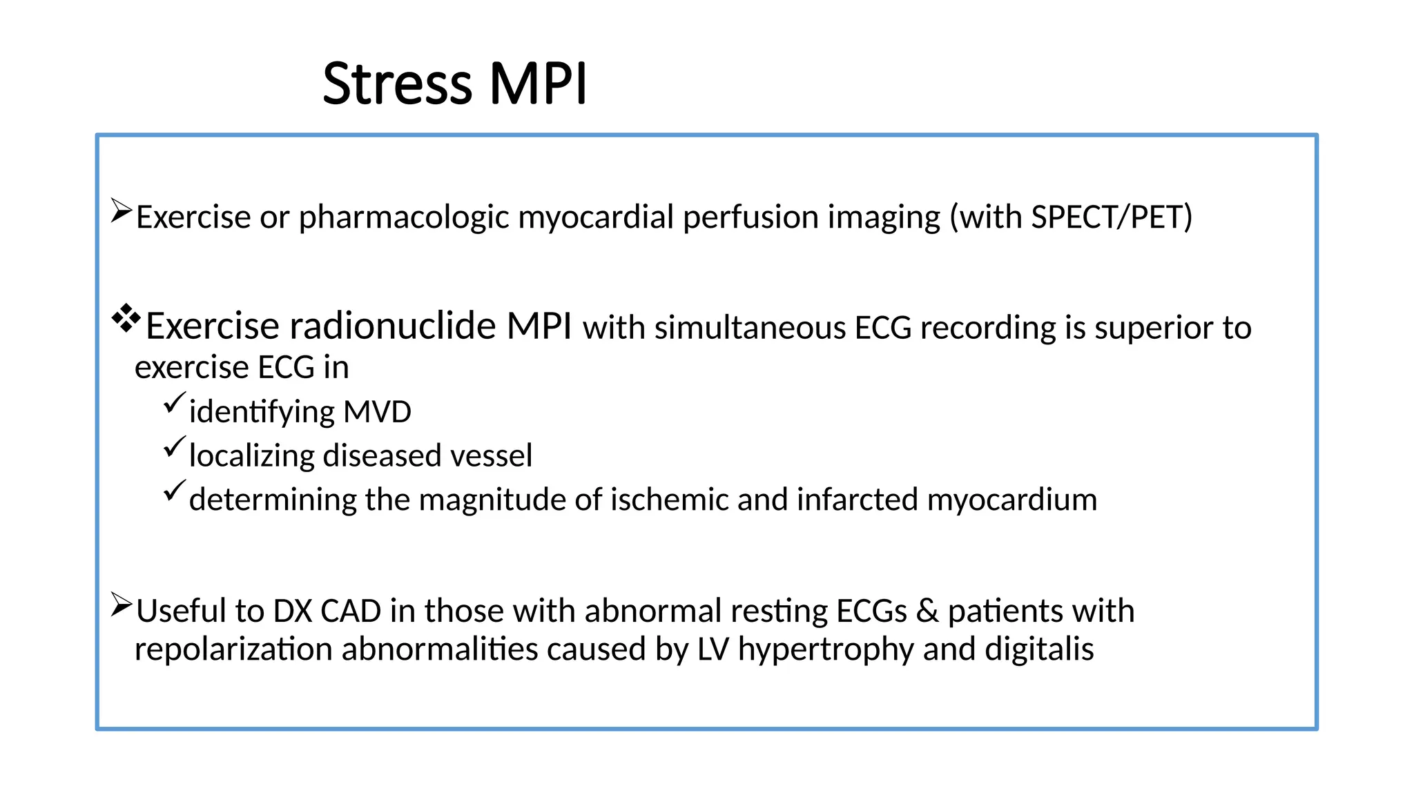
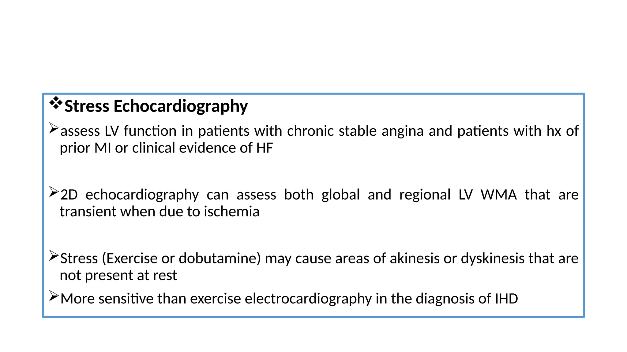
Chronic Coronary Syndrome
By Sisay Dadhi (IMR3)
Moderator: Dr. Seifu Baca (Internist , Cardiologist)
March, 2021
Outline
Introduction to CCS
Epidemiology
Pathophysiology of stable Angina Pectoris
Approach to patients with Angina/dyspnea and suspected CAD
Approach to Management
Angina without obstructive disease of epicardial artery
CCS in Specific circumstances
Introduction
The term stable CAD changed to CCS emphasizing the fact that the clinical
presentations of CAD can be categorized as either ACS or CCS
The spectrum of chronic coronary syndrome is broad and includes
 Coronary artery stenosis
 Prior MI/coronary revascularization &
 Asymptomatic ischemia
 Non-obstructive coronary disease
CCS is commonly caused by atheromatous plaque that obstructs or gradually narrow the
epicardial coronary arteries
Burden of IHD increasing even in LSE groups: Aging & ↑obesity and DM2
Pathobiological contributors of IHD
Not just epicardial stenosis!
Angina is a symptom for all cardiac ischemia regardless of the underlying mechanisms
Clinical Scenarios
oSuspected CAD and ‘stable’ angina symptoms, and/or dyspnea
oNew onset of HF or LV dysfunction and suspected CAD
oAsymptomatic/symptomatic patients <1yr after ACS or recent revascularization
oAsymptomatic/symptomatic >1 yr after initial diagnosis or revascularization
oThose with angina and suspected vasospastic or microvascular disease
oAsymptomatic subjects in whom CAD is detected at screening
All of these scenarios are classified as CCS but involve different risks
for future cardiovascular events(death or MI)
Epidemiology
It is estimated that 15.4 million Americans have IHD & 3.4 million aged ≥40 yrs have
angina pectoris
In 2013, IHD accounted for 46% of all deaths caused by CVD.
Despite decline in age-specific mortality over the past decades, IHD is the leading
cause of death worldwide.
WHO estimated by 2030, the global no of deaths from IHD will rise from 7.4 in 2012 to 9.2 million.
Lifetime risk for IHD
Major risk factors Men Women
Optimal 3.6% 1%
≥2 37.5% 18.3%
• A prospective cross-sectional, descriptive analysis done in Jan 2015
involving 6275 cardiac patients (6 referral hospital)
Stable Angina Pectoris
Results from myocardial ischemia w/c is caused by imbalance b/n
Myocardial O2 requirements
o Elevated with ↑ in HR , LV wall stress and contractility
Myocardial O2 supply
o Determined by coronary BF and arterial O2 content
Pathophysiology
Diagnostic and Management Approach: 6 Steps
Step 1: Symptoms and signs

History
Males constitute ~70% of all patients with angina pectoris
The typical patient with angina is a man >50yrs or a woman >60yrs
Typical angina described as heaviness/pressure/squeezing & rarely as frank pain
 typically lasts <10 min, and radiate to either shoulder and to both arms
 after exertion (exercise, sexual activity), heavy meal & waking up in morning
 relieved with rest or nitroglycerin, but may occur at rest
chest pain lasting for seconds is unlikely to be due to CAD
CVD symptoms or risk factors(family hx of CVD, dyslipidemia, DM, HTN, smoking)
Only 10- 15% of patients suspected of CAD present with typical angina!!!
Many patients report a fixed threshold for angina, which occurs predictably at a
certain level of activity, such as climbing two flights of stairs at a normal pace.
They are said to have Stable exertional Angina
Look for
Angina "Equivalents“ (dyspnea, syncope, fatigue, N/V)
Family hx of premature IHD (<55yr in 1st
degree M & <65yr in F relatives)
Angina as a “tip of an Iceberg!”
Canadian CV Society grading of angina pectoris
Physical Examination
Evidence of atherosclerotic disease at other sites, such as AAA, carotid bruits, and
diminished arterial pulses in the lower extremities
Evaluation for PAD should be done(ABI)
Examine the fundus
A 3rd
and/or 4th
heart sound, and, if previous infarction has impaired papillary muscle
function, an apical systolic murmur due to MR
Step 2: Comorbidities & other causes of symptoms
Before any testing considered, one must assess the patient’s general health,
comorbidities, and quality of life
If the pain is clearly non-anginal, other diagnostic testing may be indicated to
identify GI, pulmonary, or MSS causes of chest pain
Step 3: Basic testing
Basic testing in patients with suspected CAD
 laboratory biochemical testing
Resting ECG & CXR
possible ambulatory ECG monitoring
Resting Echocardiography
CBC, RFT, Lipid panel and DM2 screening in all patients suspected of CAD (Class I)
TFT in case of clinical suspicion of thyroid disorders
Biomarkers
Elevated troponin in CCS shown to have a graded relationship with the
subsequent risk of CV mortality and HF
 BNP increases in response to spontaneous or provoked ischemia
Though BNP/NT-pro BNP don’t have specificity to Dx CCS, higher values are
associated with risk for CV events in those at risk for & with established CAD
Resting & Ambulatory ECG
Normal in approximately 50% of patients with Hx of angina
Common abnormalities
nonspecific ST-T wave changes with or without abnormal Q waves
AV Conduction disturbances & LBBB
PVCs may occur, but have low sensitivity & specificity for detecting CAD
During angina episodes, abnormal ECG in 50% of cases who had normal ECG
Mostly STD
STE and normalization of resting STD or T-wave inversion may develop
Resting Echocardiography and MRI
Assessment of global LV function
RWM abnormalities is suggestive of CAD, whereas findings such as valve stenosis
or PHTN may suggest alternative diagnoses
↓ diastolic LV function reported to be early sign of ischemic myocardial
dysfunction and also be indicative of microvascular dysfunction
CMR provide information on cardiac anatomy and systolic function similar echo
Step 4: Ass’t of pre-test probability & clinical likelihood of CAD
The likelihood of obstructive CAD is influenced by the prevalence of the disease in the
population studied, as well as by clinical features of an individual patient
A simple model can be used to estimate PTP of obstructive CAD based on age, sex and
nature of symptoms
Studies have shown that outcomes in patients classified with the new PTP <15% is good
(annual risk of cardiovascular death or MI is <1%)
Diagnostic testing is most useful when the likelihood is intermediate
Different PTP scores
Diamond Forrester
Modified Diamond Forrester
CAD Consortium Basic & Clinical (Calculators)
Duke Clinical score
Acoustic CAD score
CAD Consortium Variables
Age
Sex
Chest pain
HTN
DM
Dyslipidemia
Smoking
CAC available? If available, CAC score
2016
Low CVD risk population
PTP of Obstructive CAD in 15, 815 symptomatic patients: a pooled analysis
Classic Diomond Forrester + Dyspnea
For patients with PTP of 5-15%---PTP modifiers
Step 5: Selecting appropriate testing
If the diagnosis of CAD is uncertain, establishing a diagnosis using non-invasive
functional imaging before treatment is reasonable
Direct invasive angiography (ICA) without further testing is a reasonable option
high clinical likelihood of CAD with symptoms unresponsive to medical therapy
typical angina at a low level of exercise and
initial clinical evaluation indicating high event risk
Guidelines recommend the use of either non invasive functional or anatomical
imaging using CTA as the initial test for diagnosing CAD
Functional non-invasive tests
Designed to detect myocardial ischemia & include
Exercise ECG testing
Wall motion abnormalities by stress echocardiography/CMR
Perfusion imaging by single-photon emission CT (SPECT) or PET
Myocardial contrast echocardiography or Contrast CMR
Ischemia can be provoked by exercise/pharmacological stressors (↑myocardial
work load or O2 demand)
Anatomical non-invasive evaluation
Anatomical non-invasive evaluation, by visualizing artery lumen and wall using IV
contrast: coronary CTA
Either non-invasive or invasive functional testing is recommended for further
evaluation of stenosis detected by coronary CTA or ICA, unless a very high-grade
(>90%) stenosis is detected via invasive angiography
Physiologic response to Exercise
Exercise ECG
Exercise ECG has inferior diagnostic performance compared with imaging tests
In selected patients to complement clinical evaluation for the ass’t of response to
medical therapy, exercise capacity, suspected arrhythmias and event risk (Class I)
Considered as alternative to Dx obstructive CAD if imaging tests aren’t available
(Class IIb)
Baseline ECG abnormalities interfering with ECG interpretation
LBBB, paced rhythm, WPWp, STD >1 mm or (LVH/’Digoxin/Hypokalemia) with ST-T changes
Modified Bruce protocol add 2 initial low level 3-min stages at
speed of 1.7 mph and grades 0 % and 5%, respectively
HR, BP should be monitored & ST changes and symptoms followed
Exercise ECG testing in healthy individual
Variable Normal response
Peak Heart Rate response 85% of maximal predicted value or (at least 120– 140 bpm)
Blood pressure Increase
ST changes Normal or Rapid up-sloping
Heart rate Recovery(HRR) > 12 bpm after 1 min rest, > 18 bpm in supine/sitting position, >22 after
2 min rest
Symptoms No significant symptoms
Rhythm changes Infrequent PVCs can be seen
Indications for Terminating the Exercise Test
Stress MPI
Exercise or pharmacologic myocardial perfusion imaging (with SPECT/PET)
Exercise radionuclide MPI with simultaneous ECG recording is superior to
exercise ECG in
identifying MVD
localizing diseased vessel
determining the magnitude of ischemic and infarcted myocardium
Useful to DX CAD in those with abnormal resting ECGs & patients with
repolarization abnormalities caused by LV hypertrophy and digitalis
MPI with Pharmacologic Vasodilator Stress
For patients unable to exercise, especially elderly and those with PAD, pulmonary
disease, arthritis, orthopedic problems, severe obesity, or a previous stroke
Pharmacologic vasodilator stress with dipyridamole or adenosine
Stress Echocardiography
assess LV function in patients with chronic stable angina and patients with hx of
prior MI or clinical evidence of HF
2D echocardiography can assess both global and regional LV WMA that are
transient when due to ischemia
Stress (Exercise or dobutamine) may cause areas of akinesis or dyskinesis that are
not present at rest
More sensitive than exercise electrocardiography in the diagnosis of IHD
Stress CMR
Cardiac magnetic resonance (CMR) stress testing is also evolving as an alternative
to radionuclide, PET, or echocardiographic stress imaging
CMR stress testing with dobutamine can be used to assess RWMA accompanying
ischemia, as well as myocardial perfusion and viability
Cardiac CT
A noninvasive approach to imaging atherosclerosis and its consequences
Non-contrast CT is highly sensitive for detecting coronary calcification
Also provide angiography of coronary aa & quantification of ventricular function
CCTA is reasonable in symptomatic patients at intermediate risk, particularly, in those
with indeterminate stress testing
Has limited accuracy in those with uncontrolled tachycardia, heavy calcification, or in
the region of prior stenting
The impact of clinical likelihood on the selection of a diagnostic
test
Coronary CTA is the preferred test in patients with a lower clinical likelihood of
CAD, no previous diagnosis of CAD
The non-invasive functional tests for ischemia typically have better rule-in power
Functional non-invasive testing may be preferred in those at the higher end of
the range of clinical likelihood if revascularization is likely or the patient has
previously diagnosed CAD
Invasive testing
Invasive anatomical or functional(FFR) testing
ICA is only necessary when CAD is suspected & inconclusive non-invasive testing.
High clinical likelihood of CAD & symptoms unresponsive to medication or typical
angina at low level of exercise and initial clinical evaluation indicates a high event
risk, early ICA is reasonable to identify lesions amenable to revascularization
Integration of ICA with FFR has been shown to result in changes to the management
strategies of 30- 50% of patients undergoing elective ICA
The rate of major complications associated with routine femoral diagnostic
catheterization—mainly bleeding requiring blood transfusions—is still 0.5- 2%
Indications of ICA
Chronic stable angina severely symptomatic despite medical therapy
Patients with troublesome symptoms that present diagnostic difficulties in whom
there is a need to confirm or rule out the diagnosis of IHD
Patients with known or possible AP who have survived cardiac arrest
Repeatedly admitted for possible ACS
Angina in AS, HCMP
M >45 and F >55 years who will undergo cardiac surgery for any reason
Post MI if there is recurrence of angina, HF, frequent PVCs
If non-atherosclerotic cause of angina suspected
Chest discomfort suggestive of angina, but non-invasive tests are non-diagnostic
Findings
In patients with chronic stable angina
20% SVD
20% DVD
35% TVD
10% obstruction of the left main coronary artery
15%, no critical obstruction
Anatomically significant CAD is when luminal diameter narrowing is >70%
Limitations of Angiography
It is not a reliable indicator of the functional significance of stenosis
Underestimation of the severity and extent of atherosclerosis
Its inability to identify which coronary lesions can be considered to be at high
risk, or vulnerable, for future events, such as MI or sudden cardiac death
It is not helpful in predicting sites of subsequent plaque rupture or erosion
Advanced structural coronary imaging
Intravascular US:- vulnerability of coronary atheroma to rupture
Intravascular optical coherence tomography (OCT):- measures the fibrous cap
thickness and assess coronary dissection
Step 6: Assessment of event risk
Risk stratification identify those at high event risk who will benefit from
revascularization
All should undergo CV event risk stratification using clinical evaluation, LV
function with resting Echo and non-invasive ass’t of ischemia or coronary
anatomy
High event risk is defined as mortality rate >3%/year and low event risk <1%/year
Stress Testing Identifying High Risk Groups
.
Definitions of high event risk for different test modalities in established CCS
Management of CCS
Lifestyle modification: ↓ risk of subsequent CV events & mortality!!
Medical management
Comprehensive management of CCS has five aspects
Rx of associated diseases that can worsen angina and ischemia
Reduction of risk factors
Pharmacologic & non-pharmacologic interventions for secondary prevention
 Management of angina
Revascularization by PCI/CABG, when indicated
Aim: to reduce angina and exercise-induced ischemia& prevent CV events
Treatment of Associated Diseases
Several common medical conditions that can increase myocardial O2 demand or
reduce O2 delivery may contribute to the onset of new angina pectoris or
exacerbation of previously stable angina
Anemia, occult thyrotoxicosis, fever, infections and tachycardia
Treatment of Risk Factors
Search for & treat coronary risk factors
Hypertension
Diabetes mellitus
Weight loss in Obese
Cessation of smoking
Aggressive control of dyslipidemia
Anti-anginal drugs
Optimal treatment: controls symptoms and prevents cardiac events in CCS
Response to initial antianginal therapy should be reassessed after 2-4 wk Rx initiation
Nitrates
 Systemic venodilation + ↓LVEDP and volume…..reduce wall tension & O2 requirements
 Dilation of epicardial coronary vessels
 Increased blood flow in collateral vessels
LAN (nitroglycerin, isosorbide dinitrate) is 2nd
line Rx for angina when BB/non-DHP CCB is c/i or
insufficient to control symptoms (nitrate tolerance due to attenuation of vascular effect of nitrates)
Beta-blockers
Reduce oxygen demand by decreasing HR and myocardial contractility
The dose should be adjusted to limit the heart rate to 55-60
Abrupt withdrawal after prolonged use can result in increased total ischemic
activity in patients with chronic stable angina
Combination of a beta-blocker with a nitrate attenuates the reflex tachycardia
BB can reduce mortality & re-infarction after MI
44,708 patients
31% had prior MI
27% had documented CAD without
MI and
42% had CAD risk factors only
Primary outcome: composite of CV
death, nonfatal MI or nonfatal stroke
Published in 2014, Involving 15,603 patients
MI cohort….4772
CAD cohort…..7804
Only risk factors…..2101
Primary outcome: composite of the 1st
occurrence of
nonfatal MI, stroke or death from CV causes
Calcium channel blockers
CCBs improve symptoms, but have not been shown to reduce major morbidity or
mortality in patients with CCS
Verapamil has a large range of approved indications, including all varieties of
angina (effort, vasospastic, and unstable), SVT and HTN
Diltiazem(low-side effect) has advantages vs verapamil in the Rx of effort angina
Amlodipine + BB: additive effect on blood supply and myocardial O2 demands
Short-acting dihydropyridines be avoided b/c it can precipitate infarction,
particularly in the absence of concomitant BB therapy
Ivabradine
Is a specific and selective inhibitor of the If ion channel, the principal determinant
of the sino atrial node pacemaker current.
It reduces the spontaneous firing rate of sino atrial pacemaker cells and thus
slows the heart rate through a mechanism that is not associated with negative
inotropic effects.
Management of uncontrolled Angina
Despite conventional antianginal drugs & revascularization some patients
continue to have symptoms, many of whom are not a candidate for
revascularization
Unsuitable coronary anatomy (eg. diffuse CAD)
≥1 prior PCI/CABG
Lack of vascular conduits
Severely impaired LV function in those with prior PCI/CABG
Comorbidities that ↑ peri-/postop mortality (stroke, complicated DM & CKD)
Newer therapies for angina pectoris
Ranolazine….500-1000 mg po bid
Prevent both Ca2+ overload & subsequent increase in diastolic tension by Rapidly
inhibit Late inward Na+ channel
Also inhibit fatty acid oxidation(Switch to glucose oxidation)
its anti-ischemic effect is achieved without a meaningful change in HR/BP
↓angina frequency & nitroglycerin use when used with a BB or CCB
Study show statistically significant 0.3% reduction in HbA1c(pronounced in Diabetics)
Side effects: dizziness, nausea, constipation & dose-dependent QT prolongation
Trimetazidine
inhibit fatty acid metabolism and reduce the frequency of angina without hemodynamic effects
in patients with chronic stable angina
Trimetazidine (35mg b.i.d) + BB improved effort-induced myocardial ischemia
TRIMPOL II trial & a metanalysis: with other anti-anginal it was associated with
smaller weekly mean number of angina attacks
lower weekly nitroglycerin use
longer time to 1 mm ST-segment depression
longer exercise duration at peak exercise
Contraindicated in PD & other movement disorders
Nicorandil
Nicorandil (20mg po bid) dilates peripheral & coronary vessels acting on ATP-
sensitive K+ channels and possesses a nitrate moiety
mimic a natural process of ischemic preconditioning, protecting the heart from
subsequent ischemic attacks
Side effects include nausea/vomiting and severe oral & intestinal mucosal
ulcerations
Antithrombotic therapy in CCS
Antiplatelet drugs
Low-dose aspirin
irreversibly inhibit COX-1 and thus prevent thromboxane production
Current evidence supports 75 - 100 mg daily for the prevention of ischemic
events in CAD patients with or without a hx of MI
P2Y12 inhibitors
Block the platelet P2Y12 receptor, which plays a key role in platelet activation and
arterial thrombus formation
Clopidogrel & prasugrel: irreversibly block P2Y12 via active metabolites
Ticagrelor reversible P2Y12 inhibitor that doesn’t need metabolic activation
Options of DAPT combined with ASA in those with high/moderate risk of
ischemic events who don’t have high bleeding risk
High ischemic risk: diffuse MVD + one of the f/g: DM on drugs, recurrent MI, PAD
or CKD with eGFR <60
Moderate ischemic risk: at least one of the f/g: MVd/diffuse CAD, DM on drugs,
recurrent MI, PAD, HF, or CKD with eGFR <60
DAPT post-PCI
After PCI for stable angina, 6 months of DAPT achieves the optimum balance
of efficacy and safety in most patients.
Premature discontinuation of a P2Y12I is associated with increased risk of
stent thrombosis and is discouraged.
Shorter duration of DAPT be considered in those at high risk of life-
threatening bleeding in view of very-low risk of stent thrombosis after 1-3
months(DES)
Anticoagulation in Afib
Anticoagulant drugs in sinus rhythm
Combination of antiplatelet therapy and standard anticoagulant doses of warfarin
or apixaban for secondary prevention after ACS was associated with an
unfavorable balance of efficacy and bleeding (APPRAISE-2 trial)
Rivaroxaban 2.5 mg b.i.d vs Placebo ↓ the composite of MI, stroke/CV death in
stabilized patients treated predominantly with aspirin and clopidogrel following
ACS with increased bleeding risk (COMPASS & ATLAS-TIMI 51 trial)
Greater absolute RR seen in higher-risk pts with DM, PAD, moderate CKD & current
smokers
Antithrombotic in post-PCI patients with AF/other indication for OAC
Proton pump inhibitors
Proton pump inhibitors reduce the risk of GIB in those treated with antiplatelet.
PPI that inhibit CYP2C19 (omeprazole and esomeprazole) may reduce the
pharmacodynamic response to clopidogrel.
Statins and other lipid-lowering drugs
Goal: lower LDL-C <55 mg/dL or at least reduce by 50% from baseline
Role of colchicine
Despite widespread use of statin and antithrombotics patient with CAD continues
to be at risk of CV death, MI & need for revascularization
Pts with Hx of ACS remains at increased risk of recurrent CV events (20% at 3 years)
Partly due to residual inflammation at a culprit/non-culprit sites
Inflammation plays a central in atheroma generation, plaque instability & rupture
Endothelial injury trigerred by LDL lead to release of proinflammatory cytokines
w/c reduce plaque stability
Colchicine has an anti-inflammatory effect
Interfere with inflammasome w/c is
responsible for cytokines generation
Prevent cell mitosis
LoDoCo2 trial (2020)…0.5mg daily
Enroll 5,522 patients with the median ffp of
28.6 months
Inclusion criteria:
• CAD on invasive/CT angio
• CAC >400
• Clinically stable condition ≥6 months
Exclusion criteria:
GFR <30, Severe HF, Severe VHD
Primary endpoint: composite of cv death,
MI, ischemic stroke or revascularization
Result: reduce primary endpoint
significantly, shown statistically significant
reduction in risk of MI & revascularization
and borderline CV mortality benefit
RAS blockers
ACEI can reduce mortality, MI, stroke and HF among patients with LV dysfunction and
high-risk diabetes
should be given to CCS patients with HTN, EF 40% or DM unless contraindicated
ACEI Rx in CCS patients without HF or high CV risk is not generally recommended,
unless needed to meet BP targets (EUROPEAN, HOPE & PEACE trial)
Spironolactone/eplerenone: post-MI who are already on ACEI and BB, have EF 35% and
have either diabetes or HF
Revascularization
Revascularization plays a central role in the management of CCS on top of
medical Rx, but always as an adjunct without supplanting it
The two objectives of revascularization:-
Symptom relief in patients with angina and
Improvement of prognosis
Considered in the presence of
Unstable disease, Intractable symptoms, severe ischemia or high-risk
coronary anatomy, Diabetes & Impaired LV function
Other features, in addition to lesion severity, also influence likelihood of
success
Vessel size, extent of calcification, tortuosity & relationships to side branches
Medical therapy vs Revascularization???
5179 patients with moderate/severe
ischemia followed for 3.2 years
Initial invasive strategy (angio and revasc
when feasible + medical Rx) VS medical
therapy alone
Exclusion: EF < 35%, LM stenosis >50%,
anatomy unsuitable to PCI/CABG, non-
obstructive CAD, class III/IV HF
Primary endpoint: Composite of death
from CV causes, MI or hospitalization
for UA, HF or resuscitated cardiac
arrest
Secondary outcome was death from
CV causes or MI
RCT, enrolling 2287 patients over
median of 4.6 years
2/3 of patients had 2 or more vessel ds
Primary endpoint: a composite of
death from any cause and nonfatal MI
over the follow up year
Inclusion criteria: 70% stenosis in at least
one prox epicardial a & evidence of
ischemia (exercise/pharmacologic) or one
coronary stenosis of at least 80% and
classic angina without provocation
Exclusion criteria: persistent class IV
angina, shock, EF <30% and coronary
anatomy not suitable for PCI
What is an annual mortality with Medical therapy?
PCI- Indications
Persistent angina despite medical therapy, accompanied by evidence of ischemia on stress test
More effective than medical therapy for the relief of angina
Primary success: increase in lumen >20% to a residual obstruction <50% with relief of angina,
is achieved in >95% of cases
Recurrent stenosis occurs in ~20% of cases within 6 months of BMS
Angina recur within 6 months in 10% of cases
Those with at least one stenosis functionally significant (FFR ≤0.80) assigned to
FFR-guided PCI plus medical therapy vs medical therapy alone
The primary end point was a composite of death, MI or urgent revascularization
Recruitment was halted prematurely after enrollment of 1220 patients because
of a significant between-group difference in the percentage of patients who had a
primary endpoint event but no difference in risk of MI & death from any cause.
CABG
The technical goal of bypass surgery is to achieve, whenever possible, complete
revascularization of significant proximal stenosis.
It has been documented to prolong survival, relieve angina and improve QOL in
specific subgroups of patients with CAD (STICH trial)
Patient selection
The decision to perform PCI Vs CABG???
coronary anatomy
 LV function
comorbidities
risk associated with either revascularization and patient preference
SYNTAX, BARI 2D, FREEDOM trial
Heart Team
PCI VS CABG…..Angiographic Syntax Score
Syntax trial: 1800 patients with 3VD or LM CAD undergo CABG or PCI
Generally, CABG preferred
Left main
Three-vessel CAD
LV systolic dysfunction
DM patients
Patients with stabilized symptoms <1 year after an ACS or with recent revascularization
It is recommended that at least two visits in the first year of follow-up
In those who had LV syst dysfunction before the revascularization or after ACS,
reassessment of LV function must be considered 2- 3 months after intervention
Patients >1 year after initial diagnosis or revascularization
It may be beneficial to assess LV function, valve status and cardiac dimensions in
apparently asymptomatic patients every 3 - 5 years
In cases of unexplained reduction in systolic LV function, especially if RWMA,
imaging of coronary artery anatomy is recommended
It may be beneficial to assess non-invasively for silent ischemia in an apparently
asymptomatic patient every 3 – 5 years
Angina without obstructive epicardial CAD
Low diagnostic yield of ICA can be explained by the presence of
Mild/moderate stenosis or diffuse coronary narrowing, with under-estimated
functional significance
Disorders affecting microcirculation that escape the resolution of angiographic and
Dynamic stenosis of caused by coronary spasm that are not evident during CTA/ICA
Vasospastic Angina
Clinical entity characterized by episodes of rest angina that respond to short-acting
nitrates and attributable to coronary artery vasospasm
Patient commonly develop STE but sometimes STD can occur.
Usually occurs at sites of atherosclerosis but may occur within normal vessels.
Cigarette smoking is a major risk factor
Common triggers
drugs like cocaine, alcohol, sumatriptan and amphetamines
at the time of PCI
Food-born botulism
Mg deficiency
Pathogenesis
Poorly undestood
Vascular smooth muscle hyperreactivity
Autonomic Nervous system: imbalance of vagal tone(angina commonly from midnight-morning) &
sympathetic tone(enhanced alpha R stimulation)
Endothelial dysfunction(eg. Increased VC endothelin level)
Acetylcholine injection causes vasodilation in healthy arteries but vasoconstriction
ensues in atherosclerotic ones
Patient had heightened vasoconstrictor response to Ach & enhanced response to
vasodilator effects of nitrates.
Clinical presentation
Episodes of angina is typically gradual in onset and offset, lasting 5-15 minutes.
If prolonged it may cause MI
Can have associated sweating, palpitation or dizziness
Clues to differentiate from stable angina
Younger patient (<50 yr) with fewer CV risk factors
Other vasospastic disorders: Raynaud’s ds, Migraine
Hx of drug abuse
Exercise doesn’t usually provoke spasm(only 20% of pts develop STE by exercise test)
Diagnosis
History is very important
Transient ST segment changes during spontaneous episode and subsequently
found without high-grade obstruction on angiography
Ambulatory ECG monitoring……..Angiography
Coronary Artery Vasospastic Disorders Summit(COVADIS)
criteria
Treatment
Control of cardiovascular risk factors and lifestyle changes.
CCBs and long-acting nitrates are the treatment of choice
Nifedipine has been shown to be effective in reducing coronary spasm associated
with stent implantation
Microvascular angina(MVA)
Patients with MVA typically have exercise-related angina, evidence of ischemia on
non-invasive tests, and either no stenosis or mild-to-moderate stenosis (40-60%),
revealed by ICA/CTA, that are functionally non-relevant
Regional LV wall motion abnormalities rarely develop during exercise or stress in
patients with microvascular angina
Pathogenesis ……2 mechanisms
1) Microvascular dysfunction
Ischemia is limited to subendocardium
3 mechanisms
1. Endothelial dysfunction impaired microcirculatory conductance
2. Abnormal cardiac adnergic tone (arterial dysregulation)
↓VD or cause paradoxical VC in response to pharmacologic agents(adenosine, Ach) &
exercise Microvascular Spasm
This 2 process can involve bronchi or cerebral microcirculation
3. Occult small vessel CAD
2) Enhanced pain sensitivity
Can’t demonstrate ischemia evidence on exercise or dobutamine testing
Clinical feature
Chronic recurrent predominantly exertional angina & may occur from midnight to
early morning
Response to nitroglycerin may not be effective b/c its VD effect in small arteriole is
less
Diabetic & female at risk
Investigation
Resting ECG normal between episode and absent ECG changes during episode
can’t r/o
Unlike VSA, STE not a feature
Perfusion defects on MPI or RWMA on dobutamine Echo may be absent
Angiography should show normal epicardial aa( <30% stenosis)
Diagnosis
Hx + non-invasive testing + non-obstructive CAD
If this is inconclusive, measure CFR or index of microvascular resistance(IMR)
CFR = CBF with Adenosine/resting CBF (normal 2.5 - 5)
If there is no significant epicardial CAD, shows degree of resistance to BF in
microcirculation
Measured with Doppler wire or non invasively by echo(LAD flow) or PET
IMR is measured invasively with Doppler wire (catheter)
CFR <2 & IMR ≥25 indicates abnormal microcirculation function
Risk stratification
Presence of microcirculatory dysfunction in CCS entails a worse prognosis
Microcirculatory dysfunction precedes the development of epicardial lesions,
particularly in women is associated with impaired outcomes.
Diabetics without obstructive disease but with abnormal CFR have similarly poor
long-term prognosis as those with obstructive epicardial disease
Treatment
In those with abnormal CFR <2.0/IMR 25 units and a -ve Ach provocation test, BB,
ACE inhibitors, and statins, along with lifestyle changes are indicated
ECG changes and angina in response to Ach testing but without severe epicardial
VC (suggestive of microvascular spasm) may be treated like vasospastic angina
Specific circumstances
Hypertension
A recent Meta-analysis
suggests that for every 10
mmHg reduction in SBP,
CAD can be reduced by
17%
VHD(including Transcatheter AV implantation)
Diabetes mellitus
Diabetic patients had 2-fold increased risk of CAD
Patients with DM & CAD are considered to be at very high risk; so, LDL should be
lowered to <70 mg/d or by 50% if the baseline LDL is between 70 - 35 mg/dL
Target HgA1C is recommended to be <7%
Management of Refractory Angina
Refractory angina refers to long-lasting symptoms (3 months) in the presence of
obstructive CAD, which cannot be controlled by escalating medical therapy with
the use of second- and third-line pharmacological agents, CABG/PCI
Patients with refractory angina best treated in dedicated ‘angina clinics’ by MDTs
experienced in selecting the most suitable therapeutic based on an accurate
diagnosis of the mechanisms of the pain syndrome
Unconventional therapies for refractory Angina
Enhanced External counterpulsation(EECP)
is a technique that increases arterial blood pressure and retrograde aortic blood
flow during diastole (diastolic augmentation)
Cuffs are wrapped around the patient’s calves, thigh and pelvis and, using
compressed air, sequential pressure (up to 300 mmHg) is applied in early diastole
to propel blood back to the heart
Increase exercise capacity
Decrease angina episodes & use of nitroglycerin
In one study MACE-free survival was 70 percent
Neuromodulatoin (SC stimulation or Sympathectomy)
Spinal cord stimulation
stimulation at T1-T2 is another approach to management of refractory angina
Spinal cord stimulation was originally developed to treat neurogenic pain
Its mechanism of action is poorly understood
May have effects on angina by suppressing capacity of cardiac neurons to
generate activity during myocardial ischemia
May ↓sympathetic tone or redistribute BF from nonischemic to ischemic areas
Coronary sinus reducing device
Balloon catheter is inserted into the coronary sinus percutaneously & the balloon
expanded to implant an hourglass-shaped metal mesh that leads, over time, to
partial obstruction of the coronary sinus
postulated that development of an upstream pressure gradient favors perfusion of ischemic
territories
Transmyocardial laser revascularization
References
Brauwald’s Heart Disease, 11th
Edition
Harrison’s Principle of Internal Medicine, 20th
Edition
2019 ESC guideline for the diagnosis and management of CCS
Uptodate, 2018
Internate sources
Ulfaadhaa!!!

Chronic Coronary Syndrome CCS Sisay.pptx

  • 1.
    Chronic Coronary Syndrome BySisay Dadhi (IMR3) Moderator: Dr. Seifu Baca (Internist , Cardiologist) March, 2021
  • 2.
    Outline Introduction to CCS Epidemiology Pathophysiologyof stable Angina Pectoris Approach to patients with Angina/dyspnea and suspected CAD Approach to Management Angina without obstructive disease of epicardial artery CCS in Specific circumstances
  • 3.
    Introduction The term stableCAD changed to CCS emphasizing the fact that the clinical presentations of CAD can be categorized as either ACS or CCS The spectrum of chronic coronary syndrome is broad and includes  Coronary artery stenosis  Prior MI/coronary revascularization &  Asymptomatic ischemia  Non-obstructive coronary disease CCS is commonly caused by atheromatous plaque that obstructs or gradually narrow the epicardial coronary arteries Burden of IHD increasing even in LSE groups: Aging & ↑obesity and DM2
  • 4.
    Pathobiological contributors ofIHD Not just epicardial stenosis! Angina is a symptom for all cardiac ischemia regardless of the underlying mechanisms
  • 5.
    Clinical Scenarios oSuspected CADand ‘stable’ angina symptoms, and/or dyspnea oNew onset of HF or LV dysfunction and suspected CAD oAsymptomatic/symptomatic patients <1yr after ACS or recent revascularization oAsymptomatic/symptomatic >1 yr after initial diagnosis or revascularization oThose with angina and suspected vasospastic or microvascular disease oAsymptomatic subjects in whom CAD is detected at screening All of these scenarios are classified as CCS but involve different risks for future cardiovascular events(death or MI)
  • 6.
    Epidemiology It is estimatedthat 15.4 million Americans have IHD & 3.4 million aged ≥40 yrs have angina pectoris In 2013, IHD accounted for 46% of all deaths caused by CVD. Despite decline in age-specific mortality over the past decades, IHD is the leading cause of death worldwide. WHO estimated by 2030, the global no of deaths from IHD will rise from 7.4 in 2012 to 9.2 million. Lifetime risk for IHD Major risk factors Men Women Optimal 3.6% 1% ≥2 37.5% 18.3%
  • 7.
    • A prospectivecross-sectional, descriptive analysis done in Jan 2015 involving 6275 cardiac patients (6 referral hospital)
  • 8.
    Stable Angina Pectoris Resultsfrom myocardial ischemia w/c is caused by imbalance b/n Myocardial O2 requirements o Elevated with ↑ in HR , LV wall stress and contractility Myocardial O2 supply o Determined by coronary BF and arterial O2 content
  • 9.
  • 10.
    Diagnostic and ManagementApproach: 6 Steps
  • 11.
    Step 1: Symptomsand signs  History Males constitute ~70% of all patients with angina pectoris The typical patient with angina is a man >50yrs or a woman >60yrs Typical angina described as heaviness/pressure/squeezing & rarely as frank pain  typically lasts <10 min, and radiate to either shoulder and to both arms  after exertion (exercise, sexual activity), heavy meal & waking up in morning  relieved with rest or nitroglycerin, but may occur at rest chest pain lasting for seconds is unlikely to be due to CAD CVD symptoms or risk factors(family hx of CVD, dyslipidemia, DM, HTN, smoking)
  • 12.
    Only 10- 15%of patients suspected of CAD present with typical angina!!!
  • 13.
    Many patients reporta fixed threshold for angina, which occurs predictably at a certain level of activity, such as climbing two flights of stairs at a normal pace. They are said to have Stable exertional Angina Look for Angina "Equivalents“ (dyspnea, syncope, fatigue, N/V) Family hx of premature IHD (<55yr in 1st degree M & <65yr in F relatives)
  • 14.
    Angina as a“tip of an Iceberg!”
  • 15.
    Canadian CV Societygrading of angina pectoris
  • 16.
    Physical Examination Evidence ofatherosclerotic disease at other sites, such as AAA, carotid bruits, and diminished arterial pulses in the lower extremities Evaluation for PAD should be done(ABI) Examine the fundus A 3rd and/or 4th heart sound, and, if previous infarction has impaired papillary muscle function, an apical systolic murmur due to MR
  • 17.
    Step 2: Comorbidities& other causes of symptoms Before any testing considered, one must assess the patient’s general health, comorbidities, and quality of life If the pain is clearly non-anginal, other diagnostic testing may be indicated to identify GI, pulmonary, or MSS causes of chest pain
  • 18.
    Step 3: Basictesting Basic testing in patients with suspected CAD  laboratory biochemical testing Resting ECG & CXR possible ambulatory ECG monitoring Resting Echocardiography
  • 19.
    CBC, RFT, Lipidpanel and DM2 screening in all patients suspected of CAD (Class I) TFT in case of clinical suspicion of thyroid disorders
  • 20.
    Biomarkers Elevated troponin inCCS shown to have a graded relationship with the subsequent risk of CV mortality and HF  BNP increases in response to spontaneous or provoked ischemia Though BNP/NT-pro BNP don’t have specificity to Dx CCS, higher values are associated with risk for CV events in those at risk for & with established CAD
  • 21.
    Resting & AmbulatoryECG Normal in approximately 50% of patients with Hx of angina Common abnormalities nonspecific ST-T wave changes with or without abnormal Q waves AV Conduction disturbances & LBBB PVCs may occur, but have low sensitivity & specificity for detecting CAD During angina episodes, abnormal ECG in 50% of cases who had normal ECG Mostly STD STE and normalization of resting STD or T-wave inversion may develop
  • 23.
    Resting Echocardiography andMRI Assessment of global LV function RWM abnormalities is suggestive of CAD, whereas findings such as valve stenosis or PHTN may suggest alternative diagnoses ↓ diastolic LV function reported to be early sign of ischemic myocardial dysfunction and also be indicative of microvascular dysfunction CMR provide information on cardiac anatomy and systolic function similar echo
  • 25.
    Step 4: Ass’tof pre-test probability & clinical likelihood of CAD The likelihood of obstructive CAD is influenced by the prevalence of the disease in the population studied, as well as by clinical features of an individual patient A simple model can be used to estimate PTP of obstructive CAD based on age, sex and nature of symptoms Studies have shown that outcomes in patients classified with the new PTP <15% is good (annual risk of cardiovascular death or MI is <1%) Diagnostic testing is most useful when the likelihood is intermediate
  • 26.
    Different PTP scores DiamondForrester Modified Diamond Forrester CAD Consortium Basic & Clinical (Calculators) Duke Clinical score Acoustic CAD score CAD Consortium Variables Age Sex Chest pain HTN DM Dyslipidemia Smoking CAC available? If available, CAC score
  • 27.
  • 28.
    Low CVD riskpopulation PTP of Obstructive CAD in 15, 815 symptomatic patients: a pooled analysis Classic Diomond Forrester + Dyspnea
  • 29.
    For patients withPTP of 5-15%---PTP modifiers
  • 30.
    Step 5: Selectingappropriate testing If the diagnosis of CAD is uncertain, establishing a diagnosis using non-invasive functional imaging before treatment is reasonable Direct invasive angiography (ICA) without further testing is a reasonable option high clinical likelihood of CAD with symptoms unresponsive to medical therapy typical angina at a low level of exercise and initial clinical evaluation indicating high event risk Guidelines recommend the use of either non invasive functional or anatomical imaging using CTA as the initial test for diagnosing CAD
  • 32.
    Functional non-invasive tests Designedto detect myocardial ischemia & include Exercise ECG testing Wall motion abnormalities by stress echocardiography/CMR Perfusion imaging by single-photon emission CT (SPECT) or PET Myocardial contrast echocardiography or Contrast CMR Ischemia can be provoked by exercise/pharmacological stressors (↑myocardial work load or O2 demand)
  • 33.
    Anatomical non-invasive evaluation Anatomicalnon-invasive evaluation, by visualizing artery lumen and wall using IV contrast: coronary CTA Either non-invasive or invasive functional testing is recommended for further evaluation of stenosis detected by coronary CTA or ICA, unless a very high-grade (>90%) stenosis is detected via invasive angiography
  • 34.
  • 35.
    Exercise ECG Exercise ECGhas inferior diagnostic performance compared with imaging tests In selected patients to complement clinical evaluation for the ass’t of response to medical therapy, exercise capacity, suspected arrhythmias and event risk (Class I) Considered as alternative to Dx obstructive CAD if imaging tests aren’t available (Class IIb) Baseline ECG abnormalities interfering with ECG interpretation LBBB, paced rhythm, WPWp, STD >1 mm or (LVH/’Digoxin/Hypokalemia) with ST-T changes
  • 37.
    Modified Bruce protocoladd 2 initial low level 3-min stages at speed of 1.7 mph and grades 0 % and 5%, respectively HR, BP should be monitored & ST changes and symptoms followed
  • 38.
    Exercise ECG testingin healthy individual Variable Normal response Peak Heart Rate response 85% of maximal predicted value or (at least 120– 140 bpm) Blood pressure Increase ST changes Normal or Rapid up-sloping Heart rate Recovery(HRR) > 12 bpm after 1 min rest, > 18 bpm in supine/sitting position, >22 after 2 min rest Symptoms No significant symptoms Rhythm changes Infrequent PVCs can be seen
  • 39.
    Indications for Terminatingthe Exercise Test
  • 41.
    Stress MPI Exercise orpharmacologic myocardial perfusion imaging (with SPECT/PET) Exercise radionuclide MPI with simultaneous ECG recording is superior to exercise ECG in identifying MVD localizing diseased vessel determining the magnitude of ischemic and infarcted myocardium Useful to DX CAD in those with abnormal resting ECGs & patients with repolarization abnormalities caused by LV hypertrophy and digitalis
  • 42.
    MPI with PharmacologicVasodilator Stress For patients unable to exercise, especially elderly and those with PAD, pulmonary disease, arthritis, orthopedic problems, severe obesity, or a previous stroke Pharmacologic vasodilator stress with dipyridamole or adenosine
  • 43.
    Stress Echocardiography assess LVfunction in patients with chronic stable angina and patients with hx of prior MI or clinical evidence of HF 2D echocardiography can assess both global and regional LV WMA that are transient when due to ischemia Stress (Exercise or dobutamine) may cause areas of akinesis or dyskinesis that are not present at rest More sensitive than exercise electrocardiography in the diagnosis of IHD
  • 44.
    Stress CMR Cardiac magneticresonance (CMR) stress testing is also evolving as an alternative to radionuclide, PET, or echocardiographic stress imaging CMR stress testing with dobutamine can be used to assess RWMA accompanying ischemia, as well as myocardial perfusion and viability
  • 45.
    Cardiac CT A noninvasiveapproach to imaging atherosclerosis and its consequences Non-contrast CT is highly sensitive for detecting coronary calcification Also provide angiography of coronary aa & quantification of ventricular function CCTA is reasonable in symptomatic patients at intermediate risk, particularly, in those with indeterminate stress testing Has limited accuracy in those with uncontrolled tachycardia, heavy calcification, or in the region of prior stenting
  • 46.
    The impact ofclinical likelihood on the selection of a diagnostic test Coronary CTA is the preferred test in patients with a lower clinical likelihood of CAD, no previous diagnosis of CAD The non-invasive functional tests for ischemia typically have better rule-in power Functional non-invasive testing may be preferred in those at the higher end of the range of clinical likelihood if revascularization is likely or the patient has previously diagnosed CAD
  • 47.
    Invasive testing Invasive anatomicalor functional(FFR) testing ICA is only necessary when CAD is suspected & inconclusive non-invasive testing. High clinical likelihood of CAD & symptoms unresponsive to medication or typical angina at low level of exercise and initial clinical evaluation indicates a high event risk, early ICA is reasonable to identify lesions amenable to revascularization Integration of ICA with FFR has been shown to result in changes to the management strategies of 30- 50% of patients undergoing elective ICA The rate of major complications associated with routine femoral diagnostic catheterization—mainly bleeding requiring blood transfusions—is still 0.5- 2%
  • 48.
    Indications of ICA Chronicstable angina severely symptomatic despite medical therapy Patients with troublesome symptoms that present diagnostic difficulties in whom there is a need to confirm or rule out the diagnosis of IHD Patients with known or possible AP who have survived cardiac arrest Repeatedly admitted for possible ACS
  • 49.
    Angina in AS,HCMP M >45 and F >55 years who will undergo cardiac surgery for any reason Post MI if there is recurrence of angina, HF, frequent PVCs If non-atherosclerotic cause of angina suspected Chest discomfort suggestive of angina, but non-invasive tests are non-diagnostic
  • 50.
    Findings In patients withchronic stable angina 20% SVD 20% DVD 35% TVD 10% obstruction of the left main coronary artery 15%, no critical obstruction Anatomically significant CAD is when luminal diameter narrowing is >70%
  • 51.
    Limitations of Angiography Itis not a reliable indicator of the functional significance of stenosis Underestimation of the severity and extent of atherosclerosis Its inability to identify which coronary lesions can be considered to be at high risk, or vulnerable, for future events, such as MI or sudden cardiac death It is not helpful in predicting sites of subsequent plaque rupture or erosion
  • 52.
    Advanced structural coronaryimaging Intravascular US:- vulnerability of coronary atheroma to rupture Intravascular optical coherence tomography (OCT):- measures the fibrous cap thickness and assess coronary dissection
  • 54.
    Step 6: Assessmentof event risk Risk stratification identify those at high event risk who will benefit from revascularization All should undergo CV event risk stratification using clinical evaluation, LV function with resting Echo and non-invasive ass’t of ischemia or coronary anatomy High event risk is defined as mortality rate >3%/year and low event risk <1%/year
  • 55.
  • 56.
  • 57.
    Definitions of highevent risk for different test modalities in established CCS
  • 58.
    Management of CCS Lifestylemodification: ↓ risk of subsequent CV events & mortality!!
  • 59.
    Medical management Comprehensive managementof CCS has five aspects Rx of associated diseases that can worsen angina and ischemia Reduction of risk factors Pharmacologic & non-pharmacologic interventions for secondary prevention  Management of angina Revascularization by PCI/CABG, when indicated Aim: to reduce angina and exercise-induced ischemia& prevent CV events
  • 60.
    Treatment of AssociatedDiseases Several common medical conditions that can increase myocardial O2 demand or reduce O2 delivery may contribute to the onset of new angina pectoris or exacerbation of previously stable angina Anemia, occult thyrotoxicosis, fever, infections and tachycardia
  • 61.
    Treatment of RiskFactors Search for & treat coronary risk factors Hypertension Diabetes mellitus Weight loss in Obese Cessation of smoking Aggressive control of dyslipidemia
  • 63.
    Anti-anginal drugs Optimal treatment:controls symptoms and prevents cardiac events in CCS Response to initial antianginal therapy should be reassessed after 2-4 wk Rx initiation Nitrates  Systemic venodilation + ↓LVEDP and volume…..reduce wall tension & O2 requirements  Dilation of epicardial coronary vessels  Increased blood flow in collateral vessels LAN (nitroglycerin, isosorbide dinitrate) is 2nd line Rx for angina when BB/non-DHP CCB is c/i or insufficient to control symptoms (nitrate tolerance due to attenuation of vascular effect of nitrates)
  • 64.
    Beta-blockers Reduce oxygen demandby decreasing HR and myocardial contractility The dose should be adjusted to limit the heart rate to 55-60 Abrupt withdrawal after prolonged use can result in increased total ischemic activity in patients with chronic stable angina Combination of a beta-blocker with a nitrate attenuates the reflex tachycardia BB can reduce mortality & re-infarction after MI
  • 65.
    44,708 patients 31% hadprior MI 27% had documented CAD without MI and 42% had CAD risk factors only Primary outcome: composite of CV death, nonfatal MI or nonfatal stroke
  • 67.
    Published in 2014,Involving 15,603 patients MI cohort….4772 CAD cohort…..7804 Only risk factors…..2101 Primary outcome: composite of the 1st occurrence of nonfatal MI, stroke or death from CV causes
  • 68.
    Calcium channel blockers CCBsimprove symptoms, but have not been shown to reduce major morbidity or mortality in patients with CCS Verapamil has a large range of approved indications, including all varieties of angina (effort, vasospastic, and unstable), SVT and HTN Diltiazem(low-side effect) has advantages vs verapamil in the Rx of effort angina Amlodipine + BB: additive effect on blood supply and myocardial O2 demands Short-acting dihydropyridines be avoided b/c it can precipitate infarction, particularly in the absence of concomitant BB therapy
  • 69.
    Ivabradine Is a specificand selective inhibitor of the If ion channel, the principal determinant of the sino atrial node pacemaker current. It reduces the spontaneous firing rate of sino atrial pacemaker cells and thus slows the heart rate through a mechanism that is not associated with negative inotropic effects.
  • 70.
    Management of uncontrolledAngina Despite conventional antianginal drugs & revascularization some patients continue to have symptoms, many of whom are not a candidate for revascularization Unsuitable coronary anatomy (eg. diffuse CAD) ≥1 prior PCI/CABG Lack of vascular conduits Severely impaired LV function in those with prior PCI/CABG Comorbidities that ↑ peri-/postop mortality (stroke, complicated DM & CKD)
  • 71.
    Newer therapies forangina pectoris Ranolazine….500-1000 mg po bid Prevent both Ca2+ overload & subsequent increase in diastolic tension by Rapidly inhibit Late inward Na+ channel Also inhibit fatty acid oxidation(Switch to glucose oxidation) its anti-ischemic effect is achieved without a meaningful change in HR/BP ↓angina frequency & nitroglycerin use when used with a BB or CCB Study show statistically significant 0.3% reduction in HbA1c(pronounced in Diabetics) Side effects: dizziness, nausea, constipation & dose-dependent QT prolongation
  • 72.
    Trimetazidine inhibit fatty acidmetabolism and reduce the frequency of angina without hemodynamic effects in patients with chronic stable angina Trimetazidine (35mg b.i.d) + BB improved effort-induced myocardial ischemia TRIMPOL II trial & a metanalysis: with other anti-anginal it was associated with smaller weekly mean number of angina attacks lower weekly nitroglycerin use longer time to 1 mm ST-segment depression longer exercise duration at peak exercise Contraindicated in PD & other movement disorders
  • 73.
    Nicorandil Nicorandil (20mg pobid) dilates peripheral & coronary vessels acting on ATP- sensitive K+ channels and possesses a nitrate moiety mimic a natural process of ischemic preconditioning, protecting the heart from subsequent ischemic attacks Side effects include nausea/vomiting and severe oral & intestinal mucosal ulcerations
  • 76.
    Antithrombotic therapy inCCS Antiplatelet drugs Low-dose aspirin irreversibly inhibit COX-1 and thus prevent thromboxane production Current evidence supports 75 - 100 mg daily for the prevention of ischemic events in CAD patients with or without a hx of MI
  • 77.
    P2Y12 inhibitors Block theplatelet P2Y12 receptor, which plays a key role in platelet activation and arterial thrombus formation Clopidogrel & prasugrel: irreversibly block P2Y12 via active metabolites Ticagrelor reversible P2Y12 inhibitor that doesn’t need metabolic activation
  • 79.
    Options of DAPTcombined with ASA in those with high/moderate risk of ischemic events who don’t have high bleeding risk High ischemic risk: diffuse MVD + one of the f/g: DM on drugs, recurrent MI, PAD or CKD with eGFR <60 Moderate ischemic risk: at least one of the f/g: MVd/diffuse CAD, DM on drugs, recurrent MI, PAD, HF, or CKD with eGFR <60
  • 80.
    DAPT post-PCI After PCIfor stable angina, 6 months of DAPT achieves the optimum balance of efficacy and safety in most patients. Premature discontinuation of a P2Y12I is associated with increased risk of stent thrombosis and is discouraged. Shorter duration of DAPT be considered in those at high risk of life- threatening bleeding in view of very-low risk of stent thrombosis after 1-3 months(DES)
  • 82.
  • 83.
    Anticoagulant drugs insinus rhythm Combination of antiplatelet therapy and standard anticoagulant doses of warfarin or apixaban for secondary prevention after ACS was associated with an unfavorable balance of efficacy and bleeding (APPRAISE-2 trial) Rivaroxaban 2.5 mg b.i.d vs Placebo ↓ the composite of MI, stroke/CV death in stabilized patients treated predominantly with aspirin and clopidogrel following ACS with increased bleeding risk (COMPASS & ATLAS-TIMI 51 trial) Greater absolute RR seen in higher-risk pts with DM, PAD, moderate CKD & current smokers
  • 84.
    Antithrombotic in post-PCIpatients with AF/other indication for OAC
  • 85.
    Proton pump inhibitors Protonpump inhibitors reduce the risk of GIB in those treated with antiplatelet. PPI that inhibit CYP2C19 (omeprazole and esomeprazole) may reduce the pharmacodynamic response to clopidogrel.
  • 86.
    Statins and otherlipid-lowering drugs Goal: lower LDL-C <55 mg/dL or at least reduce by 50% from baseline
  • 87.
    Role of colchicine Despitewidespread use of statin and antithrombotics patient with CAD continues to be at risk of CV death, MI & need for revascularization Pts with Hx of ACS remains at increased risk of recurrent CV events (20% at 3 years) Partly due to residual inflammation at a culprit/non-culprit sites Inflammation plays a central in atheroma generation, plaque instability & rupture Endothelial injury trigerred by LDL lead to release of proinflammatory cytokines w/c reduce plaque stability
  • 88.
    Colchicine has ananti-inflammatory effect Interfere with inflammasome w/c is responsible for cytokines generation Prevent cell mitosis LoDoCo2 trial (2020)…0.5mg daily Enroll 5,522 patients with the median ffp of 28.6 months Inclusion criteria: • CAD on invasive/CT angio • CAC >400 • Clinically stable condition ≥6 months Exclusion criteria: GFR <30, Severe HF, Severe VHD Primary endpoint: composite of cv death, MI, ischemic stroke or revascularization Result: reduce primary endpoint significantly, shown statistically significant reduction in risk of MI & revascularization and borderline CV mortality benefit
  • 89.
    RAS blockers ACEI canreduce mortality, MI, stroke and HF among patients with LV dysfunction and high-risk diabetes should be given to CCS patients with HTN, EF 40% or DM unless contraindicated ACEI Rx in CCS patients without HF or high CV risk is not generally recommended, unless needed to meet BP targets (EUROPEAN, HOPE & PEACE trial) Spironolactone/eplerenone: post-MI who are already on ACEI and BB, have EF 35% and have either diabetes or HF
  • 91.
    Revascularization Revascularization plays acentral role in the management of CCS on top of medical Rx, but always as an adjunct without supplanting it The two objectives of revascularization:- Symptom relief in patients with angina and Improvement of prognosis
  • 92.
    Considered in thepresence of Unstable disease, Intractable symptoms, severe ischemia or high-risk coronary anatomy, Diabetes & Impaired LV function Other features, in addition to lesion severity, also influence likelihood of success Vessel size, extent of calcification, tortuosity & relationships to side branches
  • 93.
    Medical therapy vsRevascularization??? 5179 patients with moderate/severe ischemia followed for 3.2 years Initial invasive strategy (angio and revasc when feasible + medical Rx) VS medical therapy alone Exclusion: EF < 35%, LM stenosis >50%, anatomy unsuitable to PCI/CABG, non- obstructive CAD, class III/IV HF Primary endpoint: Composite of death from CV causes, MI or hospitalization for UA, HF or resuscitated cardiac arrest Secondary outcome was death from CV causes or MI
  • 95.
    RCT, enrolling 2287patients over median of 4.6 years 2/3 of patients had 2 or more vessel ds Primary endpoint: a composite of death from any cause and nonfatal MI over the follow up year Inclusion criteria: 70% stenosis in at least one prox epicardial a & evidence of ischemia (exercise/pharmacologic) or one coronary stenosis of at least 80% and classic angina without provocation Exclusion criteria: persistent class IV angina, shock, EF <30% and coronary anatomy not suitable for PCI
  • 97.
    What is anannual mortality with Medical therapy?
  • 98.
    PCI- Indications Persistent anginadespite medical therapy, accompanied by evidence of ischemia on stress test More effective than medical therapy for the relief of angina Primary success: increase in lumen >20% to a residual obstruction <50% with relief of angina, is achieved in >95% of cases Recurrent stenosis occurs in ~20% of cases within 6 months of BMS Angina recur within 6 months in 10% of cases
  • 99.
    Those with atleast one stenosis functionally significant (FFR ≤0.80) assigned to FFR-guided PCI plus medical therapy vs medical therapy alone The primary end point was a composite of death, MI or urgent revascularization Recruitment was halted prematurely after enrollment of 1220 patients because of a significant between-group difference in the percentage of patients who had a primary endpoint event but no difference in risk of MI & death from any cause.
  • 101.
    CABG The technical goalof bypass surgery is to achieve, whenever possible, complete revascularization of significant proximal stenosis. It has been documented to prolong survival, relieve angina and improve QOL in specific subgroups of patients with CAD (STICH trial)
  • 102.
    Patient selection The decisionto perform PCI Vs CABG??? coronary anatomy  LV function comorbidities risk associated with either revascularization and patient preference SYNTAX, BARI 2D, FREEDOM trial Heart Team
  • 103.
  • 104.
    Syntax trial: 1800patients with 3VD or LM CAD undergo CABG or PCI
  • 106.
    Generally, CABG preferred Leftmain Three-vessel CAD LV systolic dysfunction DM patients
  • 107.
    Patients with stabilizedsymptoms <1 year after an ACS or with recent revascularization It is recommended that at least two visits in the first year of follow-up In those who had LV syst dysfunction before the revascularization or after ACS, reassessment of LV function must be considered 2- 3 months after intervention
  • 108.
    Patients >1 yearafter initial diagnosis or revascularization It may be beneficial to assess LV function, valve status and cardiac dimensions in apparently asymptomatic patients every 3 - 5 years In cases of unexplained reduction in systolic LV function, especially if RWMA, imaging of coronary artery anatomy is recommended It may be beneficial to assess non-invasively for silent ischemia in an apparently asymptomatic patient every 3 – 5 years
  • 109.
    Angina without obstructiveepicardial CAD Low diagnostic yield of ICA can be explained by the presence of Mild/moderate stenosis or diffuse coronary narrowing, with under-estimated functional significance Disorders affecting microcirculation that escape the resolution of angiographic and Dynamic stenosis of caused by coronary spasm that are not evident during CTA/ICA
  • 110.
    Vasospastic Angina Clinical entitycharacterized by episodes of rest angina that respond to short-acting nitrates and attributable to coronary artery vasospasm Patient commonly develop STE but sometimes STD can occur. Usually occurs at sites of atherosclerosis but may occur within normal vessels. Cigarette smoking is a major risk factor Common triggers drugs like cocaine, alcohol, sumatriptan and amphetamines at the time of PCI Food-born botulism Mg deficiency
  • 111.
    Pathogenesis Poorly undestood Vascular smoothmuscle hyperreactivity Autonomic Nervous system: imbalance of vagal tone(angina commonly from midnight-morning) & sympathetic tone(enhanced alpha R stimulation) Endothelial dysfunction(eg. Increased VC endothelin level) Acetylcholine injection causes vasodilation in healthy arteries but vasoconstriction ensues in atherosclerotic ones Patient had heightened vasoconstrictor response to Ach & enhanced response to vasodilator effects of nitrates.
  • 112.
    Clinical presentation Episodes ofangina is typically gradual in onset and offset, lasting 5-15 minutes. If prolonged it may cause MI Can have associated sweating, palpitation or dizziness Clues to differentiate from stable angina Younger patient (<50 yr) with fewer CV risk factors Other vasospastic disorders: Raynaud’s ds, Migraine Hx of drug abuse Exercise doesn’t usually provoke spasm(only 20% of pts develop STE by exercise test)
  • 113.
    Diagnosis History is veryimportant Transient ST segment changes during spontaneous episode and subsequently found without high-grade obstruction on angiography Ambulatory ECG monitoring……..Angiography
  • 115.
    Coronary Artery VasospasticDisorders Summit(COVADIS) criteria
  • 117.
    Treatment Control of cardiovascularrisk factors and lifestyle changes. CCBs and long-acting nitrates are the treatment of choice Nifedipine has been shown to be effective in reducing coronary spasm associated with stent implantation
  • 118.
    Microvascular angina(MVA) Patients withMVA typically have exercise-related angina, evidence of ischemia on non-invasive tests, and either no stenosis or mild-to-moderate stenosis (40-60%), revealed by ICA/CTA, that are functionally non-relevant Regional LV wall motion abnormalities rarely develop during exercise or stress in patients with microvascular angina
  • 119.
    Pathogenesis ……2 mechanisms 1)Microvascular dysfunction Ischemia is limited to subendocardium 3 mechanisms 1. Endothelial dysfunction impaired microcirculatory conductance 2. Abnormal cardiac adnergic tone (arterial dysregulation) ↓VD or cause paradoxical VC in response to pharmacologic agents(adenosine, Ach) & exercise Microvascular Spasm This 2 process can involve bronchi or cerebral microcirculation 3. Occult small vessel CAD
  • 120.
    2) Enhanced painsensitivity Can’t demonstrate ischemia evidence on exercise or dobutamine testing Clinical feature Chronic recurrent predominantly exertional angina & may occur from midnight to early morning Response to nitroglycerin may not be effective b/c its VD effect in small arteriole is less Diabetic & female at risk
  • 121.
    Investigation Resting ECG normalbetween episode and absent ECG changes during episode can’t r/o Unlike VSA, STE not a feature Perfusion defects on MPI or RWMA on dobutamine Echo may be absent Angiography should show normal epicardial aa( <30% stenosis)
  • 122.
    Diagnosis Hx + non-invasivetesting + non-obstructive CAD If this is inconclusive, measure CFR or index of microvascular resistance(IMR) CFR = CBF with Adenosine/resting CBF (normal 2.5 - 5) If there is no significant epicardial CAD, shows degree of resistance to BF in microcirculation Measured with Doppler wire or non invasively by echo(LAD flow) or PET IMR is measured invasively with Doppler wire (catheter) CFR <2 & IMR ≥25 indicates abnormal microcirculation function
  • 123.
    Risk stratification Presence ofmicrocirculatory dysfunction in CCS entails a worse prognosis Microcirculatory dysfunction precedes the development of epicardial lesions, particularly in women is associated with impaired outcomes. Diabetics without obstructive disease but with abnormal CFR have similarly poor long-term prognosis as those with obstructive epicardial disease
  • 124.
    Treatment In those withabnormal CFR <2.0/IMR 25 units and a -ve Ach provocation test, BB, ACE inhibitors, and statins, along with lifestyle changes are indicated ECG changes and angina in response to Ach testing but without severe epicardial VC (suggestive of microvascular spasm) may be treated like vasospastic angina
  • 125.
    Specific circumstances Hypertension A recentMeta-analysis suggests that for every 10 mmHg reduction in SBP, CAD can be reduced by 17%
  • 126.
  • 127.
    Diabetes mellitus Diabetic patientshad 2-fold increased risk of CAD Patients with DM & CAD are considered to be at very high risk; so, LDL should be lowered to <70 mg/d or by 50% if the baseline LDL is between 70 - 35 mg/dL Target HgA1C is recommended to be <7%
  • 129.
    Management of RefractoryAngina Refractory angina refers to long-lasting symptoms (3 months) in the presence of obstructive CAD, which cannot be controlled by escalating medical therapy with the use of second- and third-line pharmacological agents, CABG/PCI Patients with refractory angina best treated in dedicated ‘angina clinics’ by MDTs experienced in selecting the most suitable therapeutic based on an accurate diagnosis of the mechanisms of the pain syndrome
  • 130.
    Unconventional therapies forrefractory Angina Enhanced External counterpulsation(EECP) is a technique that increases arterial blood pressure and retrograde aortic blood flow during diastole (diastolic augmentation) Cuffs are wrapped around the patient’s calves, thigh and pelvis and, using compressed air, sequential pressure (up to 300 mmHg) is applied in early diastole to propel blood back to the heart Increase exercise capacity Decrease angina episodes & use of nitroglycerin In one study MACE-free survival was 70 percent
  • 131.
    Neuromodulatoin (SC stimulationor Sympathectomy) Spinal cord stimulation stimulation at T1-T2 is another approach to management of refractory angina Spinal cord stimulation was originally developed to treat neurogenic pain Its mechanism of action is poorly understood May have effects on angina by suppressing capacity of cardiac neurons to generate activity during myocardial ischemia May ↓sympathetic tone or redistribute BF from nonischemic to ischemic areas
  • 132.
    Coronary sinus reducingdevice Balloon catheter is inserted into the coronary sinus percutaneously & the balloon expanded to implant an hourglass-shaped metal mesh that leads, over time, to partial obstruction of the coronary sinus postulated that development of an upstream pressure gradient favors perfusion of ischemic territories Transmyocardial laser revascularization
  • 134.
    References Brauwald’s Heart Disease,11th Edition Harrison’s Principle of Internal Medicine, 20th Edition 2019 ESC guideline for the diagnosis and management of CCS Uptodate, 2018 Internate sources
  • 135.

Editor's Notes

  • #13 Angina called unstable: 1) new onset(within 2 months) occurring at low level of activity 2) occurring at rest & prolonged(>20 min) 3) Crescendo type
  • #18 Such testing can be done on an outpatient basis
  • #20 Measuring CRP is also important, as inflammation is an important component of atherosclerosis Despite the promising results of studies to date, routine measurement of troponins and natriuretic peptides is not yet warranted in patients with CCS, because the optimal strategies to reduce levels of these biomarkers, or to lower risk for patients with elevated levels, have not been determined
  • #21 A normal resting ECG suggests the presence of normal resting LV function and is an unusual finding in a patient with an extensive previous MI. In patients with known CAD, however, the occurrence of ST-T wave abnormalities on the resting ECG (particularly if obtained during an episode of angina) can correlate with the severity of the underlying heart disease. This correlation explains the adverse association of ST-T wave changes with prognosis In contrast, a normal resting ECG is a more favorable long-term prognostic sign in patients with suspected or definite CAD.
  • #25  Based on Bayesian theorem
  • #28 Low probability <15%, Intermediate probability 15-70%, High Probability >70% The regions shaded dark green denote the groups in which non-invasive testing is most beneficial (PTP >15%). The regions shaded light green the groups with PTPs of CAD between 5-15%, in which testing for diagnosis may be considered after assessing the overall clinical likelihood based on the modifiers of PTPs
  • #30 In patients in whom revascularization is ineffective due to comorbidities and overall quality of life, the diagnosis of CAD can be made clinically and only medical therapy is required
  • #33 However, stenosis estimated to be 50% to 90% by visual inspection are not necessarily functionally significant, i.e. they don’t always induce myocardial ischemia
  • #34 Duke treadmill score
  • #35 keeping in mind the risk of false-negative and false-positive test results
  • #38 Heart rate response depends on amount of workload and whether the patient is on drugs like BB/Digoxin. Abnormal HRR, chronotropic incompetence, complex PVCs, falling SBP below baseline, STE >1mm, exercise-induced angina and post-exercise ST changes recovery >1 min are associated with poor prognosis.
  • #40 The lateral precordial leads are the best for defining positive responses The normal and rapid upsloping ST-segment responses are normal responses to exercise. The slow upsloping ST-segment pattern may suggest an ischemic response in patients with known CAD or those with a high pretest clinical risk of CAD. Classic criteria for myocardial ischemia include horizontal ST-segment Depression, Downsloping ST-segment depression(occurs when the J point and ST80 depression are 0.1 mV) in at least three consecutive beats & ST-segment elevation in a non–Q wave noninfarct lead occurs when the J point and ST60 are 1.0 mV or higher and represents a severe ischemic response Unlike ST-segment elevation, exercise-induced ST-segment depression does not localize ischemia to a precise region or vascular bed.
  • #51 Coronary angiography provides information principally about the degree of luminal stenosis of the coronary arteries. The recent evolution in invasive diagnostics that frequently include measurement of FFR to assess the functional severity of lesions and to guide revascularization is an important step addressing this limitation of coronary angiography. However, the pathophysiologic significance of coronary stenosis lies in their impact on resting and exercise-induced blood flow and their potential for plaque rupture with superimposed thrombotic occlusion
  • #54 Event risk stratification is usually based on the assessments used to make a diagnosis of CAD
  • #58 Smoking cessation: 36% risk reduction in mortality for those who quit. Exercise has been referred to as a ‘polypill’ due to its numerous beneficial effects on cardiovascular risk factors and CVS physiology & improves angina through enhanced oxygen delivery to the myocardium. Exercise-based cardiac rehabilitation has consistently demonstrated its effectiveness in reducing cardiovascular mortality and hospitalizations
  • #63 Nitrates also exert antithrombotic activity by NO-dependent activation of platelet guanylyl cyclase, impairment of intraplatelet calcium flux & platelet activation
  • #68 BB & CCB are 1st line antianginal medication
  • #71 However, ranolazine reduced the incidence of recurrent ischemia, in particular, worsening angina, in a more diverse population with CAD than studied previously. Prevent Na/Ca exchanger from being activated Modest reduction in HgA1c
  • #72 Chinese, studies consisting of 1628 patients showed that treatment with trimetazidine on top of other anti anginal drugs was associated with a smaller weekly mean number of angina attacks, lower weekly nitroglycerin use, longer time to 1 mm ST-segment depression, higher total work, and longer exercise duration at peak exercise than treatment with the other anti anginal drugs for stable angina pectoris
  • #73 Opening of these channels results in repolarizing current that reduces duration of the cardiac action potential, leading to reduced Ca2+ entry into the myocyte
  • #77 Drugs that inhibit CYP2C19, such as omeprazole, may reduce the response to clopidogrel. Prasugrel has more rapid, more predictable, and, on average, greater antiplatelet effect compared with clopidogrel, and is not susceptible to drug interactions or the effect of CYP2C19 loss-of function variants
  • #85 Long-term proton pump inhibitor use is associated with hypomagnesaemia, but the role of monitoring serum magnesium levels is uncertain.
  • #86 For patients undergoing PCI, high-dose atorvastatin has been shown to reduce the frequency of peri-procedural events in both statin-naı¨ve patients and patients receiving chronic statin therapy…….LDL can be lowered even to <40 if patients had at least 2 CV events within 2 years
  • #88 A recent COLCOT trial(in those with high burden of inflammation as a function of CRP) done in patients within 30 days of AMI…………only shown significant reduction of risk of revascularization
  • #91 Revascularization by PCI or CABG may effectively relieve angina, reduce the use of anti anginal drugs, and improve exercise capacity and quality of life compared with a strategy of medical therapy alone.
  • #93 Most enrolled trial patients underwent coronary computed tomographic (CT) angiography to rule out left main coronary disease and nonobstructive coronary disease
  • #106 Other factors that must always be considered in the decision are general health and non–coronary-related comorbid conditions that influence both the risks associated with surgery and the likelihood of durable functional benefit Among patients with type 2 diabetes mellitus and multivessel coronary disease, CABG surgery plus optimal medical therapy is superior to optimal medical therapy alone in preventing major cardiovascular events, a benefit mediated largely by a significant reduction in nonfatal MI.
  • #108 It is recommended that the annual evaluation should assess the patient’s overall clinical status and medication compliance, as well as the risk profile Laboratory tests—which include a lipid profile, renal function, a complete blood count, and possibly biomarkers—should be performed every 2 years.
  • #118 Secondary microvascular angina, in the absence of epicardial obstruction, may result from cardiac or systemic conditions, including those that cause LV hypertrophy (such as hypertrophic cardiomyopathy, aortic stenosis, and hypertensive heart disease) or inflammation (such as myocarditis or vasculitis).
  • #122 Both CFR and IMR are typically measured while using intravenous vasodilators, such as adenosine or regadenoson In the presence of dysfunctional vascular endothelium or abnormal function of smooth muscle cells, acetylcholine triggers paradoxical arteriolar vasoconstriction. Thus, in patients with microvascular angina and arteriolar dysregulation, acetylcholine challenge is likely to trigger microvascular spasm
  • #129 Incidence is growing with more advanced CAD, multiple comorbidities, and aging of the population. The quality of life of patients with refractory angina is poor, with frequent hospitalization and a high level of resource utilization