Downloaded 678 times

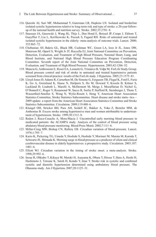
![10 R.R. Townsend and S.P. Steigerwalt
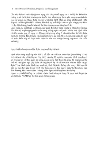
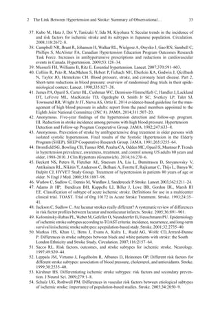
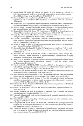
Table 1.5 thuốc liên quan đến THA
Nhóm thuốc hoặc thuốc Cơ chế tác động
NAIDS ức chế prostaglandin gây giữ natri tại thận và
làm giảm GFR
Liều cao corticosteroids Mineralocorticoid receptor (MR) kích thích giữ
Na và ức chế bơm Na-K-atpase, gây co mạch
Uống thuốc tránh thai Cơ chế chưa rõ, làm THA
Thuốc kích thích giao cảm (Meridia™,
Ritalin, Provigil™, etc.) [42, 43]
Co mạch và giữ natri
Thuốc ức chế chọn lọc serotonin (SSRIs),
ức chế chọn lọc tái hấp thu norepinephrine
(SSNIs) [44, 45]
tăng serotonin or norepinephrine qua cơ
chế tái hấp thu
Thuốc kích thích hồng cầu [46] Co mạch
Tacrolimus, cyclosporine [47] Co mạch, giữ natri, giảm GFR
Thuốc kháng retrovirus (HAART)
[48]
Chưa rõ
Cam thảo [49] Gây giữ natri, co mạch
Cocaine, thuốc lắc, methamphetamines [50,
51]
Kích thích giao cảm, co mạch
ức chế VEGF52, 53] FLT-1
VEGF yếu tố tăng trưởng nội mô mạch máu, GFR mức lọc cầu thận
Theo dõi huyết sau khám ban đầu
Sau khi kiểm tra ban đầu, theo dõi huyết áp được khuyến cáo trong khoảng thời gian
quy định. Hướng dẫn trong bảng 1.6 [3].
Một số xét nghiệm được đề nghị đánh giá thường xuyên ở các bệnh nhân có huyết áp
cao. hemoglobin hoặc hematocrit, phân tích nước tiểu với kính hiển vi, creatinin
huyết thanh và điện giải, glucose huyết thanh, lipid lúc đói và ECG. Nghiệm khác
như nồng độ hormone tuyến giáp](https://image.slidesharecdn.com/hypertensionandstroke-170712211353/85/Hypertension-and-stroke-10-320.jpg)

![12 R.R. Townsend and S.P. Steigerwalt
ở những bệnh nhân THA, 30-80% có ngưng thở khi ngủ [35]. Trong các nghiên
cứu, OSA liên quan đến suy tim sung huyết, đột quỵ, bệnh động mạch vành và
ngừng tim đột ngột. ở bệnh nhân đột quỵ, 43-91% bệnh nhân có thể OSA [21, 36].
References
1. Muldoon MF, Rutan GH. Defining hypertension: never as simple as it seems. J Hypertens.
2003;21(3):473–4.
2. Fisher JW. The diagnostic value of the sphygmomanometer in examinations for life insurance.
JAMA. 1914;63:1752–4.
3. Perera GA. Hypertensive vascular disease; description and natural history. J Chronic Dis.
1955;1:33–42.
4. Goldring W, Chasis H. Antihypertensive drug therapy: an appraisal. In: Ingelfinger FJ, Relman
AS, editors. Controversies in international medicine. Philadelphia: Saunders; 1966. p. 83.
5. Keith NM, Wagener HP, Barker NW. Some different types of essential hypertension: their
course and prognosis. Am J Med Sci. 1939;197:332–43.
6. Effects of treatment on morbidity in hypertension. Results in patients with diastolic blood
pressures averaging 115 through 129 mm Hg. JAMA 1967; 202(11):1028–34.
7. Effects of treatment on morbidity in hypertension. II. Results in patients with diastolic blood
pressure averaging 90 through 114 mm Hg. JAMA 1970;213(7):1143–152.
8. James PA, Oparil S, Carter BL, Cushman WC, Dennison-Himmelfarb C, Handler J, Lackland
DT, Lefevre ML, Mackenzie TD, Ogedegbe O, Smith Jr SC, Svetkey LP, Taler SJ, Townsend
RR, Wright Jr JT, Narva AS, Ortiz E. 2014 Evidence-based guideline for the management of
high blood pressure in adults: report from the Panel Members Appointed to the Eighth Joint
National Committee (JNC 8). JAMA. 2014;311(5):507–20.
9. Chirinos JA, Segers P,Duprez DA,Brumback L, Bluemke DA, Zamani P,Kronmal R, VaidyaD,
OuyangP,TownsendRR, Jacobs DR.Late systolic centralhypertension asapredictorofincident
heart failure:the multi-ethnic studyof atherosclerosis. JAmHeartAssoc. 2015;4(3), e001335.](https://image.slidesharecdn.com/hypertensionandstroke-170712211353/85/Hypertension-and-stroke-12-320.jpg)
![Chapter 2
Môi liên quan giữa THA và đột quỵ: tóm tắt các
nghiên cứu dịch tễ
Dilip K. Pandey, Noha Aljehani, and Youji Soga
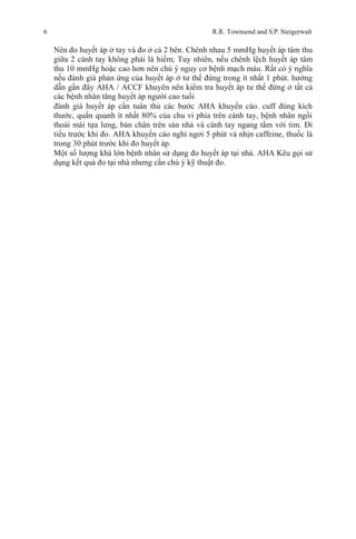
Đột quỵ là nguyên nhân gây tàn phế hàng đầu ở Mỹ. Nó xếp thứ bảy trong số 30
bệnh hàng đầu và chấn thương gây chết sớm ở Mỹ trong năm 2010, và thứ ba
trong những bệnh gây khuyết tật[1]. Trong số các yếu tố nguy cơ có thể gây biến
chứng thiếu máu cục bộ và đột quỵ do xuất huyết, tăng huyết áp là một trong
những nguyên nhân chính bất kể độ tuổi, giới tính và chủng tộc [2, 3]. Tăng huyết
áp hiện nay rất phổ biến. Khoảng 80 triệu người Mỹ trưởng thành (một trong ba)
có THA (huyết áp tâm thu ≥140 mmHg hoặc huyết áp tâm trương ≥90 mmHg)
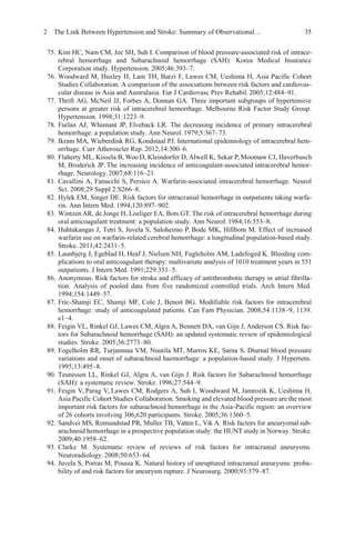
. Tỷ lệ tăng huyết áp tăng nhanh chóng trên tuổi 65. Tỷ lệ mắc THA theo tuổi
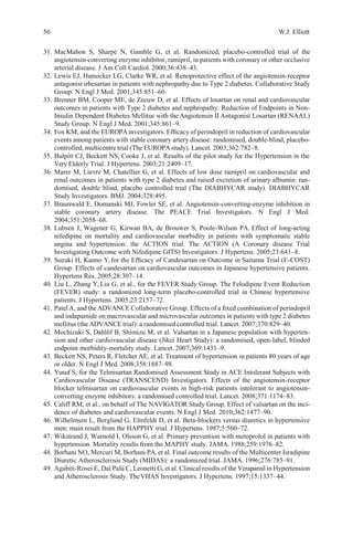
trong 2009-2012 là 80% đối với nữ giới và 76% nam giới trên 75 tuổi [8, 9]. Tỷ lệ
tăng huyết áp cũng thay đổi theo chủng tộc (Hình. 2.1). Nguy cơ tăng huyết áp là
90% với những người có huyết áp bình thường lúc 55 tuổi [10]. Một mô hình dự
báo gần đây cho thấy, cứ tăng 10% số bệnh nhân được kiểm soát huyết áp có thể
làm giảm 14.000 ca tử vong mỗi năm ở nhóm 25-79 tuổi [11]. Dự báo cho thấy
vào năm 2030, khoảng 41,4% người trưởng thành Mỹ sẽ có tăng huyết áp, tăng
8,4% ước tính từ năm 2012 (theo công bố chưa tính toán của AHA, dựa trên
phương pháp mô tả bởi Hiedenreich và cộng sự) [11].
D.K. Pandey, M.D., Ph.D. (*) • N. Aljehani, M.B.B.S. • Y. Soga, M.D., Ph.D.
Department of Neurology and Rehabilitation, University of Illinois College of
Medicine at Chicago, Chicago, IL, USA
e-mail: dpandey@uic.edu
© Springer International Publishing Switzerland 2016 17
V. Aiyagari, P.B. Gorelick (eds.), Hypertension and Stroke, Clinical
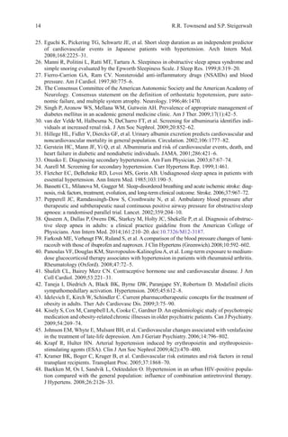
Hypertension and Vascular Diseases, DOI 10.1007/978-3-319-29152-9_2](https://image.slidesharecdn.com/hypertensionandstroke-170712211353/85/Hypertension-and-stroke-16-320.jpg)
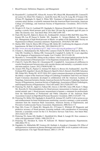
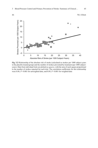
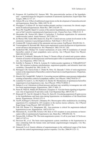
![18 D.K. Pandey et al.
Overall
Sex
Men
Women Age†
(years)
18-39
40-59
60 and over
Race and Hispanic origin
Non-Hispanic white
Non-Hispanic black
Non-Hispanic Asian
Hispanic
0 10 20 30 40
Percent
50 60 70
Fig. 2.1 Age-specific and age-adjusted prevalence of hypertension among adult aged 18 and over,
United States 2011–2012 (Source: CDC/NCHS, National Health and Nutrition Examination
Survey, 2011–2012.Accessed on 15April 2015 at http://www.cdc.gov/nchs/data/databriefs/db133.
pdf)
THA và nguy cơ đột quỵ
Nhiều bằng chứng thuyết phục từ nghiên cứu quan sát và can thiệp, cho thấy
tăng huyết áp là một yếu tố nguy cơ quan trọng và mạnh mẽ gây đột quỵ. Người ta
ước tính rằng khoảng 54% các cơn đột quỵ trên toàn thế giới là do tăng huyết áp
suất (BP) [12]. nghiên cứu tim mạch Framingham năm 1970 quan sát thấy quan hệ
rủi ro có ý nghĩa giữa đột quỵ và huyết áp ≥160 / 95 mmHg ở cả hai giới và ở mọi
lứa tuổi [13]. Người có huyết áp bình thường (<120/80 mmHg) có tỉ lệ đột quỵ
bằng ½ do với những người có huyết áp cao (≥140 / 90mmHg) [14]. Một phân tích
tổng hợp 12 nghiên cứu với 518.520 người tham gia thấy rằng cao huyết áp có liên
quan với đột quỵ
Nguy cơ đặc biệt ở những bệnh nhân chưa già những lại có huyết áp cao [15].
phân tích chi tiết của nghiên cứu thuần tập lớn đã chỉ ra rằng mối quan hệ giữa
THA và nguy cơ đột quỵ là liên tục, nhất quán và độc lập với các yếu tố nguy cơ
khác. nghiên cứu dịch tễ học trước đây đã dùng áp lực tâm trương để nghiên cứu
hơn là huyết áp tâm thu vì thấy nó có liên quan với nguy cơ đột quỵ [16, 17].
Trong một phân tích của chín nghiên cứu mô tả hồi cứu được công bố giữa năm
1958 và 1990, MacMahon kết luận rằng BP giảm làm giảm nguy cơ đột quỵ. giảm
huyết áp tâm trương 5, 7,5, và 10 mmHg có liên quan với giảm nguy cơ đột quỵ ít
nhất là 34, 46, và 56% tương ứng [16]. nghiên cứu thuần tập hợp tác châu Á Thái
Bình Dương (APCSC), cũng cho thấy mối quan hệ tích cực giữa tâm trương và
nguy cơ đột quỵ
29.1
29.7
28.5
7.3
32.4
65.0
28.0
1
42.1
2
24.7
2
26.0](https://image.slidesharecdn.com/hypertensionandstroke-170712211353/85/Hypertension-and-stroke-17-320.jpg)
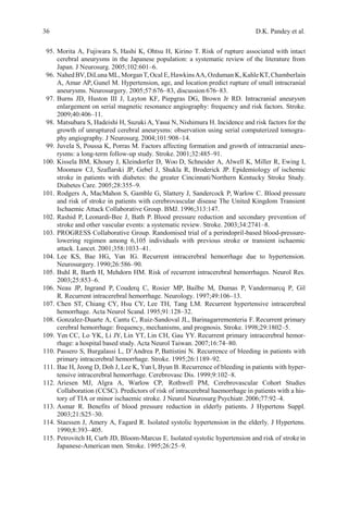
![2 The Link Between Hypertension and Stroke: Summary of Observational… 19
Bệnh nhân có tâm trương cao (DBP≥ 110) có nguy cơ đột quỵ cao hơn 13 lần so
với nhóm có mức tâm trường thấp (DBP≤ 79). Mỗi khi giảm 5 mm trong tâm
trương, nguy cơ giảm một nửa cho cả đột quỵ thiếu máu cục bộ (tỷ số chênh (OR)
0,61, 95% khoảng tin cậy (CI): 0.57- 0,66) và đột quỵ xuất huyết (OR 0,54, 95%
CI: 0,50-0,58) [18]. Huyết áp tâm thu vào những năm 1990 sau khi một số nghiên
cứu dịch tễ học cho thấy huyết áp tâm thu có thể đại diện cho một yếu tố nguy cơ
mạnh mẽ hơn cho đột quỵ so với tâm trương. Hơn nữa, huyết áp tâm thu tăng lên
cùng với tuổi tác, trong khi mức HA tâm trương tăng hơn vào khoảng tuổi 50 và
giảm sau tuổi 60. Huyết áp tâm thu đã được chứng minh là một yếu tố dự báo tốt
hơn về bệnh mạch vành sau tuổi 50 [19].
Huyết áp tâm thu tương quan mạnh mẽ hơn với nguy cơ đột quỵ sau 12 năm hơn
huyết áp theo nghiên cứu tim mạch của Framingham[20]. Ngoài ra, các nghiên
cứu tim mạch tại thành phố copenhagen cho thấy huyết áp tâm thu là yếu tố dự
báo tốt hơn so với tâm trương về nguy cơ đột quỵ [21]. Theo APCSC, phân tích 37
nghiên cứu thuần tập được tiến hành tại khu vực Châu Á Thái Bình Dương, báo
cáo có liên quan tuyến tính liên tục giữa huyết áp tâm thu và nguy cơ đột quỵ
xuống ít nhất là 115 mmHg. Sau khi chuẩn hóa theo tuổi, giảm 10 mmHg huyết áp
tâm thu (95% CI: 40-42%) làm giảm 41% nguy cơ đột quỵ ở châu Á và (95% CI:
22-37%) 30% nguy cơ đột quỵ ở Úc [22]. Trong một nghiên cứu phân tích 61
nhóm của PSC, giảm 20 mmHg huyết áp tâm thu làm giảm 1 nửa nguy cơ tử vong
ở nhóm bệnh nhân 40-69 tuổi (xem Bảng 2.1) [ 23, 24].
Một phát hiện quan trọng từ các nghiên cứu trên là mối liên hệ giữa BP và nguy
cơ đột quỵ là liên tục và tuyến tính ở tất cả các lứa tuổi, và không có bằng chứng
cho thấy BP làm giảm nguy cơ đột quỵ khi tâm thu giảm đến khoảng 115 mmHg
và 75 mmHg với tâm trương [25].
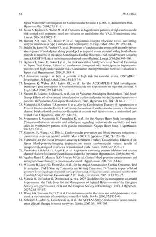
Tuổi tác là một đồng yếu tố quan trọng của mối quan hệ đột quỵ và cao huyết áp.
APCSC báo cáo rằng trong các nhóm tuổi <60, 60-69, và ≥70, giảm 10 mmHg
huyết áp tâm thu làm giảm 54, 36, và 25% nguy cơ đột quỵ, tương ứng (Bảng 2.1)
[22 ]. Kết quả tương tự trong nghiên cứu của Rochester (Hình. 2.2, Bảng 2.1) [23,
28]. Dữ liệu từ NHANES 2005-2010 cho thấy 76,5% người trưởng thành Mỹ ≥80
tuổi có tăng huyết áp. 43,9% tăng huyết áp tâm thu (ISH) và 2,0% có tăng huyết
áp tâm thu và huyết áp tâm trương [29].](https://image.slidesharecdn.com/hypertensionandstroke-170712211353/85/Hypertension-and-stroke-18-320.jpg)
![Table 2.1 nguy cơ đột quỵ liên quan THA
MacMahon et al. [11] APCSC [17] PSC [18]
Rochester
Epidemiology
Project [22]
Study type Meta-analysis of 9
prospective cohort studies
published between 1963
and 1989
Meta-analysis of 37
prospective cohort
studies conducted
between 1961 and 1992
Meta-analysis of 61
prospective cohort
studies conducted
between 1958 and 1990
Nested case–control
study
Number of
participants
418,343 425,325 958,074 1862 (931 cases)
Cases of
stroke
843 strokes of all type 5178 strokes of all type 11,960 strokes of all
type
931 ischemic strokes
Age at
baseline
25–84 20–107 NR Age matched
controls
Follow-up
period
(mean)
6–25 years (10 years) 2–27 years (7 years) 4–25 years (12 years) 15 yearsa
Sex Male 96 % Male 57 % NR Sex matched
controls
Study
population
USA, Europe, Puerto Rico China, Japan, Hong
Kong, Taiwan,
Singapore, South
Korea, New Zealand,
Australia
Europe, USA, Japan,
China, Australia
USA
Results 5 mmHg ↓
DBP
34 % ↓ risk Age 10 mmHg ↓
SBP
Age 20 mmHg ↓
SBP
Age OR (cases
vs.
controls)
7.5 mmHg ↓
DBP
46 % ↓ risk <60 54 % ↓
stroke risk
40–49 64 % ↓ risk 50 4.8
10 mmHg ↓
DBP
56 % ↓ risk 60–69 36 % ↓
stroke risk
50–59 62 % ↓ risk 60 3.2
≥70 25 % ↓
stroke risk
60–69 57 % ↓ risk 70 2.2
70–79 50 % ↓ risk 80 1.5
80–89 33 % ↓ risk 90 1.0
Age 10 mmHg ↓
DBP
40–49 65 % ↓ risk
50–59 66 % ↓ risk
60–69 60 % ↓ risk
70–79 52 % ↓ risk
80–89 37 % ↓ risk
APCSC Asia Pacific Cohort Studies Collaboration, PSC Prospective Studies Collaboration, SBP systolic blood pressure,
DBP diastolic blood pressure, OR odds ratio, NR not reported
a
Ischemic strokes identified from 15 years follow-up of Rochester Epidemiology Project](https://image.slidesharecdn.com/hypertensionandstroke-170712211353/85/Hypertension-and-stroke-19-320.jpg)
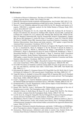
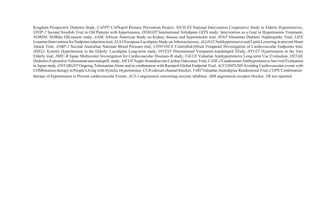
![2 mối liên quan giữa THA và đột quỵ: Summary of Observational… 21
Fig. 2.2 Stoke mortality rate in each decade of age vs. usual blood pressure at the start of that
decade. Rates are plotted on a floating absolute scale, and each square has area inversely propor-
tional to the effective variance of the log mortality rate (From Lewington S, Clarke R, Qizilbash
N, Peto R, Collins R, Prospective Studies C. Age-specific relevance of usual blood pressure to
vascular mortality: a meta-analysis of individual data for one million adults in 61 prospective stud-
ies. Lancet. Dec 14 2002; 360(9349):1903–1913, with permission)
sự khác biệt chủng tộc trong tăng huyết áp và nguy cơ đột quỵ đã được báo cáo từ
một số nghiên cứu quan sát tại Hoa Kỳ. nghiên cứu đột quỵ tại bắc Manhattan cho
thấy tăng huyết áp là một yếu tố nguy cơ độc lập với đột quỵ thiếu máu cục bộ ở
người da trắng (OR 1.8), người da đen (OR 2.0) và Caribbean gốc Tây Ban Nha (OR
1.2) [30]. Nghiên cứu hợp tác Baltimore-Washington về đột quỵ với (bệnh nhân tuổi
từ 18-44 tuổi), có mối liên quan giữa tăng huyết áp và nguy cơ đột quỵ thiếu máu cục
bộ ở người da trắng và người da đen ở cảm nam và nữ. ORS điều chỉnh theo tuổi
(95% CI) cho đột quỵ thiếu máu cục bộ với tiền sử tăng huyết áp ở đàn ông da trắng,
phụ nữ da trắng, đàn ông da đen và phụ nữ da đen 1,6 (0,7-3,2), 2,5 (1,1-5,9), 3,8
(1,8-7,9 ), và 4,2 (2,4-7,5) tương ứng [31]. Mức tăng HA tâm thu có liên quan với
nguy cơ đột quỵ cao hơn ở người da đen so với người da trắng [32].
Điều trị tăng huyết áp là một mục tiêu điều trị quan trọng trong việc ngăn ngừa đột
quỵ đã được hỗ trợ bởi một số nghiên cứu [34-38]. đột quỵ liên quan tới huyết áp hơn
so với bệnh mạch vành](https://image.slidesharecdn.com/hypertensionandstroke-170712211353/85/Hypertension-and-stroke-20-320.jpg)
![22 D.K. Pandey et al.
[39-41]. Giảm 5-6 mmHg tâm trương sau 2-3 năm điều trị liên tục làm giảm 35-
40% đột quỵ [40].
Đột quỵ thường được phân thành hai loại chính: đột quỵ thiếu máu cục bộ và đột
quỵ xuất huyết. đột quỵ xuất huyết có thể được chia thành đột quỵ do xuất huyết
nội sọ t (ICH) hoặc xuất huyết dưới nhện (SAH) . khoảng 80% các cơn đột quỵ do
thiếu máu cục bộ, 15% là ICH, và 5% là SAH [46].
Tăng huyết áp và đột quỵ thiếu máu cục bộ
Có nhiều trường phái phân loại đột quỵ thiếu máu cục bộ thành các loại phụ theo
nguyên nhân của nó [47, 48]. Trong số các phân nhóm đột quỵ, sự khác biệt về tỷ
lệ mắc, tỷ lệ tái phát, thời gian sống và theo chủng tộc đã được báo cáo [49, 50].
Một số nghiên cứu dịch tễ học cũng đã giải quyết mối quan hệ giữa các yếu tố rủi
ro và phân nhóm đột quỵ khác nhau [51-54]. BP là một yếu tố quyết định mạnh
mẽ về nguy cơ cho cả đột quỵ thiếu máu cục bộ và xuất huyết não [55]. Trong số
5017 bệnh nhân trong nghiên cứu về đột quỵ ở Đức, tăng huyết áp hay gây tổn
thương mạch nhỏ (79,4%) hơn so với mạch lớn (70,0%) [58].](https://image.slidesharecdn.com/hypertensionandstroke-170712211353/85/Hypertension-and-stroke-21-320.jpg)
![2 The Link Between Hypertension and Stroke: Summary of Observational… 23
Tuy nhiên, một số nghiên cứu xuất hiện mâu thuẫn với kết quả này. Ohira và cộng
sự báo cáo rằng tác động của tăng huyết áp gây đột quỵ thiếu máu cục bộ không
phụ thuộc vào tình trạng xơ vữa động mạch trong nghiên cứu (ARIC) [59]. Tương
tự như vậy, nghiên cứu của Lai và cộng sự cho thấy trong khi tăng huyết áp là yếu
tố nguy cơ phổ biến nhất gây đột quỵ thì tiền sử THA không lien quan về mặt
thống kê với các phân nhóm đột quỵ [60].
Đột quỵ do xuất huyết
Xuất huyết nội sọ ICH
Bằng chứng từ một số nghiên cứu dịch tễ cho thấy có mối liên quan lớn giữa ICH
và cao huyết áp [61-69]. Một nghiên cứu bệnh chứng của 331 trường hợp ICH ở
Melbourne báo cáo nguy cơ tăng gấp đôi ICH do tăng huyết áp [70]. Một nghiên
cứu bệnh chứng bởi Feldmann quan sát thấy rằng huyết áp cao là một yếu tố độc
lập đem lại nguy cơ cao gấp sáu lần (OR 5.71, 95% CI: 3,61-9,05) ICH ở nam giới
và phụ nữ tuổi từ 18-49 (Bảng 2.2) [71]. Trong nghiên cứu thuần tập, tất cả các
nghiên cứu cho thấy mối liên quan giữa tăng huyết áp và ICH, và hai nghiên cứu
cho thấy nguy cơ ngày càng tăng của ICH với t mức độ tăng huyết áp (Bảng 2.2)
[72]. tăng nguy cơ ICH với sự gia tăng mức độ tăng huyết áp này đã được nhìn
thấy trong các nghiên cứu dịch tễ học khác. Một phân tích gộp của ARIC và các
dữ liệu nghiên cứu sức khỏe tim mạch (CHS) cũng báo cáo xu hướng này. So với
bình thường, nguy cơ tương đối (RR) (95% CI) của ICH là 1,43 (0,90-2,26) cho
BP 140-159 / 90-99 mmHg, 2,71 (1,58-4,67) cho BP 160-179 / 100-109 mmHg,
và 5,55 (3,07-10,03) cho BP≥ 160/110 mmHg (Bảng 2.2) [73].](https://image.slidesharecdn.com/hypertensionandstroke-170712211353/85/Hypertension-and-stroke-22-320.jpg)
![Table 2.2 nguy cơ xuất huyết nội sọ với THA
Ariesen et al. [59] Sturgeon et al. [60] Suh et al. [61] Leppälä et al. [40] Feldmann et al. [58]
Study type Systematic Review of
11 case–control and 3
cohort studies from
1966 to 2001
Pooled analysis of ARIC and
CHS
Prospective cohort
study
Prospective cohort
study
Case–control study
Number of
participants
Case–control studies 21,680 (15,792 ARIC, 5888
CHS)
114,793 28,519 636a
72–662
Cohort studies
28,519–114,793
Cases of ICH Case–control studies 135 (61 ARIC, 74 CHS) 372 112 217
24–331
Cohort studies
112–386
Study population USA South Korea Finland USA
Age Age matched on most
of case–control
studies
Mean age 54 (ARIC) 35–59 50–69 18–49
Mean age 73 (CHS)
24D.K.Pandeyetal.](https://image.slidesharecdn.com/hypertensionandstroke-170712211353/85/Hypertension-and-stroke-23-320.jpg)
![Ariesen et al. [59] Sturgeon et al. [60] Suh et al. [61] Leppälä et al. [40] Feldmann et al. [58]
Sex Sex matched on most
of case–control
studies
44.8 % male (ARIC) Male only Male only 56 % male
42.4 % male (CHS)
Results Case–control studies BP RR BP RR SBP RR Adjusted OR 5.71
Overall crude OR
3.68
SBP <140/DBP< 90 1.0 SBP< 130/
DBP< 85
1.0 ≤139 1.0
Cohort studies SBP140–159/
DBP90–99
1.43 SBP130–139/
DBP85–89
2.16 140–159 2.20
Adjusted RR 1.14–33
by different levels of
blood pressure
SBP160–179/
DBP100–109
2.71 SBP140–159/
DBP90–99
5.32 ≥160 3.78
SBP≥ 160/DBP≥ 110 5.55 SBP160–179/
DBP100–109
10.44 DBP RR
BP as continuous measure SBP≥ 180/
DBP≥ 110
33.32 ≤89 1.0
10 mmHg ↑ SBP: 25 % ↑ risk 90–99 2.10
10 mmHg ↑ DBP: 47 % ↑ risk ≥100 4.17
ICH intracerebral hemorrhage, SBP systolic blood pressure, DBP diastolic blood pressure, BP blood pressure, OR odds ratio, RR relative risk, ARIC
Atherosclerosis Risk in Communities Study, CHS Cardiovascular Health Study
a
Cases and controls matched on race, age, and gender
2TheLinkBetweenHypertensionandStroke:SummaryofObservational…25](https://image.slidesharecdn.com/hypertensionandstroke-170712211353/85/Hypertension-and-stroke-24-320.jpg)
![26 D.K. Pandey et al.
Các nghiên cứu đã chỉ ra rằng huyết áp tăng lên trong phạm vi bình thường cũng có
liên quan đến sự gia tăng tuyến tính với nguy cơ ICH [52]. Ngoài ra, nguy cơ ICH
tăng cùng với tăng huyết áp, người điều trị tăng huyết áp không thường xuyên, 55
tuổi hoặc trẻ hơn, hoặc người hút thuốc lá [77]. Cải thiện kiểm soát tăng huyết áp có
thể làm giảm tỷ lệ mắc ICH [78]. kiểm soát huyết áp được coi là lựa chọn chính cho
công tác phòng chống ICH [79].
Bệnh nhân đang dùng thuốc chống đông đường uống lâu dài cũng có nguy cơ gia
tăng về ICH [80]. Một số yếu tố nguy cơ chảy máu do chống đông đã được nghiên
cứu: tuổi cao, tiền sử nhồi máu cơ tim hoặc bệnh tim thiếu máu cục bộ, bệnh tiểu
đường, bệnh mạch máu não, đồng thời sử dụng thuốc kháng tiểu cầu, liều cao các
thuốc chống đông máu và tăng huyết áp [81, 82]. Một nghiên cứu hồi cứu của
Wintzen thấy rằng tăng huyết áp có dùng chống đông gặp ở 80% bệnh nhân ICH
[83]. Việc sử dụng warfarin đã tăng lên nhanh chóng trong những thập kỷ qua, các
nghiên cứu cũng đã chỉ ra tăng tỉ lệ ICH liên quan đến việc sử dụng warfarin [80,
84]. Launbjerg và cộng sự thấy rằng huyết áp cao là một yếu tố nguy cơ độc lập trong
cháy máu do chống đông trong 1 phân tích đa biến ở 551 bênhh nhân dùng chống
đông trong 10 năm theo dõi điều trị [85]. Phân tích các dữ liệu tổng hợp từ năm thử
nghiệm ngẫu nhiên cho thấy những bệnh nhân ICH khi dùng warfarin có huyết áp
tâm thu và HA tâm trương cao hơn so với những bệnh nhân được điều trị warfarin-
không bị ICH [86]. Mặt khác, một nghiên cứu bệnh chứng, so sánh 170 bệnh nhân bị
ICH trong khi điều trị warfarin và 1020 bệnh nhân dùng chống đông mà không bị
ICH, thấy không có sự khác biệt thống kê về tỷ lệ tăng huyết áp được chẩn đoán [87].
Tác động của tăng huyết áp ở bệnh nhân tử vong do ICH có dùng chống đông cũng
đã được nghiên cứu. Một nghiên cứu hồi cứu của Fric-Shamji báo cáo rằng áp lực
động mạch trung bình ban đầu cao tương quan với xu hướng khối máu tụ lan rộng
hơn so với ban đầu [87].
xuất huyết dưới màng nhện SAH
Hút thuốc, cao huyết áp và uống rượu quá mức là những yếu tố nguy cơ hay gặp nhất
gây SAH [88]. Một cái nhìn tổng quan của tất cả các nghiên cứu về yếu tố nguy cơ
cho SAH được xuất bản bằng tiếng Anh từ năm 1966 đến tháng 3 năm 2005 báo cáo
về quan hệ tích cực giữa tăng huyết áp và SAH trong cả hai nhóm (RR 2.5, 95% CI:
2,0-3,1) và nghiên cứu bệnh chứng ( OR 2.6, 95% CI: 2,0-3,1) (Bảng 2.3) [88]. Năm
1996, một đánh giá của 9 nghiên cứu theo chiều dọc và 11 nghiên cứu khác xác định
tăng huyết áp là yếu tố nguy cơ lớn với SAH OR 2,8 (đối với các nghiên cứu theo
chiều dọc; KTC 95% [CI] , 2,1-3,6) và OR 2.9 (cho các nghiên cứu bệnh chứng; 95%
CI 2,4-3,7).](https://image.slidesharecdn.com/hypertensionandstroke-170712211353/85/Hypertension-and-stroke-25-320.jpg)
![2 The Link Between Hypertension and Stroke: Summary of Observational… 27
Một phân tích dữ liệu bệnh nhân trong APCSC chứng minh rằng huyết áp cao là một
yếu tố nguy cơ độc lập cho SAH (tỷ số nguy cơ (HR) 2.0, 95% CI: 1,5-2,7) (xem
Bảng 2.3). Nguy cơ SAH tăng mạnh với sự gia tăng huyết áp tâm thu [91]. Xu hướng
này cũng được nhìn thấy trong một nghiên cứu lớn giữa các nước châu Á cho cả nam
giới và phụ nữ (Bảng 2.3) [75].
vỡ phình mạch não là nguyên nhân phần lớn SAHs, nhiều nghiên cứu đã cố gắng xác
định các yếu tố nguy cơ vỡ / tăng trưởng của chứng phình mạch. Tăng huyết áp tâm
thu mạn tính đã được chứng minh là một yếu tố dự báo mạnh mẽ của vỡ phình động
mạch não. Trong một công bố mới đây của nghiên cứu Nord-Trøndelag, một nghiên
cứu trên dân số lớn ở Na Uy, tăng nhẹ (HA tâm thu 130-139 mmHg) và nặng (HA
tâm thu> 170 mmHg) HA tâm thu mạn tính có liên quan tới sự gia tăng nguy cơ
aSAH trong thời gian theo dõi 22 năm so sánh với huyết áp tâm thu dưới 130 mmHg
(tỷ số rủi ro là 2.3 và 3.3 tương ứng) [92].
Trong số những bệnh nhân bị phình động mạch nhỏ (≤7 mm), cao huyết áp, tuổi
tương đối trẻ và vị trí ở phía sau vòng tuần hoàn não là yếu tố nguy cơ đáng kể SAH
[96]. Tuy nhiên, một vài nghiên cứu, sử dụng cộng hưởng từ hoặc CT chụp động
mạch để đánh giá phình, không thấy có mối liên quan giữa mưc tiến triển phình mạch
với THA [97-99].
Tăng huyết áp và đột quỵ tái phát
tái phát đột quỵ thiếu máu cục bộ
mặc dù truyền thông rộng rãi bởi các chuyên gia trong vài thập kỷ qua, chỉ có một
phần ba số bệnh nhân tăng huyết áp có huyết áp (HA) kiểm soát ở ngưỡng<140/90
mmHg đối với huyết áp không biến chứng và <130/80 mmHg đối với bệnh nhân tiểu
đường hoặc bệnh thận. Đối với những người bị tăng huyết áp không được kiểm soát,
nguy cơ đột quỵ tăng lên đáng kể. Trong một nghiên cứu của Mỹ, ước tính rủi ro đã
tiết lộ rằng 9-16% của tất cả các trường hợp đột quỵ thiếu máu cục bộ có thể tránh
được chỉ bằng cách điều trị huyết áp [100].
Vì làm giảm huyết áp có thể làm nặng thêm tình trạng giảm tưới máu não nếu cơ chế
tự điều chỉnh rối loạn hoặc có hẹp động mạch cảnh, hạ huyết áp trong giai đoạn cấp
tính của đột quỵ thiếu máu cục bộ vẫn được tranh luận. Tuy nhiên, một vài thử
nghiệm đã xác nhận rằng sự kiểm soát HA lâu dài có thể làm giảm đột quỵ tái phát
[25, 35]. Nghiên cứu phân tích Gueyffier và cộng sự báo cáo giảm 28% nguy cơ tái
phát đột quỵ mà không có tác dụng phụ đáng kể khi điều trị bằng thuốc chống tăng
huyết áp ở bệnh nhân đột quỵ tăng huyết áp [101].](https://image.slidesharecdn.com/hypertensionandstroke-170712211353/85/Hypertension-and-stroke-26-320.jpg)
![Table 2.3 Overview of risk of subarachnoid hemorrhage associated with hypertension
KMIC Study [62] Feigin et al. [77]
Asia Pacific Cohort Studies
Collaboration [78]
Study type Prospective cohort study Systematic review of 14 cohort and 23 case–control
studies published between 1966 and 2005
Meta analysis of 26 cohort studies
Cases of SAH 308 3936 (cohort 892 case–control 3044) 236
Study population South Korea Cohort studies Japan, China, Taiwan, South Korea,
Singapore, Australia, NZUSA, Japan, UK, South Korea, Finland
Case–control studies
Finland, UK, USA, NZ, Portugal, Norway, Japan,Australia,
Germany, WHO (Africa/Asia/Europe/LatinAmerica)
Results BP RR Cohort studies BP HR
Male Sex RR SBP< 140 1.0
SBP< 120/DBP< 80 1.0 Female 3.3 SBP≥ 140 2.0
SBP 120–129/DBP 80–84 1.46 Male 2.3
SBP 130–139/DBP 85–89 2.41 Total 2.5
SBP 140–159/DBP 90–99 2.92 Case–control studies
SBP 160–179/DBP 100–109 3.66 Sex OR 10-mmHg ↑ SBP: 31 % ↑ risk
SBP≥ 160/DBP≥ 110 5.12 Female 3.3
Female Male 2.1
SBP< 120/DBP< 80 1.0 Total 2.6
SBP 120–129/DBP 80–84 1.77
SBP 130–139/DBP 85–89 2.60
SBP 140–159/DBP 90–99 3.82
SBP 160–179/DBP 100–109 9.06
SBP≥ 160/DBP≥ 110 20.49
KMIC Korea Medical Insurance Corporation, SAH subarachnoid hemorrhage, SBP systolic blood pressure, DBP diastolic blood pressure, BP blood pressure, RR relative risk,
OR odds ratio, HR hazard ratio](https://image.slidesharecdn.com/hypertensionandstroke-170712211353/85/Hypertension-and-stroke-27-320.jpg)
![2 The Link Between Hypertension and Stroke: Summary of Observational… 29
Mỗi khi giảm 5 mmHg huyết áp tâm trương và giảm 10 mmHg huyết áp tâm thu
làm giảm 34 và 28% nguy cơ đột quỵ tương ứng [101].
đột quỵ xuất huyết tái phát
Cả hai nghiên cứu châu Âu và châu Á đã cho rằng huyết áp cao là một yếu tố nguy
cơ tái phát của ICH [104-108]. Yên báo cáo tỷ lệ cao bệnh nhân tăng huyết áp bị
ICH tái phát ở Đài Loan (88,2%) [109].
Trong phần này, chúng tôi đã thảo luận về tăng huyết áp là một nguy cơ tái phát
đột quỵ thiếu máu cục bộ và xuất huyết đột quỵ 1 cách riêng biệt. Tuy nhiên, tăng
huyết áp cũng đã được báo cáo như là một yếu tố nguy cơ độc lập ICH ở những
bệnh nhân đột quỵ thiếu máu cục bộ (SBP≥ 140 HR 2,07, 95% CI: 1,23-3,83)
[112].
Tầm quan trọng của huyết áp tâm thu ở người cao tuổi
Lão hóa liên quan với tăng huyết áp tâm thu do đó, ISH (tâm thu BP≥ 140 mmHg
và huyết áp tâm trương BP <90 mmHg) là kiểu thường gặp nhất của tăng huyết áp
trong quần thể người già.
Sự gia tăng huyết áp tâm thu ở ISH chủ yếu là do độ đàn hồi giảm của các động
mạch lớn và không nhất thiết phải đi kèm với sự gia tăng huyết áp động mạch
trung bình hoặc kháng trở ngoại vi [114].](https://image.slidesharecdn.com/hypertensionandstroke-170712211353/85/Hypertension-and-stroke-28-320.jpg)
![30 D.K. Pandey et al.
Các nghiên cứu dịch tễ học đã chỉ ra rằng ISH là một yếu tố nguy cơ độc lập
đối với đột quỵ và là mục tiêu điều trị để giảm nguy cơ bị đột quỵ. Nghiên cứu
phân tích tám thử nghiệm lâm sàng bao gồm 15.693 bệnh nhân ISH cho thấy điều
trị tích cực tăng huyết áp làm giảm tỷ lệ đột quỵ 30% [117].](https://image.slidesharecdn.com/hypertensionandstroke-170712211353/85/Hypertension-and-stroke-29-320.jpg)
![Chapter 3
Kiểm soát huyết áp và dự phòng đột quỵ lần
đầu: tóm tắt các thử nghiệm lâm sàng
William J. Elliott
Tăng huyết áp hoặc huyết áp cao (BP), là yếu tố nguy cơ quan trọng nhất đối với
đột quỵ [1-5], chiếm 54% nguy cơ dân số phân bổ trên toàn thế giới trong một mô
hình y tế toàn cầu gần đây [6]. Đột quỵ là nguyên nhân thứ hai dẫn đến tử vong
trên toàn thế giới (mặc dù gần đây xếp thứ 5, sau bệnh tim, ung thư, các bệnh hô
hấp dưới mạn tính và chấn thương ở Mỹ [7]) và là nguyên nhân hàng đầu gây tàn
tật ở tất cả các nước. Mục đích của chương này là xem xét các bằng chứng thử
nghiệm lâm sàng hiện có hỗ trợ việc sử dụng các loại thuốc điều trị hạ huyết áp để
ngăn ngừa đột quỵ lần đầu.
W.J. Elliott, M.D., Ph.D. (*)
Department of Biomedical Sciences, The Pacific Northwest University of Health Sciences,
Yakima, WA, USA
e-mail: wj.elliott@yahoo.com
© Springer International Publishing Switzerland 2016 39
V. Aiyagari, P.B. Gorelick (eds.), Hypertension and Stroke, Clinical
Hypertension and Vascular Diseases, DOI 10.1007/978-3-319-29152-9_3](https://image.slidesharecdn.com/hypertensionandstroke-170712211353/85/Hypertension-and-stroke-37-320.jpg)
![40 W.J. Elliott
Các thử nghiệm lâm sàng
Đã có 35 nghiên cứu thấy rằng so sánh đột quỵ ở nhóm dùng giả dược (không điều trị) với nhóm có dùng hạ áp tích cực (Bảng 3.1). Nhiều nghiên cứu
được thực hiện trong thiên niên kỷ trước, khi dùng giả dược hoặc không điều trị vẫn được chấp nhận tính đạo đức trong nghiên cứu kết quả; hầu hết
các thử nghiệm gần đây đã so sánh kết quả dùng thuốc hạ áp được tăng cường bằng cách cho thêm một giả dược hoặc một hay nhiều hoạt chất chống
cao huyết áp để kiểm soát huyết áp là bắt buộc [9-12, 31-34, 36 -42, 44, 45].](https://image.slidesharecdn.com/hypertensionandstroke-170712211353/85/Hypertension-and-stroke-38-320.jpg)
![Table 3.1 Placebo-controlled trials of primary stroke prevention involving antihypertensive drugs
Trial acronym, year
Years of
follow-up
Subjects with
HTN (%)
∆SBP
(mmHg)
Active arm Control arm
Comments (#
with prior
strokes)Agent
# of First
strokes/# of
subjects Agent
# of First
strokes/# of
subjects
VA I, 1967 [13] 1.5 100 30 Diuretic + others 1/73 Placebo + “rescue” 3/70 (6/5)
VA II, 1970 [14] 3.3 100 31.4 Diuretic + others 5/186 Placebo + “rescue” 20/194 (NR/NR)
USPHS, 1977 [15] 7 100 16 Diuretic + others 1/193 Placebo 6/196 (0/0)
Oslo, 1980 [16] 5.5 100 17 Diuretic 0/406 No treatment 5/379 (0/0)
ANBP-1, 1980 [17] 3 100 NR Diuretic 13/1721 Placebo 22/1706 (0/0)
Kuramoto, 1981 [18] 4 100 20 Diuretic 3/44 Placebo 4/47 (0/0)
HDFPa
, 1982 [19] 5 100 10 Diuretic 87/5364 Placebo 142/5333 (N/A)
EWPHEa
, 1985 [20] 4.6 100 21 Diuretic 16/386 Placebo 22/405 (N/A)
MRC-1, 1985 [21] 5.5 100 ~13 Diuretic or β-blocker 18/4297 Placebo 109/8654 (32 or 31/61)
~9.5 42/4203
IPPPSH, 1985 [22] 4 100 3.8 β-Blocker 45/3185 Placebo 46/3172 (0/0)
Coope & Warrendera
,
1986 [23]
4.4 100 18.0 β-Blocker 18/410 No treatment 38/460 (N/A)
SHEP Pilot, 1989 [24] 2.8 100 15 Diuretic 11/443 Placebo + “rescue” 6/108 (8)
SHEPa
, 1991 [25] 4.5 100 11.1 Diuretic 95/2314 Placebo + “rescue” 152/2338 (N/A)
STOP-1, 1991 [26] 2.1 100 19.5 Diuretic or β-blocker 28/782 Placebo 49/784 (32/36)
MRC-E, 1992 [27] 5.7 100 15 Diuretic or β-blocker 45/1081 Placebo 134/2213 (NR or NR/NR)
15 56/1102
STONE, 1996 [28] 2.5 100 9.5 CCB 16/817 Placebo 36/815 (NR/NR); Not
randomized
Syst-EUR, 1997 [29] 2.5 100 10.7 CCB + other 49/2398 Placebo + other 80/2297 (103)
(continued)](https://image.slidesharecdn.com/hypertensionandstroke-170712211353/85/Hypertension-and-stroke-39-320.jpg)
![Table 3.1 (continued)
Trial acronym, year
Years of
follow-up
Subjects with
HTN (%)
∆SBP
(mmHg)
Active arm Control arm
Comments (#
with prior
strokes)Agent
# of First
strokes/# of
subjects Agent
# of First
strokes/# of
subjects
Syst-China, 1998 [30] 2.8 100 8.0 CCB + other 45/1253 Placebo + other 59/1141 (45), Not
randomized
HOPEa
, 2000 [9, 10] 4.5 46 3 Other +ACE-I 113/4188 Other + placebo 175/4190 Add-on (N/A)
PART2, 2000 [31] 4.7 ? 6.0 Other +ACE-I 7/308 Other + placebo 4/309 Add-on (34/28)
IDNT, 2001 [32] 2.6 100 3 (ARB or CCB) + other 28/579 Placebo + other 26/569 (NR/NR)
4 15/567
RENAAL, 2001 [33] 3.4 100 2 ARB + other 47/751 Placebo + other 50/762 (0/1)
EUROPA, 2003 [34] 4.2 27?
(BP> 160/95)
5.0 Other +ACE-I 98/6110 Other + placebo 102/6108 Add-on
(210/199)
HY-VET Pilot, 2003
[35]
1.1 100 23.0 ACE-I or diuretic 6/426 Placebo + Rescue 18/426 (18 or 18/22)
23.0 12/431
SCOPEa
, 2003 [11, 12] 3.5 100 3.2 ARB + other 83/2386 Placebo + other 100/2378 (N/A)
DIAB-HYCAR, 2004
[36]
3.3 55 1.3 Other +ACE-I 118/2443 Other + placebo 116/2469 Add-on
(107/100)
PEACE, 2004 [37] 4.8 45 3.0 Other +ACE-I 71/4158 Other + placebo 92/4132 Add-on
(291/248)
ACTION, 2005 [38] 4.9 100 6.6 Other + CCB 50/1975 Other + placebo 75/2002 Add-on (NR/
NR)
E-COST, 2005 [39] 3.0 100 1.7 ARB + other 47/1053 No ARB + other 77/995 (23/69)
FEVER, 2005 [40] 3.3 100 3.5 Diuretic + CCB 177/4841 Diuretic + placebo 251/4870 Second-line
(685/753)
ADVANCE, 2007 [41] 4.3 68 5.6 Other + diuretic +ACE-I 215/5569 Other + placebo 218/5571 Combination
(502/520)](https://image.slidesharecdn.com/hypertensionandstroke-170712211353/85/Hypertension-and-stroke-40-320.jpg)
![Trial acronym, year
Years of
follow-up
Subjects with
HTN (%)
∆SBP
(mmHg)
Active arm Control arm
Comments (#
with prior
strokes)Agent
# of First
strokes/# of
subjects Agent
# of First
strokes/# of
subjects
Jikei, 2007 [42] 3.1 88 1.0 Other +ARB 25/1541 Other 43/1540 Add-on (NR/
NR)
HYVET, 2008 [43] 1.8 100 15.0 Diuretic 51/1933 Placebo 69/1912 (130/131)
TRANSCEND, 2008
[44]
4.7 76 4.0 Other +ARB 112/2842 Other + placebo 136/2836 Add-on
(648/654)
NAVIGATOR, 2010
[45]
5.0 78 2.8 Other +ARB 105/4631 Other + placebo 132/4675 Add-on
(143/132)
HTN hypertension, SBP systolic blood pressure
a
Denotes study for which the number of observed primary (as opposed to both primary and secondary) strokes can be calculated. (N/A) indicates that the number of subjects
with prior stroke has been reported and subtracted from the total number of subjects in the trial. VA I First VeteransAdministration Cooperative Study Group on Antihypertensive
Agents, VA II Veterans Administration Cooperative Study Group on Antihypertensive Agents, USPHS United States Public Health Service trial, ANBP-1 First Australian
National Blood Pressure trial, HDFP Hypertension Detection and Follow-up Program, EWPHE European Working Party on Hypertension in the Elderly, MRC-1 First Medical
Research Council trial (in “mild” hypertensives), IPPPSH International Prospective Primary Prevention Study in Hypertension, SHEP Systolic Hypertension in the Elderly
Program, STOP-1 First Swedish Trial in Old Patients with Hypertension, MRC-E Medical Research Council trial (in elderly hypertensives), STONE Shanghai Trial of Nifedipine
in the Elderly trial, Syst-EUR Systolic Hypertension in Europe trial, Syst-China Systolic Hypertension in China trial, HOPE Heart Outcomes Prevention Evaluation, PART2
Prevention of Atherosclerosis with Ramipril study #2, IDNT Irbesartan Diabetic Nephropathy Trial, RENAAL Reduction of Endpoints in Non-Insulin Dependent Diabetes
Mellitus with the Angiotensin II Antagonist Losartan, EUROPA EUropean trial on Reduction of cardiac events with Perindopril in patients with stable coronary Artery disease,
HYVET Hypertension in the Very Elderly Trial, SCOPE Study on Cognition and Prognosis in the Elderly trial, DIAB-HYCAR non-insulin-dependent DIABetes, HYpertension,
microalbuminuria or proteinuria, Cardiovascular events And Rampril study, PEACE Prevention of Events with Angiotensin Converting Enzyme inhibition trial, ACTION a
Coronary disease Trial Investigating Outcome with Nifedipine GITS trial, E-COST Efficacy of Candesartan on Outcome in Saitama Trial, FEVER Felodipine EVEnt Reduction
study, ADVANCE Action in Diabetes and Vascular disease: preterAx and diamicroN-MR Controlled Evaluation, TRANSCEND Telmisartan Randomised AssessmeNt Study in
ACE iNtolerant subjects with cardiovascular Disease trial, NAVIGATOR Nateglinide and Valsartan in Impaired Glucose Tolerance Outcomes Research, CCB calcium channel
blocker, ACE-I angiotensin converting-enzyme inhibitor, ARB angiotensin receptor blocker, BP blood pressure, NR not reported](https://image.slidesharecdn.com/hypertensionandstroke-170712211353/85/Hypertension-and-stroke-41-320.jpg)
![Table 3.2 Actively controlled trials of primary stroke prevention involving initial antihypertensive drugs
Trial acronym, year
Years of
follow-up
Subjects with
HTN (%)
∆SBP
(mmHg)
Arm 1 Arm 2 (or Arm 3)
Comments (# with
prior strokes)Agent
# of First
strokes/#
of subjects Agent
# of First
strokes/#
of subjects
MRC-1, 1985 [21] 5.5 100 4.5 Diuretic 18/4297 β-Blocker 42/4203 (32/31)
HAPPHY, 1987 [46] 3.8 100 0 Diuretic 41/3297 β-Blocker 32/3276 (0/0)
MAPHY, 1988 [47] 5.0 100 0.3 Diuretic 25/1625 β-Blocker 23/1609 (0/0)
MRC-E, 1992 [27] 5.7 100 0 Diuretic 45/1081 β-Blocker 56/1102 (NR/NR)
MIDAS, 1996 [48] 3.0 100 3.5 Diuretic 3/441 CCB 6/442 (NR/NR)
VHAS, 1997 [49] 2.0 100 1.0 Diuretic 4/707 CCB 5/707 (NR/NR)
ABCD, 1998 [50] 5.0 100 0 ACE-I 7/235 CCB 11/235 (2/3)
FACET, 1998 [51] 2.5 100 -4 ACE-I 4/189 CCB 10/191 (0/0)
UKPDS, 1998 [52] 8.4 100 -1 β-Blocker 17/358 ACE-I 21/400 (0/0)
CAPPP, 1999 [53] 6.1 100 2 β-Blocker/diuretic 148/5493 ACE-I 189/5492 (39/50)
NICH-ES, 1999 [54] 4.2 100 0 Diuretic 8/215 CCB 8/214 (0/0)
STOP-2, 1999 [55] 5.0 100 1 β-Blocker/diuretic 237/2213 ACE-I 215/2205 (86/86
1 or CCB 207/2196 or 83)
INSIGHT, 2000 [56] 3.5 100 0 Diuretic 74/3164 CCB 67/3157 (NR/NR)
NORDIL, 2000 [57] 4.5 100 -3 β-Blocker/diuretic 196/5471 CCB 159/5410 (88/74)
AASK, 2001, 2002 [58,
59]
4.4 100 0 β-Blocker 23/441 ACE-I 23/436 (NR/NR)
or 3.6 2 or CCB 9/217
IDNT, 2001 [35] 2.6 100 1 CCB 28/579 ARB 15/567 (NR/NR)
LIFE, 2002 [60] 4.7 100 -1.1 β-Blocker 309/4588 ARB 232/4605 (359/369)
ELSA, 2002 [61] 3.8 100 -0.2 β-Blocker 14/1157 CCB 9/1177 (NR/NR)
ALLHAT, 2002 [62] 4.9 100 2 Diuretic 675/15255 ACE-I 457/9054 (NR/NR
1 or CCB 377/9048 or NR)
(continued)](https://image.slidesharecdn.com/hypertensionandstroke-170712211353/85/Hypertension-and-stroke-44-320.jpg)
![Table 3.2 (continued)
Trial acronym, year
Years of
follow-up
Subjects with
HTN (%)
∆SBP
(mmHg)
Arm 1 Arm 2 (or Arm 3)
Comments (# with
prior strokes)Agent
# of First
strokes/#
of subjects Agent
# of First
strokes/#
of subjects
ANBP-2, 2003 [63] 4.1 100 1 Diuretic 107/3039 ACE-I 112/3044 (~152/~122)
CONVINCE, 2003 [64] 3.0 100 0.1 Diuretic or β-blocker 58/3831 CCB 79/3986 (393/370)
60/4466 54/4393
SHELL, 2003 [65] 3.6 100 -1.6 Diuretic 38/940 CCB 37/942 (NR/NR)
INVEST, 2003 [66] 2.7 100 0.3 β-Blocker 201/11309 CCB 176/11267 (567/595)
HYVET-Pilot, 2003 [35] 1.1 100 0 Diuretic 6/426 ACE-I 12/431 (18/18)
JMIC-B, 2004 [67] 3.0 100 -2 ACE-I 16/822 CCB 16/828 (NR/NR)
VALUE, 2004 [68] 4.2 100 2.23 CCB 281/7596 ARB 322/7649 (1501/1513)
DETAIL, 2004 [69] 5.0 100 -4 ACE-I 6/130 ARB 6/120 (NR/NR)
ASCOT, 2005 [70] 5.5 100 1.6 β-Blocker 422/9618 CCB 327/9639 (1063/1050)
CASE-J, 2008 [71] 3.2 100 1.7 CCB 50/2349 ARB 61/2354 (225/248)
ONTARGET, 2008 [72] 4.7 69 0.9 ACE-I 405/8576 ARB 369/8642 (1805/1758)
ACCOMPLISH, 2008
[73]
3.0 100 0.9 ACE-I + diuretic 133/5762 ACE-I + CCB 112/5744 (736/762)
VART, 2010 [74] 3.4 100 0.0 ARB + other 10/510 CCB + other 10/511 (NR/NR)
COPE, 2011 [75] 3.2 100 −0.8 CCB +ARB 17/1110 CCB + β-blocker 27/1089
−0.7 CCB + diuretic 12/1094
Nagoya Heart Study,
2012 [76]
3.2 100 1.0 ARB 13/575 CCB 16/575 (24/30)
HTN hypertension, SBP systolic blood pressure
a
Denotes study that has reported the number of observed primary (as opposed to both primary and secondary) strokes. MRC-1 Medical Research Council trial (in “mild”
hypertensives), HAPPHY Heart Attack Primary Prevention in Hypertensives study, MAPHY Metoprolol Atherosclerosis Prevention in Hypertensives trial, NR not
reported,MRC-EMedicalResearchCounciltrial(inelderlyhypertensives),MIDASMulticenterIsradipineDiureticAtherosclerosis Study,VHASVerapamilHypertension
Atherosclerosis Study, ABCD Appropriate Blood pressure Control in Diabetes study, FACET Fosinopril Amlodipine Cardiac Events randomized Trial, UKPDS United](https://image.slidesharecdn.com/hypertensionandstroke-170712211353/85/Hypertension-and-stroke-45-320.jpg)
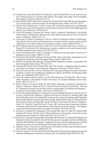
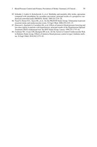
![52 W.J. Elliott
1.50
1.25
1.00
0.75
0.50
–5 0 5 10 15
Difference in Systolic Blood Pressure (mm Hg)
Fig. 3.4 Meta-regression plot of the relationship between the difference in achieved systolic
blood pressure between randomized arms vs. the odds ratio for stroke for the larger trials in
Tables 3.1 and 3.2. Note that trials with fewer than 58 strokes (5 % of those observed in the
chlorthalidone-lisinopril comparison in the Antihypertensive and Lipid-Lowering to prevent Heart
Attack Trial) are not shown, as their symbols are below the resolution of the figure. Trials involv-
ing an angiotensin receptor blocker are denoted by a triangle, calcium antagonists by squares,
ACE-inhibitors by circles, and both of the latter by an octagon. Open symbols denote placebo-
controlled trials. The area of each symbol is proportional to the number of strokes observed in each
trial. The identity of each symbol can be ascertained by reference to Tables 3.1 and 3.2. Note that
91 % of the area for all symbols falls within the dark, curved, dotted lines, representing the upper
and lower 95% confidence limits for the significant (P<0.0001) meta-regression analysis that was
based on the results of placebo-controlled trials of diuretic and/or β-blocker reported before the
year 2000. (Data from Staessen JA, Wang J-G, Thijs L. Cardiovascular prevention and blood pres-
sure reduction: A quantitative overview updated until 01 March 2003. J Hypertens.
2003;21:1055-1076)
Tuy nhiên, một phân tích 22 thử nghiệm đo những thay đổi trong độ dày động mạch cảnh liên
quan thử nghiệm dùng chẹn kênh calci thấy đièu này không đúng sự thật
Hiện cũng nhiều quan điểm cho rằng ARB có thể bảo vệ chống đột quỵ, đặc biệt là đột quỵ lần
thứ hai [39, 84, 85].
OddsRatioforStroke](https://image.slidesharecdn.com/hypertensionandstroke-170712211353/85/Hypertension-and-stroke-49-320.jpg)
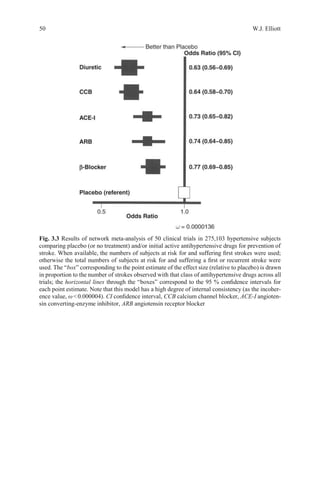
![3 Blood Pressure Control and Primary Prevention of Stroke: Summary of Clinical… 53
Trong phòng ngừa đột quỵ lần đầu, cho dùng Losartan để làm giảm tỷ lệ đột quỵ
[60], và các nghiên cứu khác liên quan đến ARB cho thấy không thể phòng ngừa đột
quỵ [11, 12, 44, 45]. Nhiều nghiên cứu gần đây và lớn hơn của ARB để phòng ngừa
đột quỵ lần đầu hoặc lần 2 đã không chứng minh được những quan sát trước đó [44,
68, 72, 74, 75, 86].
17.980 đối tượng tăng huyết áp trong nghiên cứu điều trị cao huyết áp tối ưu được
chọn ngẫu nhiên để huyết áp tâm trương ≤80, ≤85, hoặc ≤90 mmHg, điều trị chuyên
sâu với các phác đồ nhiều loại thuốc (thuốc đối kháng canxi ban đầu, theo sau là một
ACE-inhibitor), và sau đó theo dõi trong 3,8 năm, không có khác biệt về đột quỵ trên
ba nhóm ( P = 0,74) [87]. Mặc dù sự khác biệt thực tế trong HA tâm trương trên các
nhóm đều ít hơn nhiều so với kế hoạch ban đầu (~ 2 vs 5 mmHg), kết luận rõ ràng là
hạ huyết áp hơn những gì hiện nay đang khuyến cáo sẽ không có tác dụng hoặc đôi
khí có hại với những bệnh nhân THA [87]. Ngược lại, các nghiên cứu của HOT cho
thấy rõ khi giảm huyết áp thấp hơn bình thường ở bệnh nhân tiểu đường (tâm
trương≤ 80 mmHg) làm giảm 51% biến cố tim mạch, so với những người điều trị với
mục tiêu "thông thường" (tâm trương BP≤ 90 mmHg) [87]. kết luận tương tự cũng
được khuyến cáo để kiểm soát nguy cơ tim mạch ở bệnh nhân tiểu đường (nghiên
cứuACCORD) [88]. huyết áp tâm thu thấp hơn (< 119,3 mmHg) không khác gì so
với những người điều trị huyết áp tâm thu bình thường (tầm 133,5 mmHg) về nguy
cơ đột quỵ, nhưng giảm 12% tổng thể các biến cố tim mạch (P = 0,20). Những số liệu
này là một phần của lý do tại sao hướng dẫn tăng huyết áp gần đây [1-4] đã từ bỏ các
mục tiêu BP thấp hơn bình thường mà trước đó vẫn đề nghị cho các nhóm nguy cơ
cao (ví dụ, bệnh nhân bị bệnh tiểu đường, bệnh thận mãn tính và bệnh mạch vành).](https://image.slidesharecdn.com/hypertensionandstroke-170712211353/85/Hypertension-and-stroke-50-320.jpg)
![3 Blood Pressure Control and Primary Prevention of Stroke: Summary of Clinical… 57
50. Schrier RW, Estacio RO. Additional follow-up from the ABCD Trial in patients with Type 2
diabetes and hypertension [letter]. N Engl J Med. 2000;343:1969.
51. Tatti P, Pahor M, Byington RP, et al. Outcome results of the Fosinopril Amlodipine
Cardiovascular Events Randomized Trial (FACET) in patients with hypertension and
NIDDM. Diabetes Care. 1998;21:1779–80.
52. Efficacy of atenolol and captopril in reducing the risk of macrovascular and microvascular
complications in type 2 diabetes: UKPDS 39. UK Prospective Diabetes Study Group. BMJ.
1998;317:713–20.
53. Hansson L, Lindholm LH, Niskanen L, et al. Effect of angiotensin-converting-enzyme inhibi-
tion compared with conventional therapy on cardiovascular morbidity and mortality inhyper-
tension: the Captopril Prevention Project (CAPPP) randomised trial. Lancet. 1999;353:611–6.
54. National Intervention Cooperative Study in Elderly Hypertensives Study Group. Randomized
double-blind comparison of a calcium-antagonist and a diuretic in elderly hypertensives.
Hypertension. 1999;34:1129–33.
55. Hansson L, Lindholm LH, Ekbom T, et al. Randomised trial of old and new antihypertensive
drugs in elderly patients: cardiovascular mortality and morbidity. The Swedish Trial in Old
Patients with Hypertension-2 study. Lancet. 1999;354:1751–6.
56. Brown MJ, Palmer CR, Castaigne A, et al. Morbidity and mortality in patients randomised to
double-blind treatment with a long-acting calcium-channel blocker or diuretic in the
International Nifedipine GITS study: Intervention as a Goal in Hypertension Treatment
(INSIGHT). Lancet. 2000;356:366–72.
57. Hansson L, Hedner T, Lund-Johansen P, et al. for the NORDIL Study Group. Randomised trial
of effects of calcium antagonists compared with diuretics and β-blockers on cardiovascular
morbidity and mortality in hypertension: the Nordic Diltiazem (NORDIL) Study. Lancet.
2000;356:359–65.
58. Agodoa LY,Appel L, Bakris GL, et al. Effect of ramipril vs. amlodipine on renal outcomes in
hypertensive nephrosclerosis: a randomized controlled trial. African American Study of
Kidney Disease and Hypertension (AASK) Study Group. JAMA. 2001;285:2719–28.
59. Wright Jr JT, Bakris GL, Greene T, et al. Effect of blood pressure lowering and antihyperten-
sive drug class on progression of hypertensive kidney disease: results from the AASK Trial.
JAMA. 2002;288:2421–31.
60. Dahlöf B, Devereux RB, Kjeldsen SE, et al. for the LIFE Study group. Cardiovascular morbid-
ity and mortality in the Losartan Intervention For Endpoint reduction in hypertension study
(LIFE): a randomised trial against atenolol. Lancet. 2002;359:995–1003.
61. Zanchetti A, Bond M, Hennig M, et al. Calcium-antagonist lacidipine slows down progression
of asymptomatic carotid atherosclerosis. Circulation. 2002;106:2422–7.
62. Major outcomes in high-risk hypertensive patients randomized to angiotensin-converting
enzyme inhibitor or calcium channel blocker vs. diuretic: the Antihypertensive and Lipid
Lowering Treatment to Prevent Heart Attack Trial (ALLHAT). The ALLHAT Officers and
Coordinators for the ALLHAT Collaborative Research Group. JAMA. 2002;288:2981–97.
63. Wing LMH, Reid CM, Ryan P,et al. A comparison of outcomes with angiotensin-converting-
enzyme inhibitors and diuretics for hypertension in the elderly. Second Australian National
Blood Pressure Study Group. N Engl J Med. 2003;348:583–92.
64. Black HR, Elliott WJ, Grandits G, et al., for the CONVINCE Research Group. Principal results
of the Controlled ONset Verapamil INvestigation of Cardiovascular Endpoints (CONVINCE)
Trial.JAMA.2003;289:2073–82.
65. Malacco E, Marcia G, Rappelli A, et al. Treatment of isolated systolic hypertension: the
SHELL study results. The SHELL Investigators. Blood Press. 2003;12:160–7.
66. Pepine CJ, Handberg EM, Cooper-DeHoff RM, et al. A calcium antagonist vs. a non-calcium
antagonist hypertension treatment strategy for patients with coronary artery disease: the
International Verapamil-Trandolapril Study (INVEST): a randomized controlled trial. The
INVEST Investigators. JAMA. 2003;290:2805–16.
67. Yui Y, Sumiyoshi T, Kodama K, et al. Comparison of nifedipine retard with angiotensin con-
verting enzyme inhibitors in Japanese hypertensive patients with coronary artery disease: the](https://image.slidesharecdn.com/hypertensionandstroke-170712211353/85/Hypertension-and-stroke-54-320.jpg)
![Chapter 4
Cơ chế bản chất THA: liên quan
thần kinh và không do thần kinh
Scott H. Carlson, Sean Stocker, and J. Michael Wyss
Đột quỵ là nguyên nhân đứng hàng thứ tư gây tử vong ở Mỹ và là nguyên nhân hàng
đầu gây tàn tật, thường để lại cho bệnh nhân sự suy giảm vĩnh viễn và không thể làm
việc hoặc sống một cuộc sống độc lập. Một trong những yếu tố nguy cơ hàng đầu đối
với đột quỵ là tăng huyết áp và nguy cơ đột quỵ là tỷ lệ thuận với độ tăng và thời
gian bị THA [1-3]. Hơn nữa, tăng huyết áp cũng góp phần đáng kể vào bệnh tim
mạch, mà chính nó làm tăng nguy cơ đột quỵ. gần một thế kỷ nghiên cứu, các cơ chế
cơ bản làm tăng mạn tính áp lực động mạch ở hầu hết các bệnh nhân tăng huyết áp
vẫn khó nắm bắt. bước đầu được sáng tỏ bởi Guyton và những người khác, các yếu tố
tại thận có vai trò chính gây tăng huyết áp ở nhiều bệnh nhân, nhưng số lượng ngày
càng tăng các nghiên cứu chỉ ra rằng hệ thống thần kinh giao cảm và tương tác của
nó với hormone vận mạch và các chất trong tế bào được tạo ra cũng góp phần vào
bệnh lý tăng huyết áp. Chương này xem xét các bằng chứng, cho thấy có cơ chế thần
kinh mãn tính có thể làm tăng sức cản ngoại vi và huyết áp, sự tương tác giữa hệ
thống thần kinh giao cảm và hormone (ví dụ, angiotensin và nitric oxide) phối hợp
hiệu quả để làm tăng huyết áp. Cuối cùng, nó đánh giá vai trò của gốc tự do oxy hóa
S.H. Carlson, Ph.D.
Department of Biology, Luther College, Decorah, IA, USA
e-mail: carlsosc@luther.edu
S. Stocker, Ph.D.
Departments of Cellular and Molecular Physiology and Neural and Behavioral Sciences,
Penn State College of Medicine, Hershey, PA, USA
e-mail: sstocker@hmc.psu.edu
J.M. Wyss, Ph.D. (*)
Departments of Cell Developmental and Integrative Biology and Medicine, University of
Alabama at Birmingham, Birmingham, AL, USA
e-mail: jmwyss@uab.edu
© Springer International Publishing Switzerland 2016 63
V. Aiyagari, P.B. Gorelick (eds.), Hypertension and Stroke, Clinical
Hypertension and Vascular Diseases, DOI 10.1007/978-3-319-29152-9_4](https://image.slidesharecdn.com/hypertensionandstroke-170712211353/85/Hypertension-and-stroke-58-320.jpg)
![64 S.H. Carlson et al.
(ROS) trong điều hòa hoạt động giao cảm và vai trò của eicosanoid 20-
hydroxyeicosatetraenoic acid (20-HETE) điều chỉnh cơ trơn mạch máu trong
THA và co thắt mạch do đột quỵ
Hệ thống thần kinh giao cảm và tăng huyết áp
Trong 40 năm qua, các nghiên cứu lâm sàng và nghiên cứu trên động vật đã xác định
rằng hệ giao cảm góp phần vào bản chất của THA [4, 5]. Tuy nhiên, những gì dẫn
đến tăng hoạt động giao cảm cao này thì chưa rõ ràng. Tủy vùng bụng mỏ (RVLM)
là nhân chính trong não dẫn truyền xung cho tế bào thần kinh trước hạch trong tủy
sống nhân trước bên để hỗ trợ hoạt động của hệ thần kinh giao cảm. Các tế bào thần
kinh RVLM rất nhạy cảm với bộ phận nhận cảm áp phản hồi tới thần kinh trung
ương và tự điều hòa với tần số tương ứng với hoạt động của hệ thần kinh giao cảm
[6]. Các chất hóa học kích thích tế bào thần kinh RVLM làm tăng hoạt động thần
kinh giao cảm và huyết áp, nhiều nghiên cứu chỉ ra rằng tăng huyết áp do thần kinh
xuất phát từ tính chất thần kinh RVLM bị thay đổi và / hoặc tăng kích thích đầu vào
synaptdo đó làm tăng hoạt động của hệ thần kinh giao cảm, làm tăng co mạch làm
cao huyết áp (xem [5, 8];. Hình 4.1). Tăng hướng kích thích trong tăng huyết áp do
thần kinh có thể phát sinh ở một số vùng của não: các nhân ở vùng dưới đồi như
nhân cận não thất và nhân lưng, nhân đuôi ở thân não [9, 10], receptor nhận cảm hóa
học [11], natri kích thích tế bào thần kinh trong vùng AV3V của não trước [12], và
kìm hãm receptor nhận cảm áp lực ở trung ương như vùng đuôi bụng bên. Các
nghiên cứu cho thấy tăng kích thích bộ phận nhận cảm, giảm ức chế RVLM góp phần
làm THA do thần kinh [5, 6, 12-15].
Thận có chức năng ổn định nội môi, nhiều nghiên cứu đã tập trung vào các dây thần
kinh giao cảm ở thận và quá trình tăng hoạt của chúng. Cắt dây thàn kinh ở thận làm
giảm huyết áp ở một số mô hình động vật gặm nhấm [16-19] và ở những người tăng
huyết áp không đáp ứng thuốc [17, 20]. Tuy nhiên, kiểm soát giao cảm của vùng
mạch máu khác cũng có thể kiểm soát tăng huyết áp. hoạt động thần kinh nội tạng
được nâng lên để đáp ứng với quá trình tăng huyết áp của angiotensin [21], và cắt bỏ
các dây thần kinh tạng làm giảm huyết áp](https://image.slidesharecdn.com/hypertensionandstroke-170712211353/85/Hypertension-and-stroke-59-320.jpg)
![4 Mechanisms Underlying Essential Hypertension: Neurogenic and Non-neurogenic… 65
Fig. 4.1 Arterial .pressure is a function of sympathetic nervous system (SNS) activity,circulating
endocrine factors, and renal function. SNS activity is regulated by the spontaneous discharge of
neurons in the rostral ventrolateral medulla (RVLM), which are regulated by baroreceptor and
chemoreceptor input to the nucleus of the solitary tract (NTS) and innervation from the caudal
pressor area of the medulla and from hypothalamic regions, e.g., the paraventricular (PVN), lateral
hypothalamic area (LHA), and anterior hypothalamic (AHN) nuclei
trong khi hoạt động thần kinh của thận giảm [24]. Những kết quả này gợi ý rằng hoạt
động của thần kinh giao cảm được phân bố không đồng đều cho tất cả các cơ quan,.
Khái niệm này được hỗ trợ bởi một số lượng ngày càng nhiều các nghiên cứu [25-27]
tăng hoạt động của hệ thần kinh giao cảm có thể là hậu quả của sự suy giảm các phản
xạ thần kinh. Có lẽ hệ thống thông tin phản hồi được nghiên cứu nhiều nhất là
receptor nhận cảm áp lực ở động mạch (nhằm đáp ứng với những thay đổi trong áp
lực động mạch), các thụ thể phổi tim mạch (trong đó phát hiện những thay đổi về thể
tích máu), và thụ thể nhận cảm hóa học (đáp ứng với những thay đổi khí máu và pH)
. Tất cả những cơ chế phản hồi phản ứng cấp tính thay đổi theo các tham số theo dõi
và làm thay đổi hoạt động của hệ thần kinh tự trị để duy trì áp lực động mạch cơ bản
(Hình. 4.1). Bristow và cộng sự [28] là những người đầu tiên đề xuất rằng sự mất cân
bằng bộ phận nhận cảm áp động mạch có thể làm thay đổi mạn tính quy định áp lực
động mạch; Tuy nhiên, nghiên cứu mở rộng của Cowley và Guyton đã chứng minh
rằng ở chó, việc loại bỏ phản xạ nhận áp lai làm tăng rối loạn nhận cảm huyết áp
nhưng không làm tăng huyết áp trung bình](https://image.slidesharecdn.com/hypertensionandstroke-170712211353/85/Hypertension-and-stroke-60-320.jpg)
![66 S.H. Carlson et al.
[29]. Những nghiên cứu sau đó đã chứng minh để phản ứng lại quá trình THA
bền vững, receptor nhận cảm áp lực nhanh chóng thiết lập lại một điểm mới và sau
đó, điều chỉnh hoạt động của hệ thần kinh tự trị để bảo vệ các điểm đặt áp lực
động mạch mới. Những phát hiện này dẫn đến giả thuyết rằng receptor nhận áp chỉ
điều chỉnh ngắn hạn hoạt động tự trị và không tham gia lâu dài vào tăng huyết áp
do thần kinh (Cowley [30]).
Ngược lại thập kỷ trước đó, một số nghiên cứu gần đây cho thấy receptor nhận áp
có vai trò mãn tính hơn trong việc điều tiết hoạt động của hệ thần kinh tự động, do
đó, có thể góp phần vào làm tăng giao cảm và duy trì huyết áp ở một số cá nhân.
Các nghiên cứu của Thrasher và Lohmeier thấy receptor nhận áp mạn tính có thể
ức chế hệ thần kinh giao cảm và sự mất cân bằng trong cung phản xạ có thể dẫn
đến tăng huyết áp duy trì [31-33].
điều tiết hệ thống thần kinh chi phối huyết áp
Ngoài những thay đổi trong kiểm soát phản xạ của hoạt động tự trị, tăng hoạt động
giao cảm có thể do điều tiết trung ương với áp lực động mạch. Trong khi RVLM
đã được nghiên cứu rộng rãi bởi vì vai trò của nó như là loại thuốc giao cảm và
điều chỉnh cấp tính áp lực động mạch. Vùng dưới đồi như 1 vùng chi phối chủ yếu
của hệ thần kinh tự động (Hình. 4.1).](https://image.slidesharecdn.com/hypertensionandstroke-170712211353/85/Hypertension-and-stroke-61-320.jpg)
![4 Mechanisms Underlying Essential Hypertension: Neurogenic and Non-neurogenic… 67
Quá trình deoxycorticosterone acetate steroid (quá trình khử oxy của
corticosterone acetate) và chế độ ăn nhiều muối, kích thích vùng bên và hậu đồi
làm tăng huyết áp động mạch và nhịp tim, trong khi tổn thương sau vùng dưới đồi
làm giảm áp lực động mạch [38]. Hơn nữa, khu vực bên và lưng dưới đồi đáp ứng
với nồng độ leptin có vai trò tăng hoạt động giao cảm và huyết áp, là nguyên nhân
dẫn đến tăng huyết áp kèm theo béo phì [39, 40].
Tế bào nơ ron magnocellular tổng hợp và giải phóng vasopressin vào tuần hoàn,
trong khi tế bào thần kinh parvocellular đơn hoặc đa synap kiểm soát hoạt động
tim mạch của hệ, bao gồm cả các RVLM, khu vực nhận cảm sàn não thất 4 (area
postrema), NTS và nhân sừng bên tủy sống. Thông qua sự liên quan với tế bào
parvocellular làm thay đổi chức năng tim mạch.
Vùng trước đồi có 1 số khu vực kiểm soát tim mạch như vùng trước não thất 3 góp
phần làm THA ở một số mô hình động vật. nhân trung gian trước vùng giao thoa
thị giác cũng liên quan tới 1 số tác động trên tim mạch ( [42, 43]). Nhân giao thoa
thị giác cũng điều chỉnh giải phóng vasopressin và nước, tác động gián tiếp tới bộ
phận nhận áp động mạch. Nhân trước vùng dưới đồi cùng nhân giao thoa thị giác
tác động lớn tới giao cảm.
Một số vùng vỏ não cũng ảnh hưởng tới huyết áp, đặc biệt là liên quan đến ý
thức[45] hoặc tình huống tâm lý [46]. Chúng bao gồm vùng trước vỏ thùy trán[45]
thùy đảo và hạch nhân [47]. Cho dù các khu vực này đóng góp cho tăng huyết áp
ít được biết đến.
Hệ Renin-Angiotensin và tăng huyết áp
các yếu tố nội tiết đóng góp đáng kể vào việc điều chỉnh áp lực động mạch ngoại
biên thông qua tác động trung gian và có thể là yếu tố gây bệnh trong tăng huyết
áp. Trong số này, hệ thống renin-angiotensin-aldosterone (RAAS) có lẽ được
nghiên cứu kỹ lưỡng nhất hormone, chủ yếu là do angiotensin II (AII) gây co
mạch mạnh và liên quan nhiều đến thể tích tuần hoàn](https://image.slidesharecdn.com/hypertensionandstroke-170712211353/85/Hypertension-and-stroke-62-320.jpg)
![68 S.H. Carlson et al.
trong các loại thuốc hạ huyết áp hiện nay, ACEi như 1 thuốc hạ huyết áp hiệu quả
nhất với phần lớn bệnh nhân. Hơn nữa, một số mô hình động vật gặm nhấm có tăng
huyết áp liên quan chủ yếu tới AII [48]. Trong khi AII xuất hiện để tăng áp lực động
mạch trong các nghiên cứu này, ít nhất là một phần, thông qua duy trì khối lượng
tuần hoàn hoặc tăng kháng trở mạch máu do tăng hoạt động giao cảm. Điều này đã
khiến các nhà nghiên cứu đưa ra giả thuyết rằng RAAS hoạt động quá mức có thể
gây tăng huyết áp bằng cách trực tiếp làm tăng hoạt động của hệ thần kinh giao cảm.
Một tác động trung tâm mà AII có thể thay đổi điều hòa dòng máu chảy là do ức chế
chức năng bộ phận nhận cảm áp (Hình. 4.2). Một số nghiên cứu đã chứng minh rằng
kiểm soát receptor nhận áp động mạch làm giảm huyết áp đáng kể sau khi dùng ARB
AII 1 (AT1) receptor blocker [50]. Các quan sát sự lưu hành AII ức chế hoạt động
phản xạ nhận áp do AII tác động vào các vị trí ở thần kinh trung ương để gây tác
dụng . Tuy nhiên, AII không vượt qua hàng rào máu não còn nguyên vẹn, và hoạt
động bằng cách gắn vào và kích thích các thụ thể ở khu vực quanh não thất, nơi
không có hàng rào máu não
Fig. 4.2 Circulating angiotensin (AII) can bind to circumventricular organs (e.g., the area pos-
trema (AP) or subfornical organ (SFO)), increasing neuronal activity of sympathoexcitatory nuclei
or modulating baroreceptor sensitivity. Alternatively, AII stimulation can lead to activation of the
intraneuronal renin–angiotensin–aldosterone system (RAAS) and subsequent generation of AII
and related metabolites and/or generation of reactive oxygen species (ROS), both of which may
alter the neuronal firing rate of cardiovascular regulatory nuclei and increase sympathetic nervous
system activity. AII activation of receptors in the blood vessels (BVs) of the brain stem can also
alter neuronal activity in the NTS and RVLM through generation of second messengers that cross
the blood–brain barrier (e.g., NO and superoxide)](https://image.slidesharecdn.com/hypertensionandstroke-170712211353/85/Hypertension-and-stroke-63-320.jpg)
![4 Mechanisms Underlying Essential Hypertension: Neurogenic and Non-neurogenic… 69
liên kết với các thụ thể cảm biến mạch máu não sau đó truyền tín hiệu đến não [56].
cần lưu ý rằng một nghiên cứu gần đây cho thấy rằng hàng rào máu não có thể trở
nên tăng tính thấm với AII trong tăng huyết áp [57]. Khu vực nền não thất là khu vực
như vậy. Nó tiếp giáp với nhân tractus solitarius, là vị trí của đầu vào bộ phận nhận
cảm áp đến vùng tủy, receptor AII [48].Một số quan sát hỗ trợ tiêm angiotensin vào
khu vực nền não thất có receptor nhận áp làm tăng huyết áp, cắt bỏ khu vực nền não
thất làm giảm huyết áp [59]. Hơn nữa, loại bỏ khu vực quanh nền não thất làm ngăn
tác động của ACEi lên huyết áp và phong tỏa bộ phận nhận cảm áp trong SHR [60].
Nghiên cứu về vai trò của RAAS trong tăng huyết áp tập trung vào các tác động
ngoại vi của AII (ví dụ, làm tăng giải phóng norepinephrine) hoặc tác động lên tuần
hoàn qua tác dụng trung gian. Tuy nhiên, trong vòng 30 năm qua, các nghiên cứu
chứng minh rằng tế bào thần kinh và các tế bào thần kinh đệm trong thần kinh trung
ương bao gồm tất cả thành phần cuả hệ RAAS, bao gồm angiotensinogen, renin, men
chuyển và tất cả các thụ thể angiotensin [48, 61]. gợi ý rằng angiotensin có thể hoạt
động như một tác nhân điều biến hoặc tiết ra như 1 chất dẫn truyền thần kinh. thụ thể
AII được phân bố ở hầu hết các nhân não liên quan tác động lên tim mạch [61], bao
gồm PVN, nhân parabrachial, RVLM và NTS [48].
Sau khi angiotensin giải phóng sẽ liên kết với các thụ thể thần kinh dẫn đến tăng sản
xuất vasopressin (từ PVN).](https://image.slidesharecdn.com/hypertensionandstroke-170712211353/85/Hypertension-and-stroke-64-320.jpg)
![70 S.H. Carlson et al.
Nitric Oxide và tăng huyết áp
Nitric oxide (NO) là một thuốc thứ hai liên quan mạnh mẽ tới hoạt động của hệ
thống thần kinh giao cảm và kiểm soát huyết áp. NO, được sản xuất từ các axit
amin tiền thân arginine bởi nitric oxide (NOS), là một thuốc giãn mạch mạnh và
có xu hướng làm giảm huyết áp khi được tạo ra trong các mạch máu. ức chế NOS
bởi L-NAME gây tăng huyết áp do tăng kháng trở mạch máu ngoại vi và phản ứng
mạch máu do kích thích adrenergic [72].
Điều thú vị là, L-NAME gây tăng huyết áp liên quan đến cả hệ thống renin-
angiotensin và hệ thần kinh giao cảm[72].
Mặc dù cơ chế mà NO tác động trung tâm điều biến hoạt động thần kinh không
rõ ràng, nghiên cứu cho thấy NO có thể làm thay đổi phản ứng thần kinh ở đuôi
gai ở tế bào thần kinh và các yếu tố kích thích. Một khu vực đáng lưu ý ở đây là
NTS, đó là nơi chuyển tiếp giữa đầu vào và bộ phận nhận cảm áp RVLM. Tiêm
glutamate vào RVLM làm tăng thoáng qua áp lực động mạch. Điều này thường
gây ra phản xạ giảm nhịp tim và hoạt động thần kinh giao cảm thận làm giảm áp
lực động mạch [13]. Mifflin và các đồng nghiệp đã chỉ ra rằng tiêm đối kháng L-
NAME vào RVLM trước khi tiêm glutamate làm giảm đáng kể phản xạ của huyết
áp và nhịp tim. Những kết quả này ủng hộ giả thuyết rằng trong RVLM, NO tạo
cơ chế phản hồi với glutamine trung gian qua receptor cảm áp và thụ thể ở tim
phổi.](https://image.slidesharecdn.com/hypertensionandstroke-170712211353/85/Hypertension-and-stroke-65-320.jpg)
![4 Mechanisms Underlying Essential Hypertension: Neurogenic and Non-neurogenic… 71
Phản ứng oxy hóa (ROS) và tăng huyết áp
Trong khi NO và angiotensin đóng vai trò tác động hệ thống thần kinh giao cảm
trong tăng huyết áp làm tăng huyết áp. Các nghiên cứu gần đây đã tập trung vào sự
cảm ứng với angiotensin của ROS, bao gồm các ion oxy, các gốc tự d, và peroxit, tất
cả đều là sản phẩm phụ quá trình chuyển hóa bình thường của enzyme như NADPH-
oxidase. ROS được tạo ra, chúng được chuyển đổi bởi superoxide dismutase nội bào
(SOD) thành hydro peroxide. hydrogen peroxide tự nó là một gốc tự do mạnh, nó
nhanh chóng bị phân hủy bởi các enzyme như catalase, glutathione peroxidase và
peroxiredoxins.
Các nghiên cứu gần đây chỉ ra rằng ROS có vai trò trong tăng huyết áp, ROS đã được
chứng minh góp phần tăng huyết áp do thần kinh bằng cách gây kích thích giao cảm.
ROS tăng trong SHR [75], thậm chí trước khi bắt đầu có tăng huyết áp do mạch máu
và thận, thì ROS đã được tiết ra [76, 77]. Những dữ liệu này hỗ trợ vai trò trung tâm
của ROS trong tăng huyết áp.
Cơ chế mà ROS thay đổi kiểm soát tim mạch chưa rõ ràng. Một số ảnh hưởng của
ROS xảy ra rõ ràng ở ngoại vi và thay đổi nội mô và chức năng thận. Tuy nhiên,
nghiên cứu gần đây đã chỉ ra rằng ROS làm tăng hoạt động giao cảm, dẫn đến tăng
huyết áp do thần kinh. Nhiều bằng chứng cho thấy ROS tham gia vào con đường
truyền tín hiệu nội bào, bao gồm cả con dường dẫn truyền AII. truyền tĩnh mạch AII
lăngm t áp lực động mạch và kích thích giao cảm [82]. Những kết quả này gợi ý rằng
tăng AII có thể làm tăng áp lực động mạch bằng cách tăng hệ ROS trong tế bào thần
kinh SFO, do đó làm tăng hoạt hóa của các trung tâm ở vùng dưới đồi kiểm soát hoạt
động của hệ thần kinh
20-HETE
Trong khi huyết áp có thể được điều khiển bởi các cơ chế trung ương mô tả ở trên,
các yếu tố mạch máu cũng hoạt động đồng thời làm tăng huyết áp. Một đóng góp
nghiên cứu là eicosanoid axit 20-hydroxyeicosatetraenoic (20-HETE), là một sản
phẩm phụ của quá trình chuyển hóa acid arachidonic bởi enzym cytochrome P450](https://image.slidesharecdn.com/hypertensionandstroke-170712211353/85/Hypertension-and-stroke-66-320.jpg)
![72 S.H. Carlson et al.
(CYP). 20-HETE làm tăng phản ứng co mạch do kích thích giao cảm và hormone
vận mạch như angiotensin và phenylephrine [85]. Những tác động trung gian bằng
cách chặn dẫn truyền canxi qua các kênh kali (BKCa), dẫn đến quá trình khử cực và
co cơ trơn mạch máu. 20-HETE cũng làm tăng lượng canxi bằng cách mở cổng
protein kinase C-gated L-type Ca2+ [58, 85], và tăng độ nhạy canxi với myosin trong
cơ trơn mạch máu [85].
Các hoạt động vận mạch của 20-HETE chỉ ra rằng nó là 1 nguyên nhân góp phần
tăng huyết áp. Thật vậy, CYP tăng quá mức trong một số mô hình tăng huyết áp và
góp phần vào sự phát triển và duy trì huyết áp cao ở động vật. Ví dụ, những thay đổi
trong biểu hiện của CYP tương ứng với sự phát triển của bệnh cao huyết áp trong
SHRs, và mức độ tăng huyết áp có thể hãm lại bằng cách ức chế các enzym [86].
CYP quá mức cũng gặp trong angiotensin gây tăng huyết áp [87-89]. Hơn nữa, oxit
nitric có vẻ ức chế hoạt động CYP và 20-HETE [94], cho thấy khả năng tăng huyết
áp do cạn kiệt NO dẫn đến tăng sản xuất 20-HETE và góp phần làm tăng huyết áp.
Gộp chung lại, những kết quả này cho thấy sự thay đổi CYP và tương ứng với nồng
độ 20-HETE góp phần làm đa dạng bệnh lý tăng huyết áp.
Ngoài khả năng giúp làm tăng mẫn cảm với các kích thích mạch máu ngoại sinh,
nghiên cứu cho thấy 20-HETE tham gia điều chỉnh mô cơ liên quan kháng trở của
tiểu động mạch[97]. Những thay đổi trong áp lực xuyên thành của động mạch kích
thích sinh 20-HETE gây co động mạch bằng cách tăng canxi tràn qua các receptor
vận chuyển (TRP) và đóng các kênh BKCa [97, 98]. Điều này chỉ ra vai trò liên quan
tới mô cơ của 20-HETE trong nhiều loại mạch máu (ví dụ, mạc treo ruột, thận, tim)
[99-101], và phong bế 20-HETE làm giảm cơ chế tự điều chỉnh của cả hai dòng máu
tới thận và não trong cơ thể [102].](https://image.slidesharecdn.com/hypertensionandstroke-170712211353/85/Hypertension-and-stroke-67-320.jpg)
![4 Mechanisms Underlying Essential Hypertension: Neurogenic and Non-neurogenic… 73
Có 1 chất là Eets kích hoạt kênh BKCa trong tế bào cơ trơn mạch máu, dẫn đến tăng
phân cực và giãn mạch máu[105]. Eets được tổng hợp bởi tế bào hình sao trong hệ
thống thần kinh trung ương, và những thay đổi trong hoạt động thần kinh dẫn đến
hình thành EET ở tế bào hình sao và giãn mạch tương ứng [104, 106]. Hiệu ứng ròng
của EET là để phù hợp với lưu lượng máu não tùy theo nhu cầu trao đổi chất từng
khu vực. mức độ thay đổi của Eets cũng liên quan đến tăng huyết áp và chức năng
của mạch; trong nhiều trường hợp tăng huyết áp, nồng độ của Eets giảm thông qua
giảm trao đổi chất và mức độ huyết áp (xem [105]). Như vậy, huyết áp có thể được
kiểm soát ở mức độ mạch máu bằng cán cân 20-HETE và EET.
Vai trò của CYP có nguồn gốc từ 20-HETE và Eets trong mạch máu não đã được
quan tâm đặc biệt trong đột quỵ. Tương tự như mạch khác, lưu lượng máu não được
tự điều chỉnh qua 2 quá trình kiểm soát này, trong điều kiện bình thường thì nó giúp
bảo trì lưu lượng máu não không đổi. Tuy nhiên, ở trạng thái bệnh lý như THA cao
có thể hạn chế quá trình tự điều chỉnh gây giảm tác động tới mạch máu và thay đổi
chức năng nội mô [85, 104]. Những rối loạn này có thể làm thay đổi cán cân 20-
HETE và EET, do đó tăng co thắt mô cơ và giảm lưu lượng máu não trong khi cũng
tăng nguy cơ đột quỵ thiếu máu cục bộ và suy giảm tri giác [104, 105]. 20-HETE
tăng trong dịch não tủy ở chuột bị đột quỵ xuất huyết, và tăng nồng độ 20-HETE góp
phần gây co thắt mạch [107]. dùng chất ức chế 20-HETE ngăn ngừa giảm thiếu máu
cục bộ dựa trên lượng máu tới tim [108] và não [109], trong khi cũng làm giảm kích
thước vùng nhồi máu. 20-HETE cũng tăng trong mạch máu não SHR [110] và đột
quỵ SHR [111], góp phần giảm oxy hóa, rối loạn chức năng nội mô và tăng tổn
thương do thiếu máu cục bộ.](https://image.slidesharecdn.com/hypertensionandstroke-170712211353/85/Hypertension-and-stroke-68-320.jpg)
![Chapter 5
Ảnh hưởng của THA và đột quỵ tới
tuần hoàn mạch não
Anne M. Dorrance
lưu lượng máu não phải được điều hòa chặt chẽ để đảm bảo rằng các nhu cầu trao
đổi chất của tế bào thần kinh luôn được đáp ứng [1]. Cơ chế kiểm soát tinh tế này
được hỗ trợ bởi hai cơ chế sinh lý và cơ chế tự điều hòa của bộ đôi thần kinh-
mạch máu. Chương này sẽ thảo luận về các cơ chế này và làm thế nào mà chúng
bị rối loạn bởi THA và thiếu máu cục bộ não. sức cản động mạch cũng góp phần
vào việc điều tiết lưu lượng máu não. Tăng huyết áp gây ra thay đổi cấu trúc trong
các động mạch não làm tăng sức cản động mạch và gây ra tổn thương do thiếu
máu cục bộ não. Các cơ chế liên quan vấn đề này cũng sẽ được thảo luận. đột quỵ
thiếu máu cục bộ hay gặp nhất ; do đó ở đây sẽ nói phần lớn về kiểu đột quỵ này.
Các vấn đề kiểm soát mạch máu não phụ thuộc vào thời gian và vùng thiếu máu
cục bộ trong cơn đột quỵ, do đó cấu trúc và chức năng của chúng là yếu tố quyết
định quan trọng tới độ lớn cuối cùng của vùng nhồi máu và kết quả lâm sàng [2].
Giải phẫu động mạch não
Trước khi thảo luận về những tác động của tăng huyết áp trên động mạch não cần
tạm dừng để mô tả một số sinh lý cơ bản của tuần hoàn não. Các động mạch não
cũng có cấu trúc phù hợp để ngăn chặn suy giảm lưu lượng máu đến nhu mô trong
trường hợp đột quỵ thiếu máu cục bộ [3, 4]. vòng Willis là một vòng động mạch
có các nhánh nối tại đáy não (Hình. 5.1a). Cấu trúc này cho phép tưới máu ngang
sang bán cầu não, và lưu lượng máu trong vòng tròn của Willis có thể được đảo
ngược để
A.M. Dorrance, Ph.D. (*)
Department of Pharmacology and Toxicology, Michigan State University,
East Lansing, MI, USA
e-mail: dorranc3@msu.edu
© Springer International Publishing Switzerland 2016 81
V. Aiyagari, P.B. Gorelick (eds.), Hypertension and Stroke, Clinical
Hypertension and Vascular Diseases, DOI 10.1007/978-3-319-29152-9_5](https://image.slidesharecdn.com/hypertensionandstroke-170712211353/85/Hypertension-and-stroke-75-320.jpg)
![a ACA Front b
ACA
ICA MCA
PCA
Basilar a.
ACA
Vertebral a. Back MCA
ICA
c
Glial basal lamina
Vascular
basal lamina
Perivascular space
Smooth
muscle
Astrocyte end-feet Pericyte
Endothelial
basal lamina Tight junctions
Endothelial cells
82 A.M. Dorrance
Artery Arteriole Capillary
Fig. 5.1 Cerebral blood supply. (a) Depicts the major cerebral arteries including the circle of
Willis, the middle cerebral artery, and the basilar artery. (b) Depicts the blood supply to the deep
white matter. The penetrating arterioles branch from the pial arteries and dive into the brain paren-
chyma. (c) Depicts the structure of the artery/arteriole/capillary wall. ACA anterior cerebral artery,
ICA internal carotid artery, MCA middle cerebral artery, PCA posterior cerebral artery. (From
Iadecola, C., The pathobiology of vascular dementia. Neuron, 2013. 80(4): p. 844-66, with
permission)
Cấp máu thay cho động mạch bị tắc. Các động mạch màng mềm bao phủ bề mặt của
não cũng cấp máu bảo vệ não nếu thiếu máu cục bộ. có 1 số lượng lớn các chỗ nối
kết nối các động mạch phân nhánh sau và động mạch não giữa và phía trước với
động mạch não giữa [5]. Những vòng nối này cấp máu thay thế trong trường hợp 1
động mạch nhỏ bề mặt bị tắc [6-8]. Các động mạch màng mềm được phân bố theohệ
thần kinh ngoại biên [9], và các dây thần kinh giao cảm và phó giao cảm giúp điều
chỉnh trương lực của mạch máu giúp co mạch (norepinephrine và neuropeptide Y) và
giãn mạch (acetylcholine và oxit nitric) tương ứng [9-11].
Các tiểu động mạch và tiểu động mạch từ động mạch màng mềm lại đi vào nhu mô
để điều tiết vi tuần hoàn trong nhu mô (5.1b hình.) [12].](https://image.slidesharecdn.com/hypertensionandstroke-170712211353/85/Hypertension-and-stroke-76-320.jpg)
![5 The Effects of Hypertension and Stroke on the Cerebral Vasculature 83
Fig. 5.2 Illustration of the cerebral arteries and the neurovascular unit. The diagram shows the
pial arteries on the surface of the brain including the extrinsic innervation. The penetrating arteri-
oles are also depicted; these arterioles contain endothelial cells and one layer of vascular smooth
muscle cells. The penetrating arterioles are surrounded by astrocytes, neurons, and pericytes (not
illustrated here) and they receive intrinsic innervation from within the neuropil. The capillaries
consist of a layer of endothelial cells sounded by pericytes, neurons, and astrocytes. (Adapted from
Filosa, J.A., et al., Beyond neurovascular coupling, role of astrocytes in the regulation of vascular
tone. Neuroscience, 2015., doi:10.1016/j.neuroscience.2015.03.064, with permission)
Nhưng các tiểu động mạch xâm nhập vào nhu mô lại có rất ít nhánh, do đó tắc
một tiểu động mạch đi vào nhu mô có thể gây tổn thương đáng kể tới vỏ não xung
quanh [13]. Dòng chảy của tiểu động mạch chi phối bởi tế bào thần kinh neuropil
(Hình. 5.2) [9]. Hầu hết các dây thần kinh có các tế bào hình sao xung quanh động
mạch giúp truyền tín hiệu chuyển tiếp từ tế bào thần kinh sang các tiểu động mạch
để tăng tưới máu trong thời gian cần nhu cầu trao đổi chất cao [14, 15].
Một mạng lưới dày đặc các mao mạch não phát sinh từ các tiểu động mạch xâm
nhập [16], và có khả năng dự phòng quan trọng để cung cấp máu giúp bảo vệ khi
có thiếu máu cục bộ não [17]. Các mao mạch được bao quanh bởi các tế bào
quanh mạch (pericytes) giúp điều chỉnh tưới máu [18]. Tuy nhiên, nhiều nghiên
cứu gần đây cho thấy pericytes không chứa các protein alpha gây co sợi actin cơ
trơn, và chúng không điều chỉnh lưu lượng máu để đáp ứng với kích thích thần
kinh hoặc giảm tưới máu lan rộng [19]. Tuy nhiên, mật độ tế bào pericyte cao](https://image.slidesharecdn.com/hypertensionandstroke-170712211353/85/Hypertension-and-stroke-77-320.jpg)
![84 A.M. Dorrance
Theo quan sát thấy các tế bào này đóng một vai trò sinh lý quan trọng trong não [20,
21]. Các pericytes là một phần của các đơn vị thần kinh mạch máu, mà cũng có chứa
các tế bào cơ trơn mạch máu, tế bào nội mô, tế bào hình sao và tế bào thần kinh
(Hình. 5.2). Các pericytes có thể hoạt động như một cầu nối để chuyển tiếp thông tin
giữa hệ thần kinh và mạch máu trong não [19].
Bộ đôi thần kinh mạch máu
Bội đôi thần kinh mạch máu đi với nhau phục vụ nhu cầu trao đổi chất của tế bào
thần kinh để tưới máu não [13]. Điều này đòi hỏi tín hiệu tích hợp nhanh chóng giữa
các tế bào thần kinh, thần kinh quanh mạch, thần kinh đệm và các tế bào trong mạch
máu [25, 26]. Các tế bào kích thích gây giãn tiểu động mạch và nhánh động mạch đi
vào nhu mô. Bộ đôi thần kinh mạch máu hoạt động nhờ ba con đường giãn mạch
chính: oxit nitric (NO) [29, 30], cyclooxygenase (COX) -2 [31], và axit
epoxyeicosatrienoic (Eets) [32]. mô tả chi tiết về các cơ chế truyền tín hiệu liên quan
đến bộ đôi thần kinh mạch máu xin vui lòng xem [25].
Ảnh hưởng của tăng huyết áp trên thần kinh - mạch máu
Các nghiên cứu về ảnh hưởng của tăng huyết áp trên thần kinh mạch máu ở người
còn hạn chế. 1 số các nghiên cứu cho thấy angiotensin II có thể có tác động trực tiếp
vào mạch máu thần kinh mà không liên quan đến sự gia tăng huyết áp.](https://image.slidesharecdn.com/hypertensionandstroke-170712211353/85/Hypertension-and-stroke-78-320.jpg)
![5 The Effects of Hypertension and Stroke on the Cerebral Vasculature 85
Điều trị hạ huyết áp với losartan, chẹn thụ thể angiotensin (ARB), không có tác dụng
có lợi trên bộ đôi thần kinh mạch máu [37]. Nó cũng cho thấy hiệu quả bộ đôi thần
kinh mạch máu bị suy giảm không phụ thuộc vào huyết áp. Nghiên cứu này rõ ràng
là mâu thuẫn với các nghiên cứu trước đây đã mô tả ở những con chuột bị tăng huyết
áp phụ thuộc angiotensin II. Những con chuột được điều trị bằng losartan (30 tuần)
đã tăng huyết áp ít nhất hai phần ba cuộc đời của chúng. Do đó, có thể là thuốc không
có hiệu quả vì các tổn thương mạch máu qua tác động trung gian của tăng huyết áp
kéo dài không thể đảo ngược.
Ảnh hưởng của đột quỵ trên thần kinh mạch máu
Nghiên cứu về thần kinh mạch máu ở người sau đột quỵ đã mang lại kết quả khác
nhau. Điều thú vị là khi dùng thuốc để giãn động mạch não mạnh, acetazolamide
không làm tăng lưu lượng máu trong vỏ não cùng bên. Acetazolamide được sử dụng
để đo khả năng dự trữ mạch máu não hoặc tưới máu tối đa có thể trong não. Nghiên
cứu này cho thấy rằng thiếu máu cục bộ làm giảm khả năng dự trữ mạch máu mà
không làm suy yếu thần kinh mạch máu, do đó các cơ chế làm giãn mạch máu xuất
hiện để bù trừ giảm tưới máu não sau thiếu máu cục bộ.
Các nghiên cứu sử dụng PET và kích thích thị giác ở những bệnh nhân hẹp động
mạch cảnh](https://image.slidesharecdn.com/hypertensionandstroke-170712211353/85/Hypertension-and-stroke-79-320.jpg)
![86 A.M. Dorrance
cả hai mắt thấy có tăng tưới máu cho cả vỏ não và thị giác ở tất cả các bệnh nhân.
Hầu hết bệnh nhân có giảm lưu lượng máu ở bán cầu bị tổn thương khi so với bán
cầu bên đối diện. Sự giảm lưu lượng máu trong các khu vực xung quanh vỏ não,
thị giác có thể là hậu quả của sự phân phối lại dòng máu trong não, tức là, sự gia
tăng lưu lượng máu đến vỏ não thị giác có thể ăn cắp máu từ các vùng lân cận
[39].
Các phản ứng thần kinh mạch máu bị suy giảm ở vùng nhồi máu trong cùng 1 bán
cầu. cần lưu ý rằng trong nghiên cứu chức năng của não có thể giảm sau đột quỵ vì
sử dụng glucose [40];. Ba giờ sau khi thiếu máu cục bộ gây giảm đáng kể tưới
máu não để đáp ứng với kích thích thần kinh, sự giảm tưới máu não nguy hiểm
hơn suy giảm đáp ứng thần kinh- mạch máu [43, 44].
Tự điều chỉnh mạch não
Trước khi thảo luận về ảnh hưởng của tăng huyết áp và đột quỵ với sự tự điều
chỉnh mạch não, chúng ta chú ý 2 định nghĩa quan trọng: trương lực mô cơ và
phản ứng mô cơ (myogenic). Trương lực mô cơ là khả năng nội tại của động mạch
và tiểu động mạch để duy trì một lực co bóp hoạt động trong các tế bào cơ trơn
của thành mạch máu. Trương lực cơ trơn được quy định bởi một số yếu tố như áp
lực nội mô, dẫn truyền kali, hoạt động kênh canxi và](https://image.slidesharecdn.com/hypertensionandstroke-170712211353/85/Hypertension-and-stroke-80-320.jpg)
![Force-mediated dilation
Increased blood flow
Vasogenic edema
BBB breakdown
No active dilation
Artery collapse
Hypoxia/Ischemia Autoregulatory Range
Normotensive
Lower Limit Of
Autoregulation
Normotensive
Upper Limit Of
Autoregulation
5 The Effects of Hypertension and Stroke on the Cerebral Vasculature 87
Lumen Diameter
80
70
60
50
40
30
20
10
0
0 50 100
Blood Pressure (mmHg)
150 200
Fig. 5.3 Cerebral autoregulation. The lower and upper limits of autoregulation are indicated by
the dotted vertical lines. The solid line depicts normal cerebral blood flow, the dashed and dotted
lines depict two of the proposed effects of hypertension on the autoregulatory response. The red
circles at the top of the figure provide an indication of the effects of intraluminal pressure on the
lumen diameter of the cerebral arteries. The Text boxes describe the effects of intralumenal pres-
sures that rise above or fall below the limits of autoregulation
nhạy cảm của quá trình truyền tín hiệu tới canxi. Trương lực cơ trơn phụ thuộc
bởi các tế bào cơ trơn mạch máu, nhưng nội mạc có thể điều chỉnh trương lực [45,
46] thông qua việc sản xuất NO [47], prostacyclin [48], và yếu tố phân cực có
nguồn gốc nội mô (EDHF) [49]. Các phản ứng cơ trơn thể hiện khả năng đáp ứng
của động mạch, thay đổi phù hợp với những biến động trong áp lực trong lòng
mạch để duy trì không đổi lưu lượng máu. Những cơ chế của mô cơ chịu trách
nhiệm duy trì trương lực mạch máu giúp điều chỉnh kháng mạch máu não [50].
Tự điều hòa mạch não hoặc các hiệu ứng Bayliss góp phần vào khả năng điều
chỉnh của não bộ để duy trì dòng máu liên tục trong khi áp lực tưới máu đang dao
động (Hình. 5.3). động mạch não tự điều chỉnh áp lực động mạch trung bình trong
khoảng 60-150 mmHg [51]. Khi huyết áp tăng lên trong phạm vi có thể tự điều
chỉnh của động mạch não thì động mạch não co lại, khi giảm huyết áp thì động
mạch lại giãn ra. Sự co thắt và giãn nở này cho phép duy trì không đổi lượng máu
tới não [52, 53]. khi áp lực tưới máu cao có sự phản ứng của mô cơ để tự điều tuần
hoàn não, điều này đặc biệt quan trọng vì động mạch phải co lại để ngăn chặn tình
trạng chảy máu [52]. Ở áp suất trên và dưới các giới hạn của cơ chế tự điều chỉnh
dòng chảy tỷ lệ thuận với áp lực trong lòng mạch. Vượt quá giới hạn trên của cơ
chế tự điều chỉnh sẽ gây phù nề giãn mạch
CerebralBloodFlow(ml/100mhg/min)](https://image.slidesharecdn.com/hypertensionandstroke-170712211353/85/Hypertension-and-stroke-81-320.jpg)
![88 A.M. Dorrance
áp lực trong động mạch giảm xuống dưới giới hạn dưới của sự tự điều chỉnh, sẽ giảm
tưới máu và tổn thương thiếu máu cục bộ xảy ra sau đó [6]. Một số yếu tố có liên
quan đến việc kiểm soát tự điều chỉnh của não do các tế bào thần kinh sản xuất NO
[51, 54-56], tự cầm máu [57], co mạch qua dẫn truyền thần kinh [58] và giãn mạch
cholinergic [59].
Ảnh hưởng của tăng huyết áp trên hệ trương lực mô cơ Myogenic (SHRSPs) tự điều
chỉnh
Các nghiên cứu về ảnh hưởng của tăng huyết áp lên sự tự điều chỉnh và phản ứng của
mô cơ trong mô hình thực nghiệm trên động vật gặm nhấm cótăng huyết áp đã mang
lại kết quả khác nhau.
Các nghiên cứu gần đây tập trung vào các tiểu động mạch xuyên tâm thây trương lực
cơ nội mô giảm khi được điều trị bằng thuốc hạ áp (hydralazine, hydrochloro thiazide
và reserpin) sau khi tăng huyết áp.
Quá nhiều tế bào mô cơ trong các động mạch não lớn có thể cản trở lưu lượng máu
não, nhưng mất hoàn toàn trương lực của chúng cũng bất lợi. Mất trương lực cơ trong
các động mạch làm các tiểu động mạch nhỏ và mao mạch có nguy cơ bị vỡ nếu huyết
áp tăng lên. SHRSPs có thể gây mất trương lực cơ trơn khi cho ăn nhiều muối [67].
SHRSP thường tiến triển sau đột quỵ xuất huyết trong vòng 10 tuần, lúc này các
động mạch não đã mất khả năng để tạo ra trương lực và tưới máu não phần lớn là do
huyết áp [68]. Điều này có thể dẫn đến tăng huyết áp ác tính trong SHRSP do ăn
muối [69, 70]. Hiện nay tăng huyết áp ác tính không kiểm soát được liên quan tới suy
giảm trương lực hệ mô cơ (cơ trơn)
Lão hóa ảnh hưởng tới các tế bào cơ trơn và phản ứng của chúng với tăng huyết áp.](https://image.slidesharecdn.com/hypertensionandstroke-170712211353/85/Hypertension-and-stroke-82-320.jpg)
![5 The Effects of Hypertension and Stroke on the Cerebral Vasculature 89
Lão hóa làm mất các phản ứng co bóp để tăng áp lực lòng mạch có liên quan với
giảm sản xuất axit icosatetraenoic co mạch 20-hydroxye- (20-HETE) [71] và giảm
nhạy cảm của các tế bào cơ trơn 20-HETE [72]. Các hoạt động của các thụ thể
trên kenh (TRPC6) cũng bị giảm ở những con chuột cao huyết áp nuôi lâu năm
[71]. TRPC6 đã được công nhận như cảm biến trong động mạch não về nhận áp
và co thắt [73].
Ảnh hưởng của đột quỵ trên hệ cơ trơn và cơ chế tự điều chỉnh
Những ảnh hưởng của thiếu máu cục bộ não lên cơ chế tự điều chỉnh của động
mạch não đã được nghiên cứu [90]. thường được chấp nhận rằng chức năng của
động mạch não bị suy yếu do thiếu máu cục bộ não và điều này có thể làm tăng
tổn thương thiếu máu cục bộ ban đầu và góp phần chấn thương thứ phát như phù
và gây xuất huyết [90]. động mạch não dãn, hoặc mất trương lực mạch để đáp ứng
với tổn thương thiếu máu cục bộ; đây là hậu quả của sự tích lũy các chất chuyển
hóa vận mạch như acid lactic và carbon dioxide trong nhu mô [2]. giãn nở cơ trơn
nội mạch](https://image.slidesharecdn.com/hypertensionandstroke-170712211353/85/Hypertension-and-stroke-83-320.jpg)
![90 A.M. Dorrance
Rất quan trọng sau đột quỵ. Khi có bất kỳ động mạch nào tắc làm áp lực tưới máu
trong động mạch hạ lưu bị giảm sẽ kích thích gây giãn cơ trơn. Nói chung các phản
ứng mạch máu khi có thiếu máu cục bộ / tái tưới máu là giãn mạch [91-94]; điều này
xảy ra ở hầu hết 50% số bệnh nhân đột quỵ trong vòng 3 ngày sau đột quỵ [94, 95].
Trong các nghiên cứu ở động vật, việc giãn mạch này làm tăng kich thước vùng nhồi
máu do thiếu máu cục bộ và làm xấu đi các dấu hiệu thần kinh khu trú sau đột quỵ
[97, 98]. phản ứng giãn mạch động mạch; trong tình huống này đường kính lòng
động mạch là một yếu tố quyết định quan trọng của tưới máu não. Những tác động
của tăng huyết áp trên đường kính lòng động mạch não sẽ được thảo luận nhiều hơn
ở những chương sau. Điều quan trọng cần lưu ý rằng sự giãn nở động mạch không
xảy ra nếu áp lực trong lòng mạch giảm xuống dưới giới hạn dưới của quá trình tự
điều chỉnh. Trong tình huống này, lưu lượng máu sẽ tỷ lệ thuận với áp lực lòng mạch
và trong một số trường hợp, các động mạch sẽ xẹp, làm nặng tổn thương thiếu máu
cục bộ. Ở chuột huyết áp bình thường, các chỗ nối giữa động mạch màng mềm sẽ
giãn ra một cách nhanh chóng để đáp ứng với thiếu máu cục bộ; cơ chế này suy giảm
trong SHRSP [99]. Không có khả năng làm tăng tưới máu đến vùng thiếu máu cục bộ
qua các động mạch màng mềm làm tăng tổn thương thiếu máu cục bộ quan sát thấy
trong trường hợp đột quỵ SHRSP (đột quỵ có THA) [100].
Mất trương lực và đáp ứng cơ trơn cũng góp phần gây giãn mạch sau đột quỵ. Sau 2
h thiếu máu cục bộ và sau 1 phút tái tưới trương lực cơ trơn vẫn bình thường trong
MCA, nhưng sau khi tái tưới máu kéo dài (24 h) thì trương lực và đáp ứng cơ trơn bị
suy giảm đáng kể [101]. Thời gian thiếu máu cục bộ có tác dụng tương tự trên trương
lực cơ trơn từ 6 đến 24 giờ sau tổn thương thiếu máu cục bộ ban đầu [102, 103].
MCA ở phía bên đối diện của não cũng bị suy yếu do thiếu máu cục bộ não / tái tưới
máu, cho thấy có một yếu tố được sản sinh ra sau đột quỵ có thể có những tác động
bất lợi tới toàn bộ các động mạch não để tự điều chỉnh [103]. yếu tố tuần hoàn này
tác dụng như chất oxy hóa và không thể đảo ngược tình trạng tổn thương do thiếu
máu cục bộ /tái tưới máu trên cơ trơn [98, 104]. Những nghiên cứu khác đã báo cáo
tác động tương tự của đột quỵ ở bán cầu bên đối diện. Trong một nghiên cứu trương
lực cơ trơn giảm liên quan tới co động mạch trong 1h và 15’ sau thiếu máu cục bộ
ban đầu. Các tác giả này thấy có sự liên quan giữa tăng nitrat hóa protein sau tái tưới
máu [105
Có một sự đồng thuận chung rằng thiếu máu cục bộ sau tái tưới máu gây giảm trương
lực cơ trơn ở chuột có huyết áp bình thường. ở chuột bị cao huyết áp, 2 h thiếu máu
cục bộ sau 22 h tái tưới máu không có tác dụng trên đường kính mạch do MCA tắc
thấy không ảnh hưởng tới trương lực cơ trơn [106]. Điều này phù hợp với các nghiên
cứu rằng không có hiện tượng giãn mạch màng mềm sau đột quỵ SHRSP.](https://image.slidesharecdn.com/hypertensionandstroke-170712211353/85/Hypertension-and-stroke-84-320.jpg)
![5 The Effects of Hypertension and Stroke on the Cerebral Vasculature 91
có vẻ như liên quan tới quá trình mất sợi actin trong các tế bào cơ trơn mạch máu
[102].
Các nghiên cứu đã mô tả ở trên đã được tiến hành trong động mạch não giữa, và các
tiểu động mạch xuyên nhu mô sau tổn thương thiếu máu cục bộ. Các nghiên cứu gần
đây đã cố gắng để hiểu được cơ chế chịu trách nhiệm cho việc duy trì trương lực
trong các tiểu động mạch xuyên nhu mô sau đột quỵ. Nghiên cứu này cho thấy rằng
sự cân bằng giữa co mạch và giãn mạch rất quan trọng, và cả hai đều bị thay đổi
trong các tiểu động mạch xâm nhập khi tái tưới máu.
Ở người tái tưới máu có thể tự phát hoặc sau kích thích hóa học. tác nhân hóa học là
yếu tố hoạt hóa plasminogen mô (rTPA), và rTPA có tác dụng có hại cho động mạch
não. Trong động mạch não giữa đã bị thiếu máu cục bộ sau tái tưới máu, rTPA gây
giảm đáng kể trương lực cơ trơn và đáp ứng của cơ trơn. Thiếu máu cục bộ và rTPA
giảm độ giãn nở của tế bào cơ trơn. Điều thú vị là, thiếu máu cục bộ và rTPA cũng
làm giảm phản ứng co các động mạch với serotonin, mà có thể làm tăng tưới máu
não và có khả năng gây giãn mạch](https://image.slidesharecdn.com/hypertensionandstroke-170712211353/85/Hypertension-and-stroke-85-320.jpg)
![92 A.M. Dorrance
Sử dụng nicardipin hoặc labetalol để giảm huyết áp. Người ta thấy trong 24 giờ đầu
sau xuất huyết não cơ chế điều chỉnh còn nguyên vẹn [117]. các giao thức điều trị
tương tự đã được sử dụng để nghiên cứu sự ảnh hưởng của thiếu máu cục bộ với cơ
chế tự điều chỉnh.
Tăng huyết áp làm giảm số lượng các mao mạch trong não [119, 120]. Trong SHR
việc giảm số lượng mao mạch phụ thuộc huyết áp;
Những tác động của tăng huyết áp trên động mạch màng mềm còn đang gây nhiều
tranh cãi.
.](https://image.slidesharecdn.com/hypertensionandstroke-170712211353/85/Hypertension-and-stroke-86-320.jpg)
![5 The Effects of Hypertension and Stroke on the Cerebral Vasculature 93
Fig. 5.4 Hypertensive artery remodeling. The figure depicts most commonly observed effects of
hypertension on cerebral artery structure, a normotensive artery is shown on the left, and hyperten-
sive artery undergoing hypertrophic inward remodeling is shown on the right. The text boxes
describe some of the clinically relevant mechanisms to prevent or reverse cerebral artery
remodeling
Thích nghi của động mạch não
Phục hồi động mạch là quá trình đường kính lòng mạch giảm xuống và đường kính
bên ngoài tăng lên(Hình. 5.4)
Năm 1973 lần đầu tiên đưa ra giả thuyết rằng quá trình phục hồi động mạch là quá
trình có hại, nhưng nó là quá trình thích ứng để bảo vệ mạch nhỏ của não với huyết
áp cao, ngăn xuất huyết, phù giãn mạch [13, 136-138]. quá trình thích nghi bảo vệ
này trở nên không tốt khi tăng huyết áp được duy trì và chức năng của động mạch
não bị suy yếu. Ban đầu phục hồi động mạch bằng cách tăng kháng trở mạch máu
[33, 139]. Tuy nhiên, với bệnh nhân cao tuổicos tăng huyết áp thì quá trình phục hồi
trở thành có hại; bệnh nhân tăng huyết áp cao tuổi quá trình này làm giảm lưu
lượng máu ở vỏ não trước trán và vùng chẩm, thái dương và vùng hải mã](https://image.slidesharecdn.com/hypertensionandstroke-170712211353/85/Hypertension-and-stroke-87-320.jpg)
![94 A.M. Dorrance
Lão hóa là một yếu tố nguy cơ độc lập với bệnh mạch máu não tăng huyết áp, nhưng
70% dân số cao tuổi có tăng huyết áp [142. bản thân lão hóa gây ra quá trinh thay đổi
động mạch và điều này có liên quan đến tiến triển của chứng mất trí [143, 144].
Quá trình thay đổi động mạch có liên quan tới sự tăng độ dày thành động mạch và
giảm đường kính lòng mạch [131, 147].
lão hóa có tác động đáng kể về cấu trúc động mạch ở SHRSP. các nghiên cứu cho
thấy có sự gia tăng kháng trở mạch máu trong các động mạch nhỏ khi tăng huyết áp
mạn tính [159].](https://image.slidesharecdn.com/hypertensionandstroke-170712211353/85/Hypertension-and-stroke-88-320.jpg)
![5 The Effects of Hypertension and Stroke on the Cerebral Vasculature 95
Hệ Renin-Angiotensin-Aldosterone
Những loại thuốc ức chế hệ renin-angiotensin-aldosterone (RAAS) được sử dụng
rộng rãi trên lâm sàng và trở thành tâm điểm của nhiều nghiên cứu về cấu trúc động
mạch trong tăng huyết áp. Nghiên cứu so sánh hiệu quả của (ACEi) ức chế men
chuyển, ARB và β-blockers dẫn đến việc khám phá ra hạ huyết áp không thôi thì
không đủ để ngăn chặn những tác động của tăng huyết áp trên động mạch màng
mềm. Tất cả các loại thuốc hạ huyết ức chế tác động của RAAS đồng thời ngăn tăng
huyết áp trên động mạch màng mềm [163-166] phục hồi động mạch MCA [74, 167]
trong SHR hoặc SHRSP. Việc thiếu tác dụng của β-blockers đang gây ngạc nhiên vì
các thuốc này làm giảm hoạt động renin huyết tương và angiotensin II thứ phát [168].
các nghiên cứu cho thấy tưới máu não tăng khi đường kính lòng động mạch não tăng
[166, 169, 170]. Điều thú vị là một nghiên cứu gần đây đã chỉ ra rằng phục hồi động
mạch trong SHRSP có thể được cải thiện với điều trị hạ huyết áp mà không can thiệp
trực tiếp vào hệ thống RAAS [66].
Không tác động vào hệ RAAS thấy sự phục hồi động mạch sau tăng huyết áp diễn ra
khá nhanh ở chuột. Các tác giả đề xuất rằng ảnh hưởng của telmisartan (ARB) xảy ra
thông qua một cơ chế phụ thuộc peroxisome proliferator kích hoạt thụ thể γ
(PPARg). Candesartan cũng gây hạ huyết áp trong nghiên cứu này, nhưng không có
ảnh hưởng đến cấu trúc động mạch [171]. Những tác động của PPARg trên quá trình
phục hồi động mạch sẽ mô tả chi tiết hơn ở những phần sau. telmisartan không ảnh
hưởng đến cấu trúc của động mạch não giữa [172]. Điều này nói lên ảnh hưởng khác
nhau của ARB trên động mạch giữa não và động mạch màng mềm nhưng hiện này
chưa giải thích được cơ chế
Một vai trò của angiotensin II trong phục hồi động mạch được hỗ trợ bởi các nghiên
cứu trên chuột cho sản xuất angiotensinogen mà không cho sản xuất angiotensin II.
Nghiên cứu này tập trung vào các động mạch nhánh thay thế và động mạch não giữa.
Đường kính lòng mạch của các động mạch tăng. Những con chuột dùng
angiotensinogen cũng có huyết áp thấp hơn và do đó có thể có cấu trúc động mạch bị
ảnh hưởng [173]](https://image.slidesharecdn.com/hypertensionandstroke-170712211353/85/Hypertension-and-stroke-89-320.jpg)
![96 A.M. Dorrance
Do đó, ACEi chỉ giúp ngăn tăng huyết áp mà không đảo ngược được quá trình phục
hồi động mạch.
Aldosterone, kích hoạt các thụ thể mineralocorticoid và ảnh hưởng của nó trên động
mạch não có thể bị ức chế với thuốc đối kháng thụ thể mineralocorticoid và
Spironolactone cũng làm giảm tổn thương do thiếu máu cục bộ não [175]. Các
nghiên cứu gần đây đã chỉ ra rằng các chất đối kháng thụ thể mineralocorticoid cải
thiện cấu trúc của các tiểu động mạch xâm nhập ở chuột bị tăng huyết áp. Eplerenone
tăng đường kính động mạch thâm nhập SHRSP [66]. kích hoạt thụ thể
Mineralocorticoid cũng liên quan đến các rối loạn chức năng nội mô xảy ra ở động
mạch não ở chuột có tăng huyết áp. Tác dụng này của aldosterone trên động mạch
thân nền dường như là hậu quả gia tăng quá trình oxy hóa [176].
Angiotensin II và aldosteron làm tăng NADPH oxidase qua các phản ứng trung gian
[177, 178]. Điều này làm tăng nồng độ superoxide và có thể điều khiển quá trình
phục hồi động mạch não. Điều trị SHRSP với tempol, một superoxide giúp ngăn sự
giảm đường kính lòng mạch MCA hay gặp trong SHRSP [179].
Thuốc kháng sinh tetracycline
Phục hồi động mạch cần có sự tham gia của matrix metalloproteinase (MMP). Quá
trình phục hồi này cho phép các tế bào cơ trơn sắp xếp lại. Ức chế MMP với
doxycycline làm giảm sự phát triển của tăng huyết áp liên quan phục hồi động mạch
MCA. Doxycycline không có ảnh hưởng trên huyết áp nhưng lại giúp ngăn chặn tái
tạo động mạch xảy ra. Điều quan trọng là những cải thiện trong cấu trúc động mạch
có liên quan với việc giảm các tổn thương gây ra do thiếu máu cục bộ và cải thiện
tưới máu động mạch màng mềm [158]. Trong nghiên cứu này](https://image.slidesharecdn.com/hypertensionandstroke-170712211353/85/Hypertension-and-stroke-90-320.jpg)
![5 The Effects of Hypertension and Stroke on the Cerebral Vasculature 97
Kết quả cho thấy thuốc kháng sinh tetracycline có tác dụng có lợi sau đột quỵ cấp
[181, 182]; những tác động này dường như không tự nhiên có trong mạch máu. Điều
thú vị là tác động lên MMP được điều tiết bởi RAAS [174, 183], vì vậy điều này có
thể là một cơ chế tiềm năng cho những tác động của các thuốc ức chế men chuyển,
ARB và thuốc chẹn thụ thể mineralocorticoid.
Tác động nhanh của thụ thể γ Peroxisome và HMG-CoA reductase
Ức chế sản xuất NO dẫn đến tăng huyết áp và gây phục hồi các động mạch não lớn
(hẹp lòng mạch và tăng đường kính ngoài). Rosiglitazone, một activator PPARg, đảo
ngược quá trình phục hồi động mạch này mà không làm giảm huyết áp [184]. Nghiên
cứu lớn ở New Zealand trên chuột tăng huyết áp thấy động mạch thân nền khi dùng
pioglitazone, một activator PPARg, làm giảm độ dày thành mạch. Pioglitazone cũng
làm hạ huyết áp [185].
HMG-CoA reductase (statin) cũng cải thiện cấu trúc động mạch thân nền ở chuột
tăng huyết áp ở New Zealand bằng cách giảm độ dày thành và tăng đường kính lòng
mạch[186]. Vai trò có lợi này vẫn chưa rõ ràng bởi vì liệu pháp statin gây ra giảm
huyết áp đáng kể.
Quá trình viêm
Tăng huyết áp và bệnh tim mạch có liên quan với quá trình viêm [187-189], các tế
bào T [189-191] và đại thực bào [192-194] có liên quan tổn thương mạch máu ngoại
vi do tăng huyết áp. Các tác động của các tế bào T vào động mạch não ít được mô tả,
nhưng đại thực bào là yếu tố quan trọng quyết định quá trình phục hồi MCA trong
SHRSP. Giảm đại thực bào ngoại vi, trong khi huyết áp tăng nhanh, giảm số lượng
đại thực bào mạch máu trong não còn 50%. Đường kính lòng mạch MCA đã tăng lên
và độ dày động mạch đã giảm. suy giảm đại thực bào không ảnh hưởng đến trương
lực cơ trơn nhưng cải thiện giãn mạch trung gian qua NO. Những thay đổi này xảy ra
mà không làm giảm đáng kê huyết áp [195].](https://image.slidesharecdn.com/hypertensionandstroke-170712211353/85/Hypertension-and-stroke-91-320.jpg)
![98 A.M. Dorrance
Các tác giả thấy khi có THA tiến triển, việc các đại thực bào tích tụ quanh mạch
trong não và góp phần vào quá trình tu sửa động mạch.
Các đại thực bào giải phóng cytokine như yếu tố hoại tử khối u (TNF) -α liên quan
đến tổn thương thận do tăng huyết áp [196, 197]. Etanercept, một chất ức chế TNF-α,
cải thiện cấu trúc MCA trong SHRSP. Chuột được điều trị 6-12 tuần bằng thuốc ức
chế TNF-α đã làm tăng đáng kể lòng mạch và giảm độ dày thành động mạch, cải
thiện tưới máu màng mềm [198].
Ảnh hưởng của thiếu máu cục bộ lên cấu trúc động mạch
ngày càng trở nên rõ ràng rằng thiếu máu cục bộ cũng làm thay đổi cấu trúc của các
động mạch não. Hiện chỉ có các nghiên cứu trong phòng thí nghiệm trên chuột, và tất
cả các nghiên cứu dựa trên các mô hình liên quan đến thiếu máu cục bộ sau tái tưới
máu.
Sau 24 h tái tưới máu, độ dày thành mạch tăng lên để thích nghi với quá trình này
[101]. Điều thú vị là, thiếu máu cục bộ sau 24 h tái tưới máu làm tăng độ cứng của
MCA [199].
Quá trình oxy hóa xảy ra do tổn thường thiếu máu cục bộ / tưới máu lại có thể có một
số tác động vào cấu trúc động mạch. CR-6 là một dẫn xuất của vitamin E và chất
chống oxy hóa mạnh. CR-6 được dùng cho những con chuột sau khi thiếu máu cục
bộ tại thời điểm tái tưới máu và điều này làm giảm diện tích tổn thương gây ra do
thiếu máu cục bộ. Nó cũng làm giảm quá trình dày lên của thành động mạch sau
thiếu máu cục bộ [104]. Các nghiên cứu gần đây đã xác nhận sử dụng chât một chất
oxy hóa khác, acid uric [98]. Nghiên cứu này cũng nghiên cứu những tác động sau
thiếu máu cục bộ trên cấu trúc động mạch. Như đã đề cập ở trên, 50% bệnh nhân đột
quỵ có giãn mạch sau thiếu máu cục bộ[94, 95]. Acid uric giảm kích thước vùng nhồi
máu não ở chuột có THA](https://image.slidesharecdn.com/hypertensionandstroke-170712211353/85/Hypertension-and-stroke-92-320.jpg)
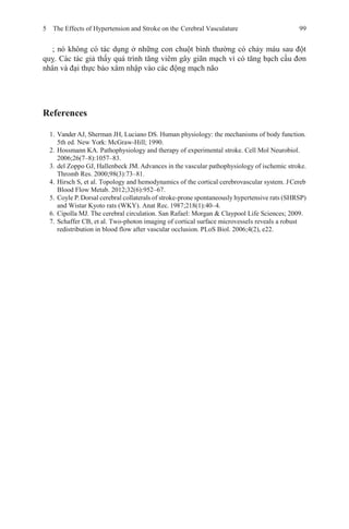
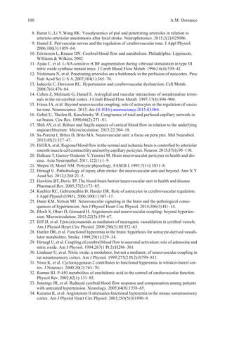
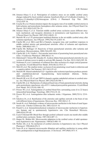
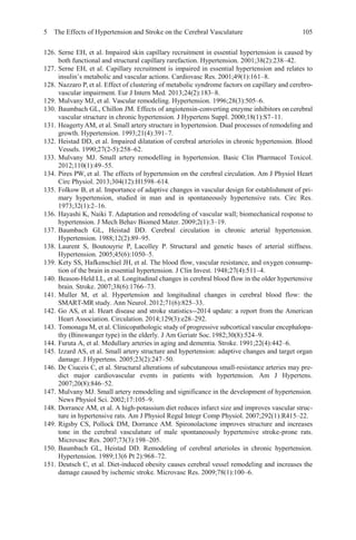
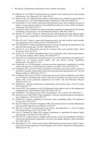
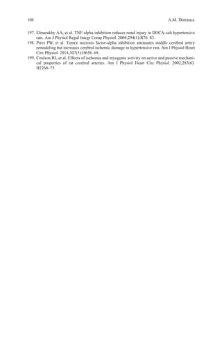
![Chapter 6
Sinh lý bệnh và cơ chế có thể gây đột quỵ do
THA
Beom Joon Kim, Hee-Joon Bae, and Lawrence K.S. Wong
Mối liên hệ giữa tăng huyết áp và đột quỵ đã được biết đến từ thế kỷ mười
chín. Mặc dù một số yếu tố nguy cơ tim mạch cũng có liên quan, người ta ước tính
rằng 51% tử vong do đột quỵ là do tăng huyết áp. [1].
Tăng huyết áp là yếu tố nguy cơ quan trọng nhất đối với đột quỵ. tỷ lệ tăng
huyết áp ở bệnh nhân đột quỵ thiếu máu cục bộ tới 80% [2]. Các mức độ tăng
huyết áp có mối tương quan chặt chẽ với nguy cơ đột quỵ. Tăng huyết áp đóng
một vai trò quan trọng trong bệnh sinh của xơ vữa động mạch động mạch lớn, nó
sẽ gây ra đột quỵ thiếu máu cục bộ do tắc huyết khối động mạch, động mạch đến
động mạch tắc mạch hoặc một sự kết hợp của những yếu tố này. Ở cấp độ vi mô
của các động mạch nhỏ hoặc tiểu động mạch, tăng huyết áp cũng gây bệnh lý
mạch máu như vỡ mạch nhỏ trong não và gây nhồi máu lỗ khuyết. Tăng huyết áp
mạn tính có thể gây bệnh cơ tim thiếu máu cục bộ hoặc rung nhĩ, dễ tạo huyết khối
trong tim và đi vào tuần hoàn. Ngoài ra, tăng huyết áp là một yếu tố nguy cơ chính
gây xuất huyết nội sọ (ICH) và xuất huyết dưới màng nhện (SAH), hai phân nhóm
chính của đột quỵ xuất huyết.
B.J. Kim, M.D., Ph.D. • H.-J. Bae, M.D., Ph.D. (*)
Department of Neurology, Seoul National University Bundang Hospital,
Gyeonggi-do, South Korea
e-mail: braindoc@snu.ac.kr
L.K.S. Wong, M.D.
Departments of Medicine and Therapeutics, Chinese University of Hong Kong,
Hong Kong Special Administrative Region, Hong Kong, China
© Springer International Publishing Switzerland 2016
V.Aiyagari, P.B. Gorelick (eds.), Hypertension and Stroke, Clinical
Hypertension and Vascular Diseases, DOI 10.1007/978-3-319-29152-9_6
109](https://image.slidesharecdn.com/hypertensionandstroke-170712211353/85/Hypertension-and-stroke-103-320.jpg)
![110 B.J. Kim et al.
Trong chương này, chúngât sẽ thảo luận về các vấn đề lâm sàng liên quan đến huyết
áp cao mãn tính liên quan đến đột quỵ thiếu máu cục bộ hoặc xuất huyết.
Cơ chế của đột quỵ thiếu máu cục bộ hoặc Tia với tăng huyết áp
Bệnh động mạch lớn
Xơ vữa động mạch có thể gặp ở nhiều động mạch của cơ thể, bao gồm động mạch
chủ, động mạch vành, mạch máu ngoại biên và mạch máu não. tổn thương xơ vữa
động mạch bắt đầu với phản ứng viêm theo sau là quá trình tăng sinh cơ trơn và dày
lên của thành động mạch. Tăng huyết áp, rối loạn chức năng nội mô, lipoprotein cao,
gốc tự do và phản ứng viêm mạn tính có liên quan chặt chẽ với quá trình xơ vữa động
mạch [6]. Các vị trí thường xơ vữa động mạch bao gồm các nhánh của động mạch
cảnh chung, phần gốc và xoang cảnh của động mạch cảnh trong (ICA), đoạn đầu tiên
của động mạch não giữa (MCA), gốc và phần xa của động mạch đốt sống, đoạn giữa
của động mạch thân nền. kết quả khám nghiệm tử thi thấy xuất huyết mảng bám tiểu
cầu, tái tạo mạch và loét là những yếu tố dẫn đến sự bất ổn của mảng bám động mạch
cảnh [7-9]. Xơ vữa động mạch MCA hay ảnh hưởng nhất ở vị trí M1, kéo dài từ gốc
động mạch tới thùy đảo. Các động mạch nhân đậu- thể vân bắt nguồn từ vị trí này và
dễ bị ảnh hưởng do sự phát triển của các mảng xơ vữa động mạch, có thể dẫn đến bị
cô lập vùng nhồi máu dưới vỏ nhỏ. Xuất huyết, loét và vôi hóa ít gặp trong mảng xơ
vữa động mạch nội sọ so với ngoài sọ. Một nghiên cứu khám nghiệm tử thi từ Hồng
Kông cho thấy hẹp lòng động mạch do mảng bám xơ vữa, tỷ trọng của lipid trong các
tổn thương và sự xuất hiện mạch tân tạo trong MCA là những yếu tố nguy cơ độc lập
cho nhồi máu MCA [10].
sự kết hợp của PET-MRI cung cấp những hiểu biết sâu sắc hơn về các đặc trưng của
mảng xơ vữa động mạch. mảng xơ vữa động mạch chủ yếu được chia thành mảng
bám ổn định hoặc dễ bị tổn thương. Tuy nhiên, các mảng bám dễ bị tổn thương, đặc
trưng bởi nắp sợi mỏng trên lõi hoại tử với mật độ cao của tình trạng viêm, có](https://image.slidesharecdn.com/hypertensionandstroke-170712211353/85/Hypertension-and-stroke-104-320.jpg)
![6 Pathophysiology and Mechanisms Whereby Hypertension May Cause Stroke 111
xu hướng bị vỡ và hình thành đột ngột huyết khối lòng mạch và thuyên tắc xa
hoặc tại chỗ [11].
Tuổi tác, tăng huyết áp, đái tháo đường, hút thuốc lá, tăng lipid máu là những
yếu tố nguy cơ xơ vữa động mạch. xơ vữa động mạch động mạch lớn là loại hay
gặp nhất của bệnh lý mạch máu, trong đó mô xơ và cơ sinh sôi nảy nở dưới màng
trong của thành mạch máu, và lipid hình thành mảng bám có thể đụng chạm đến
lòng mạch. Tiểu cầu sau đó mắc ở mảng bám và hình thành huyết khối do lắng
động fibrin tạo huyết khối, cục máu đông [12]. huyết khối cấp tính bắt đầu với tình
trạng nứt của nắp xơ của mảng bám xơ vữa động mạch, và sản sinh các yếu tố
hình thành cục máu đông tiếp giáp với mảng bám. sau đó gây tắc tại chỗ làm giảm
đáng kể lưu lượng máu và giảm cung cấp oxy, có thể gây tổn thương não do thiếu
máu cục bộ.
Tính đặc thù của xơ vữa động mạch nội sọ
hẹp động mạch nội sọ là một chủ đề thú vị. bệnh nhân Châu Á và người Mỹ
gốc Phi có tỷ lệ hẹp xơ vữa động mạch nội sọ hơn dân da trắng [23-25] trong thập
niên 60 và 70, khoảng 50% bệnh nhân cao tuổi ở độ tuổi ≥80
hai đặc điểm chính phân biệt xơ vữa động mạch nội sọ và ngoài sọ là ở ngoại
sọ khởi phát muộn hơn và mảng bám ổn định hơn trong các động mạch nội sọ
[18].](https://image.slidesharecdn.com/hypertensionandstroke-170712211353/85/Hypertension-and-stroke-105-320.jpg)
![112 B.J. Kim et al.
Hai đặc tính trên có thể được giải thích bởi các đặc tính riêng biệt của các động mạch
nội sọ, bao gồm (1) dày hơn; (2) các động mạch nội sọ lớn bao quanh bởi dịch não
tủy; (3) chuyển hóa thành mạch rõ rệt; (4) có mối liên quan chặt chẽ giữa các tế bào
nội mô; (5) giảm tính thấm màng trong; (6) tương đối nhạy cảm với kích thích thần
kinh giao cảm và histamine; (7) tăng cường cơ chế bảo vệ chống lại quá trình giảm
oxy hóa; và (8) đặc điểm lưu thông phụ thuốc vòng Willis [18].
vấn đề lâm sàng liên quan đến xơ vữa động mạch nội sọ có thể được tóm tắt bởi
những điểm sau đây: (1) tắc nhánh bên của các động mạch não hoặc động mạch thân
nền. đột quỵ thiếu máu cục bộ do tắc động mạch nhánh có thể xảy ra rất lâu trước khi
có sự tiến triển hẹp lòng mạch. (2) Khi vùng cắt ngang của động mạch nội sọ trở nên
nhỏ hơn, suy giảm tình trạng huyết động và biến đổi bệnh lý liên quan có thể phát
triển trước khi xuất hiện bệnh lý xơ vữa động mạch điển hình. (3) các động mạch nội
sọ liên tục gây khó khăn, tạo ra những rào cản kỹ thuật đối với các can thiệp nội
mạch
tắc động mạch - động mạch
tắc động mạch - động mạch là cơ chế đột quỵ quan trọng ở những bệnh nhân có tắc
động mạch một cơ chế đột quỵ quan trọng ở những bệnh nhân bị bệnh động mạch lớn
ngoài sọ. Thuyên tắc do cục máu đông, khối tiểu cầu hoặc bong các mảng bám từ các
mạch gần đó [27].
Tắc động mạch - động mạch cũng là một cơ chế quan trọng nhưng ít được công nhận
gây đột quỵ ở những bệnh nhân bị bệnh động mạch nội sọ. Wong và cộn sự [22] đã
báo cáo nhiều bệnh nhân đột quỵ cấp có MCA hẹp, 1 bên hay gặp nhất.
Bệnh xơ vữa động mạch
mảng bám xơ vữa động mạch, thường được gọi là xơ vữa động mạch nhỏ, có thể gây
tắc các chỗ giao nhau với động mạch đi vào nhu mô và gây tắc, nhồi máu dưới vỏ
nhỏ. Biểu hiện của xơ vữa động mạch nhỏ có thể bao gồm bóc tách nhỏ](https://image.slidesharecdn.com/hypertensionandstroke-170712211353/85/Hypertension-and-stroke-106-320.jpg)
![6 Pathophysiology and Mechanisms Whereby Hypertension May Cause Stroke 113
Fig. 6.1 Classification of SSSI according to the lesion extension and the presence of PAD. (a)
Distal SSSI without PAD; (b) proximal SSSI without PAD; (c) SSSI associated with PAD. SSSI
indicates single small subcortical infarction and PAD parent artery disease. (From Nah H-W, Kang
D-W, Kwon SU, Kim JS. Diversity of Single Small Subcortical Infarctions According to Infarct
Location and Parent Artery Disease Analysis of Indicators for Small Vessel Disease and
Atherosclerosis. Stroke; a journal of cerebral circulation. 2010;41(12):2822-7, with permission)
Mảng bám xuất huyết và lắng đọng tiểu cầu, fibrin [28]. các nghiên cứu gần đây đã
chứng minh rằng ở những bệnh nhân có hẹp MCA, tắc động mạch xâm nhập hay gặp
nhất là gây nhồi máu dưới vỏ[32, 33].](https://image.slidesharecdn.com/hypertensionandstroke-170712211353/85/Hypertension-and-stroke-107-320.jpg)
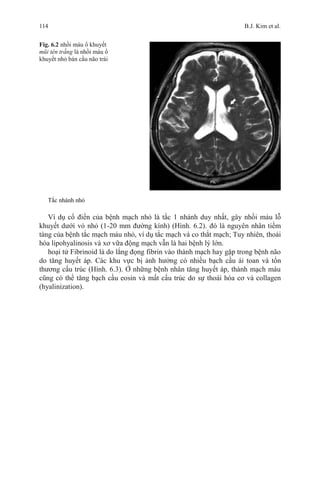
![6 Pathophysiology and Mechanisms Whereby Hypertension May Cause Stroke 115
Fig. 6.3 Hyaline arteriosclerosis, roughly concentric vessel wall thickening by hyaline collagenous
material (asterisk), with occasional surviving smooth muscle cell nuclei (arrow). (From Lammie
GA. Pathology of small vessel stroke. Br Med Bull. 2000;56:296–306, with permission [39])
Fig. 6.4 Lipohyalinosis, an asymmetrically thickened, disorganized vessel wall with focal fibrosis
(asterisk) and foam cell infiltration (thick arrow). (From Lammie GA. Pathology of small vessel
stroke. Br Med Bull. 2000;56:296–306, with permission.)](https://image.slidesharecdn.com/hypertensionandstroke-170712211353/85/Hypertension-and-stroke-109-320.jpg)
![116 B.J. Kim et al.
Tắc mạch tim
Trong đột quỵ do tắc tim mạch, nhồi máu thường bắt nguồn từ các van tim, màng
trong tim và tâm nhĩ hoặc tâm thất. cục máu đông khác có thể bắt nguồn từ tĩnh mạch
hệ thống, sau đó đi đến não. Rung nhĩ là thuyên tắc não do tim hay gặp nhất. Rung
nhĩ có nhiều khả năng tiến triển ở bệnh nhân tăng huyết áp có phì đại thất trái và tăng
kích thước nhĩ trái. Một nghiên cứu gần đây cũng cho thấy ở bệnh nhân phì đại thất
trái bằng điều trị hạ huyết áp làm giảm nguy cơ tiến triển thành rung nhĩ [46]. Do đó,
kiểm soát huyết áp tốt ngoài việc dùng chống đông có thể làm giảm nguy cơ đột quỵ
tắc mạch.
Đột quỵ - huyết động
suy tim và hạ huyết áp hệ thống là hai nguyên nhân chính gây giảm tưới máu. Giảm
tưới máu hệ thống hay gặp trong huyết khối động mạch não hoặc tắc mạch và thường
ảnh hưởng đến cả hai bán cầu não.](https://image.slidesharecdn.com/hypertensionandstroke-170712211353/85/Hypertension-and-stroke-110-320.jpg)
![6 Pathophysiology and Mechanisms Whereby Hypertension May Cause Stroke 117
Các nghiên cứu đã chứng minh mối liên hệ giữa huyết áp và suy tim. Trong
một nghiên cứu của Mỹ trên 48.000 bệnh nhân nhập viện do suy tim cấp tính,
chúng tôi cho rằng kiểm soát tốt huyết áp có thể giảm nguy cơ phát triển suy tim
và do đó làm giảm nguy cơ hypoperfusion não.
Ngoài ra, cao huyết áp mãn tính dẫn đến xơ vữa động mạch và tăng sức đề
kháng mạch máu ngoại biên [49].
Giảm tưới máu từ tim hoặc động mạch ngoài sọ hiếm khi gây nhồi máu não
lớn. Ngược lại, giảm lưu lượng máu do tổn thương trực tiếp xuất phát từ động
mạch của mô não thường gây nhồi máu lớn. Ngoài ra, bệnh động mạch nội sọ
nặng cũng có vẻ nhiều khả năng gây ra nhồi máu não hơn so với bệnh tắc động
mạch ngoài não [50]. Trước đây, giảm tưới máu và tắc mạch được coi là cơ chế
độc lập của đột quỵ ở bệnh nhân bị bệnh tắc động mạch. Caplan đề xuất rằng
chúng thường cùng tồn tại ở những bệnh nhân bị bệnh tắc động mạch nghiêm
trọng [51, 52]. Hẹp lòng động mạch và những bất thường nội mạc thúc đẩy sự
hình thành cục máu đông và thuyên tắc tiếp đó [51, 52].
Đột quỵ và những biến đổi của huyết áp
Huyết áp có xu hướng dao động theo thời gian. biến đổi như vậy được cho là
phản ánh sinh lý bình thường của hệ thống thần kinh tự trị, chu kỳ tim và những
thay đổi trong tư thế cơ thể cũng như sự thay đổi môi trường bên ngoài, stress và
nhịp sinh học [53].
Đáng ngạc nhiên, thay đổi huyết áp biến thiên nhỏ, chủ yếu bị ảnh hưởng bởi
sự thay đổi của môi trường giữa mỗi lần đo và do tác dụng của các thuốc hạ áp
[56-58].](https://image.slidesharecdn.com/hypertensionandstroke-170712211353/85/Hypertension-and-stroke-111-320.jpg)
![118 B.J. Kim et al.
1 số nghiên cứu năm 2010 đã chứng minh rằng sự thay đổi huyết áp rất quan trọng và
liên quan tới mạch máu sau đột quy. Rothwell và các đồng nghiệp chọn đột quỵ thiếu
máu cục bộ gần đây hoặc các bệnh nhân đột quỵ nhỏ đo huyết áp cho bệnh nhân tại
phòng khám trong ít nhất là 4 tháng. Họ phát hiện ra có sự khác biệt đáng kể trong
việc đo huyết áp trong thời gian dài theo dõi, và sự thay đổi huyết áp cho thấy mối
quan hệ đáng chú ý với nguy cơ đột quỵ tái phát [59]. Trong một bài báo song song,
họ thông báo rằng điều trị bằng beta-blocker tốt hơn so với nhóm điều trị bằng chẹn
kênh canxi ở bệnh nhân sau đột quỵ [56].
Cơ chế của xuất huyết nội sọ với tăng huyết áp
xuất huyết nội sọ liên quan đến nhu mô não hoặc khoang dưới nhện hoặc cả hai.
Khoảng 15% các cơn đột quỵ là do xuất huyết. Trong khi chiếm tỷ lệ nhỏ của đột
quỵ, đột quỵ xuất huyết có tỷ lệ tử vong cao hơn so với đột quỵ thiếu máu cục bộ.
Cao huyết áp và chứng phình động mạch não bị vỡ là hai nguyên nhân chính của xuất
huyết nội sọ, mà sẽ thảo luận sau đó.](https://image.slidesharecdn.com/hypertensionandstroke-170712211353/85/Hypertension-and-stroke-112-320.jpg)
![6 Pathophysiology and Mechanisms Whereby Hypertension May Cause Stroke 119
Fig. 6.5 Intracerebral
hemorrhage. A small
hypertensive intracerebral
hemorrhage in left basal
ganglia
Có hai cơ chế quan trọng dẫn đến tăng huyết áp ICH: (a) vỡ động mạch xâm nhập
nhỏ do tăng huyết áp mãn tính và lão hóa và (b) tăng huyết áp dẫn đến vỡ các tiểu
động mạch và mao mạch bình thường.
Tăng huyết áp mãn tính
cao huyết áp mạn tính làm thay đổi cấu trúc động mạch như gồm hoại tử fibrinoid,
lipohyalinosis, thoái hóa trung gian và sự hình thành các phình mạch nhỏ, tất cả
đều làm mạch máu dễ bị vỡ. Các điểm vỡ thường ở gốc các động mạch xâm nhập
hoặc gần nhánh. Hậu quả của phình mạch nhỏ trong gây ra xuất huyết nội sọ
(ICH) đề xuất lần đầu tiên bởi Charcot và Bouchard vào năm 1868, nhưng đã được
tranh luận hơn một thế kỷ. Có nhiều bằng chứng chống lại lý thuyết cho rằng ICH
tự phát là do vỡ phình mạch nhỏ. Challa và cộng sự[65] không chứng minh được
vai trò của phình mạch nhỏ ở bệnh nhân tăng huyết áp có ICH tự phát. Một nghiên
cứu bằng kính hiển vi điện tử của động mạch bị vỡ trong THA gây ICH cho thấy
những thoái hóa nghiêm trọng ở 46 trong 48 động mạch bị vỡ, và vỡ phình mạch
nhỏ chỉ được tìm thấy trong hai trường hợp [66]. Những nghiên cứu chỉ ra rằng
những thay đổi thoái hóa do tuổi và cao huyết áp có thể dẫn đến ICH, nhưng
không phải chắc chắn rằng vỡ phình mạch nhỏ là nguyên nhân gây ICH](https://image.slidesharecdn.com/hypertensionandstroke-170712211353/85/Hypertension-and-stroke-113-320.jpg)
![120 B.J. Kim et al.
Tăng huyết áp cấp tính
Trong thực hành lâm sàng, nhiều bệnh nhân ICH không có tiền sử tăng huyết áp.
Ngoài ra, không có bằng chứng bệnh lý của tăng huyết áp mãn tính, như phì đại thất
trái hoặc rối loạn chức năng tim thận khác. Bahemuka và cộng sự [67] thấy chỉ có
46% số ca tử vong do ICH tự phát bị tăng huyết áp mạn tính hoặc phì đại tâm thất
trái. Trong nghiên cứu này, vị trí tụ máu, tăng huyết áp lúc nhập viện, và sự vắng mặt
của các nguyên nhân khác cho rằng ICH thường được gây ra do tăng huyết áp cấp
tính. Bằng chứng cũng cho thấy sự gia tăng cấp tính áp lực và lưu lượng máu có thể
làm vỡ động mạch và mao mạch bình thường.
Sự kết hợp gia tăng đáng kể lưu lượng máu não và huyết áp cũng có thể dẫn đến ICH
sau cắt bỏ lớp áo trong mạch cảnh hoặc stent động mạch. Một đánh giá hồi cứu 4494
bệnh nhân đã cắt lớp áo trong đm cảnh hoặc đặt stent động mạch thấy có liên quan
giữa ICH và hội chứng tái tưới máu sau can thiệp [69].
ICH cũng hay gặp ở bệnh nhân lạm dụng cocaine và amphetamine, có tác dụng
cường giao cảm. tăng huyết áp do Cocaine là một yếu tố nguy cơ từ lâu được công
nhận của ICH. Một số bệnh nhân cũng xuất hiện bệnh não do tăng huyết áp có ICH
và phù nề não [71]. Cơ chế chính xác mà các thuốc này gây ra ICH là chưa rõ ràng.
Phình mạch dưới nhện xuất huyết
SAH (Hình. 6.6) xảy ra khi một mạch máu bề mặt não vỡ dẫn đến thoát máu vào
khoang dưới nhện. SAH thường do vỡ phình túi. phình túi thường gặp nhất tại ICA-
sau ngã ba động mạch, đỉnh của động mạch thân nền, và phân nhánh MCA.](https://image.slidesharecdn.com/hypertensionandstroke-170712211353/85/Hypertension-and-stroke-114-320.jpg)
![122 B.J. Kim et al.
Fig. 6.7 White matter
lesions. Extensive white
matter changes
(leukoaraiosis) are
observed in both
periventricular and
subcortical white matter
Áp lực trên thành mạch tăng khi bán kính của phình to ra. Bằng chứng cho thấy
phình có đường kính lớn hơn 10 mm có nhiều khả năng bị vỡ [78]. Phình động
mạch có thể vỡ bất cứ lúc nào, khi huyết áp hoặc lưu lượng máu tăng lên trong
hoạt động gắng sức.
tổn thương chất trắng (WMLs) (Hình. 6.7) WMLs thường liên quan tuổi tác.
Tuy nhiên, trong hầu hết các nghiên cứu, thay đổi chất trắng hay gặp hơn trong
tăng huyết ở những người huyết áp bình thường, đặc biệt là người trẻ. Tầm quan
trọng của WMLs là một yếu tố dự báo nguy cơ đột quỵ [79, 81] và chứng mất trí
[80] đã được chứng minh trong các nghiên cứu trước đó.
Các bệnh lý của WML không đồng nhất, bao gồm cả nhồi máu nhỏ, mất bao
myelin và dãn mạch máu [82]. Các cơ chế chính xác của WMLs chưa rõ ràng,
nhưng xơ hóa tiểu động mạch trong tăng huyết áp dường như là yếu tố gây bệnh
quan trọng nhất, và mức độ WMLs được cho là phản ánh mức độ xơ cứng tiểu
động mạch não [83].](https://image.slidesharecdn.com/hypertensionandstroke-170712211353/85/Hypertension-and-stroke-116-320.jpg)
![6 Pathophysiology and Mechanisms Whereby Hypertension May Cause Stroke 123
Nhồi máu im lặng: nhồi máu ổ khuyết cũ và nhồi máu nhỏ
nhồi máu im lặng có thể được chia thành hai loại: nhồi máu ổ khuyết cũ và nhồi máu
nhỏ. nhồi máu ổ khuyết cũ được định nghĩa là tăng cường độ trên T2W, 3 mm hoặc
hơn, tương ứng với giảm cường độ trên T1W [79]. nhồi máu ổ khuyết Cũ và WMLs
được cho là có nguồn gốc mạch máu tương tự. Tuy nhiên, phần lớn các nhồi máu im
lặng là nhồi máu ổ khuyết có thể do tổn thương mạch lớn hoặc nhỏ trong khi WMLs
liên quan mạch nhỏ. Nhồi máu nhỏ thường không phát hiện trên MRI cấu trúc thông
thường [86, 87].
Xuất huyết não vi thể Microbleeds
Microbleeds (MBS) (Hình. 6.8) có hình ảnh dấu lấm chấm, đồng nhất, tròn, các tổn
thương kích thước dưới 0,5 cm, với mất tín hiệu hoặc giảm tín hiệu trên MRI echo.
Các bệnh lý của xuất huyết vi mạch nhỏ có tích tụ hemosiderin quanh mạch não
Fig. 6.8 Microbleeds. Several microbleeds are seen in both thalamus and basal ganglia. The black
arrow indicates one of them. (Sources: Figs. 5, 6, 7, and 8 from Dr Bae’scollections)](https://image.slidesharecdn.com/hypertensionandstroke-170712211353/85/Hypertension-and-stroke-117-320.jpg)
![124 B.J. Kim et al.
Do đó, thuốc kháng tiểu cầu nên được sử dụng thận trọng ở những bệnh nhân
có MBS lan tỏa. Nhiều nghiên cứu đã chỉ ra rằng xuất huyết não vi thể có liên
quan với chảy máu não sau này ở những bệnh nhân bị đột quỵ thiếu máu cục bộ
bao gồm cả bệnh nhân xuất huyết cấp tính sau dùng thuôc làm tan huyết khối [93],
References
1. Gaciong Z, Siński M, Lewandowski J. Blood pressure control and primary prevention of
stroke: summary of the recent clinical trial data and meta-analyses. Curr Hypertens Rep. 2013;
15(6):559–74.
2. Kim BJ, Park JM, Kang K, Lee SJ, Ko Y, Kim JG, et al. Case characteristics, hyperacute treat-
ment, and outcome information from the clinical research center for stroke-fifth division reg-
istry in South Korea. J Stroke. 2015;17(1):38–53.](https://image.slidesharecdn.com/hypertensionandstroke-170712211353/85/Hypertension-and-stroke-118-320.jpg)
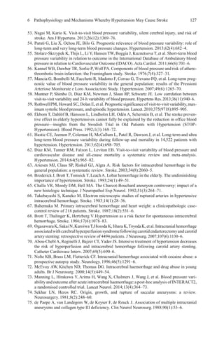
![Chapter 7
Đánh giá nguy cơ tim mạch, Tóm tắt Hướng dẫn
việc điều trị tăng huyết áp và đánh giá quan trọng
của năm 2014
Luke J. Laffin and George L. Bakris
Trên thế giới, 54% bệnh tim thiếu máu cục bộ và 47% đột quỵ là do cao huyết áp [3].
Sự gia tăng tỷ lệ tăng huyết áp là do một số yếu tố, bao gồm cả sự lão hóa của dân số,
béo phì, và giảm hoạt động thể chất. THA là một yếu tố nguy cơ độc lập đối với sự
phát triển của tất cả các biểu hiện lâm sàng của bệnh tim mạch (CVD), bao gồm bệnh
động mạch vành (CAD), bệnh động mạch ngoại biên (PAD), suy tim sung huyết
(CHF) và đột quỵ. Tại Hoa Kỳ, 69% bệnh nhân đột quỵ có nhồi máu cơ tim (MI),
77% bị đột quỵ lần đầu, và 74% có CHF với huyết áp> 140/90 mmHg [4].
L.J. Laffin, M.D. (*) • G.L. Bakris, M.D. (*)
ASH Comprehensive Hypertension Center, University of Chicago Medical Center,
Chicago, IL, USA
e-mail: luke.laffin@uchospitals.com; gbakris@gmail.com
© Springer International Publishing Switzerland 2016
V.Aiyagari, P.B. Gorelick (eds.), Hypertension and Stroke, Clinical
Hypertension and Vascular Diseases, DOI 10.1007/978-3-319-29152-9_7
131](https://image.slidesharecdn.com/hypertensionandstroke-170712211353/85/Hypertension-and-stroke-124-320.jpg)
![7 Cardiovascular Risk Assessment, Summary of Guidelines… 133
1 số yếu tố nguy cơ tim mạch, trong đó có tăng huyết áp và hội chứng chuyển hóa.
Tiêu chuẩn của ATPIII thuộc chương trình giáo dục về cholesterol quốc gia của Hoa
Kỳ(NCEP- National Cholesterol Education Program) hội chứng chuyển hóa khi có sự
xuất hiện của ba hoặc hơn ba dấu hiệu: (a)huyết áp tăng (≥130 / 85 mmHg), (b) giảm
dung nạp đường huyết lúc đói (> 100 mg / dL), (c) tăng chu vi vòng eo (> 102 cm
đối với nam và 88 cm đối với nữ), (d) HDL-C thấp (<40 mg / dL), và (e) tăng
triglyceride (> 150 mg / dL) [23].](https://image.slidesharecdn.com/hypertensionandstroke-170712211353/85/Hypertension-and-stroke-125-320.jpg)
![134 L.J. Laffin and G.L. Bakris
Table 7.1 Cardiovascular
risk factors
Hypertension
Diabetes mellitus
Tobacco use
Age (>55 years for men, >65 years for women)
Family history of premature cardiovascular disease
(<55 years for men, <65 years for women)
CKD
Obesity
Dyslipidemia
Physical inactivity
Target organ damage
Heart
Left ventricular hypertrophy
Angina pectoris or myocardial infarction
Coronary revascularization
Heart failure
Brain
Stroke or transient ischemic attack
Chronic kidney disease
Peripheral arterial disease
Retinopathy
Phần lớn sự gia tăng nguy cơ liên quan với hội chứng chuyển hóa xảy ra ở bệnh
nhân béo phì. Béo phì là một trong những nguyên nhân chính của bệnh cao huyết
áp, tỷ lệ đó đang gia tăng ở mức báo động. béo phì định nghĩa là khi (BMI)> 30 kg
/ m2 [4]. Béo phì dẫn đến một số thay đổi sinh lý, bao gồm tăng insulin và đề
kháng insulin, rối loạn chức năng nội mô, tăng hoạt hóa hệ thống thần kinh giao
cảm, tích tự natri và tăng nhu cầu oxy hóa. Béo phì là một trong những yếu tố
nguy cơ tim mạch chính được nhấn mạnh trong Bảng 7.1.
Song song với sự gia tăng của bệnh béo phì, cao huyết áp, bệnh thận mãn tính
(CKD) [30ư khi mức lọc cầu thận (eGFR) <60 ml / phút, là một yếu tố nguy cơ
độc lập cho biến cố tim mạch gây tử vong [31–33].](https://image.slidesharecdn.com/hypertensionandstroke-170712211353/85/Hypertension-and-stroke-126-320.jpg)
![7 Cardiovascular Risk Assessment, Summary of Guidelines… 135
eGFR giảm dẫn đến nhiều nguy cơ tim mạch như tăng huyết áp không được
kiểm soát và rối loạn lipid máu. Một số nghiên cứu lớn đã chỉ ra rằng những bệnh
nhân eGFR giảm có huyết áp cao và tăng cholesterol toàn phần, giảm HDL-C, và
có nhiều khả năng bị bệnh thiếu máu cục bộ tim, phì đại thất trái, tiểu đường và
suy tim [35-37]. Kết quả là, nó đã được mặc nhiên công nhận rằng giảm eGFR có
thể là một dấu hiệu nguy cơ của bệnh tim mạch [39]. chức năng thận giảm cũng có
liên quan với một số bất thường, bao gồm tăng mức độ viêm, rối loạn đông máu,
tăng xơ cứng mạch, rối loạn chức năng nội mô …
Một ví dụ minh họa mối liên quan giữa CKD và tăng nguy cơ của bệnh tim
mạch được cung cấp trong dữ liệu từ nghiên cứu Kaiser của hơn 1 triệu người lớn.
Bệnh nhân có eGFR 40-59 mL / phút tăng 40% bệnh mạch vành hơn người có
chức năng thận bình thường. giảm eGFR, albumin niệu cao có liên quan với rối
loạn chức năng nội mô, tính thấm thành mạch, tăng tình trạng viêm và bất ổn rối
loạn đông máu (Hình. 7.1) [39]. albumin niệu cao trên 300 mg / ngày cho thấy có
biểu hiện suy thận [41]](https://image.slidesharecdn.com/hypertensionandstroke-170712211353/85/Hypertension-and-stroke-127-320.jpg)
![136 L.J. Laffin and G.L. Bakris
Fig. 7.1 Interaction of microalbuminuria with other factors that affect atherosclerosis develop-
ment. NO nitric oxide, AGEs advanced glycation end products, TC triglycerides, LDL low density
lipoproteins
Hướng dẫn điều trị tăng huyết áp
Mối liên hệ giữa tăng huyết áp và các yếu tố nguy cơ tim mạch khác, chẳng hạn như
đái đường, rối loạn lipid máu, suy thận và béo phì, là cơ sở của các kiến nghị của tất
cả các hướng dẫn xử trí tăng huyết áp quốc gia và quốc tế được viết năm 2014 bởi
nhóm chuyên gia của Viện Y tế quốc gia [42], Hiệp hội THA Châu Âu - Hội tim
mạch Châu Âu (ESH-ESC) [43], thội THA Mỹ/quốc tế (ASH / ISH) [44], và NICE
[45] .
Các phân loại tăng huyết áp đã được điều chỉnh trong năm 2014 sau báo cáo từ JNC
7 trước đó [46]. Sự thay đổi trọng yếu nhất đã tăng mục tiêu chiến lược điều trị THA
ở người dưới 60 tuổi với mục tiêu <140/90 mmHg và cho người lớn hơn 60 tuổi ở
mức <150/90 mmHg. Với bệnh nhân ĐTĐ, suy thận đề nghị ở mức <140/90 mmHg.
Những chiến lược này được cập nhật cho các phân loại và điều trị mục tiêu tăng
huyết áp ở người lớn thể hiện trong Bảng 7.2.
Chỉ dùng thuốc huyết áp khi chẩn đoán xác định, ngoài ra nếu THA không biến
chứng có thể điều chỉnh lối sống trước khi dùng thuốc khi huyết áp ở mức giữa 140-
159 mmHg huyết áp tâm thu và huyết áp tâm trương 90-100. Bệnh nhân có huyết áp
cao hơn 160/100 mmHg](https://image.slidesharecdn.com/hypertensionandstroke-170712211353/85/Hypertension-and-stroke-128-320.jpg)
![7 Cardiovascular Risk Assessment, Summary of Guidelines… 137
Table 7.2 Goal blood pressure by Guideline Committees (all expressed in mmHg)
Year of
publication
<60 yo no
additional
risk factors
60–79 yo no
additional
risk factors
>80 yo no
additional
risk factors
DM (any
age)
CKD (any
age)
014 Expert
Panel (JNC 8)
[38]
2014 <140/90 <150/90a
<150/90 <140/90 <140/90
ESH-ESC [39] 2013 <140/90 <140/90 <150/90 <140/90 <140/90
ISH/ASH [40] 2014 <140/90 <140/90 <150/90 <140/90 <140/90
BHS/NICE
[41]
2011 <140/90 <140/90 <150/90 <140/90 <140/90
DM diabetes mellitus, CKD chronic kidney disease, YO years old
a
If treatment leads to a blood pressure <140/<90 and is well tolerated, then treatment does not need
to be adjusted
nên được bắt đầu dùng thuốc. Điều quan trọng cần lưu ý là thay đổi lối sống kèm
theo điều trị thuốc
khi đánh giá ban đầu để xác định chẩn đoán cần chú ý loại trừ các nguyên nhân có
thể gây THA khi khám như: rối loạn giấc ngủ, ngưng thở khi ngủ và tăng huyết áp do
thuốc. Gần đây trong một tuyên bố dự thảo, tổ chức USPTF khuyến cáo rằng cần
theo dõi huyết áp 24 h tại nhà(ABPM) trước khi chẩn đoán THA. Điều này vẫn chưa
được công bố chính thức bởi USPTF, nhưng nhiều khả năng sẽ theo các hướng dẫn
năm 2011 của NICE có đề nghị tương tự [47]. Đáng chú ý, ABPM tiên lượng tốt về
tỷ lệ tử vong hơn đo huyết áp tại phòng khám [48].
Mục tiêu điều trị
hướng dẫn hiện nay nhấn mạnh rằng nguy cơ tim mạch tổng thể của bệnh nhân là cơ
sở để quyết định mục tiêu điều trị và khi nào bắt đầu bằng thuốc. Tuy nhiên, có sự
khác biệt giữa các nhóm hướng dẫn bằng văn bản đối với các nhóm bệnh nhân để bảo
đảm mục tiêu điều trị tích cực hơn.
Đối với bệnh nhân dưới 60 tuổi, năm 2014 JNC đề nghị nên bắt đầu bằng thuốc nếu
can thiệp lối sống không ổn định được THA giai đoạn 1 với mục tiêu huyết áp
<140/90 mmHg và <150/90 mmHg đối với bệnh nhân hơn 60 tuổi. sự thay đổi trọng
yếu nhất trong hướng dẫn hiện này là loại bỏ các mục tiêu huyết áp thấp hơn ở những
bệnh nhân ĐTĐ hoặc suy thận](https://image.slidesharecdn.com/hypertensionandstroke-170712211353/85/Hypertension-and-stroke-129-320.jpg)
![138 L.J. Laffin and G.L. Bakris
Trước đây mục tiêu điều trị với nhóm này ≤130 / 80 mmHg; Tuy nhiên, do sự
thiếu bằng chứng cho những mục tiêu thấp hơn, khuyến nghị đã được sửa đổi. Bây
giờ mục tiêu ở bệnh nhân dưới 60 tuổi là <140/90 mmHg [42].
Sự đồng thuận từ AHA, ACC và ASH đã thấy trong hướng dẫn mới công bố
với nguy cơ CAD [49]. Điều này cập nhật các khuyến nghị thực hiện trong năm
2007 của AHA, trong đó ghi nhận các mục tiêu thấp hơn ở những bệnh nhân
CAD. những khuyến cáo hiện nay một lần nữa cho thấy rằng hầu hết các bệnh
nhân CAD nên hướng tới mục tiêu HA<140/90 mmHg, nhưng có thể cố gắng đạt
ngưỡng 130/80 mmHg ở bệnh nhân có nhiều nguy cơ tương đương CAD (bệnh
động mạch ngoại biên, bệnh động mạch cảnh, phình động mạch chủ), hoặc tiền sử
TIA, đột quỵ. [50]. Đối với những bệnh nhân bị đột quỵ, các hướng dẫn cập nhật
báo cáo rằng bắt đầu điều trị hạ huyết áp được chỉ định, nếu trước đó không điều
trị đột quỵ thiếu máu cục bộ hoặc TIA, cần điều trị khi huyết áp ≥140 / 90mmHg.
Nếu <140/90 mmHg lợi ích khi điều trị chưa rõ ràng. Nói chung, mục tiêu giữ
huyêt áp sau đột quỵ <140/90 mmHg [51]
Trong lịch sử, đã có lo ngại rằng giảm huyết áp, đặc biệt là huyết áp tâm
trương, có thể gây hại, cái gọi là giả thuyết đường cong J. Một lý do là huyết áp
tâm trương giảm sẽ gây tổn thương do giảm tưới máu vành trong thì tâm trương
dẫn đến thiếu máu cục bộ cơ tim. Dữ liệu gần đây cho thấy rằng ở những người
> 65 tuổi, những người có bệnh tiểu đường, CAD, giảm huyết áp tâm trương
<60 mmHg có liên quan với tăng nguy cơ bệnh tim mạch [52, 53]. Lưu ý rằng
áp lực tâm trương thấp dưới 60 mmHg nguy cơ cao ở bệnh nhân suy thận giai
đoạn cuối và tử vong [54].](https://image.slidesharecdn.com/hypertensionandstroke-170712211353/85/Hypertension-and-stroke-130-320.jpg)
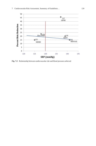
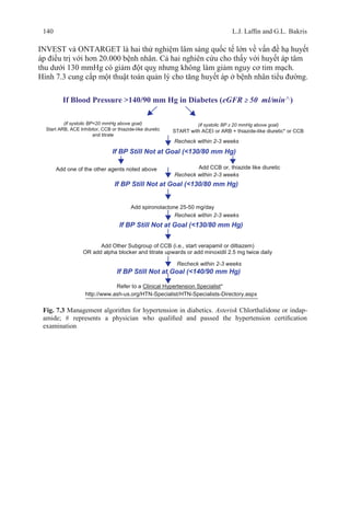
![7 Cardiovascular Risk Assessment, Summary of Guidelines… 141
Đối với bệnh nhân có bệnh thận, tất cả những thử nghiệm ngẫu nhiên có thấy huyết
áp thấp không làm giảm tiến triển của bệnh [70]. Ở những bệnh nhân suy thận, năm
2014 hướng dẫn khuyến cáo nên dùng thuốc ức chế hệ thống renin-angiotensin.
Thuốc thứ 2 thêm vào để kiểm soát HA phải là một thuốc chẹn kênh canxi (CCB)
[71], tuy nhiên gần đây dữ liệu cho thấy việc bổ sung các thuốc lợi tiểu hoặc CCB là
thuốc thứ hai thích hợp.
Liệu pháp dược lý
Một nghiên cứu phân tích 29 thử nghiệm lâm sàng gồm 162.341 người tham gia.Đối
với những người huyết áp lớn hơn 20/10 mmHg trên mục tiêu điều trị của họ, tức là,
giai đoạn 2 tăng huyết áp, tất cả các hướng dẫn khuyên bạn nên bắt đầu điều trị hai
thuốc.
Điều trị lợi tiểu, thuốc lợi tiểu thiazide, không còn là thuốc đầu tiên cho tăng huyết áp
không biến chứng (JNC 7). Hướng dẫn hiện nay điều trịgiảm huyết áp <140/90
mmHg sử dụng hoặc là CCB, ACEi hoặc thuốc lợi tiểu. Beta-blockers không phải
thuốc điều trị đầu tiên với hầu hết các bệnh nhân theo JNC và các hướng dẫn gần đây
2014. Mặc dù thuốc chẹn beta dùng điều trị tốt cho các bệnh nhân có biểu hiện thiếu
máu cục bộ tim, suy tim, loạn nhịp. Ví dụ, trong nghiên cứu, losartan ARB có hiệu
quả hơn so với atenolol trong phòng đột quỵ ở bệnh nhân tăng huyết áp có điện
tâmđồ phì đại thất trái [59]. Hơn nữa, trong các đối tượng tăng huyết áp ở nghiên cứu
ASCOT chọn ngẫu nhiên thấy amlodipine (và nếu cần thiết perindopril) có tai biến
tim mạch ít hơn so với những người ngẫu nhiên dùng atenolol (và nếu cần
bendrofluazide)](https://image.slidesharecdn.com/hypertensionandstroke-170712211353/85/Hypertension-and-stroke-133-320.jpg)
![142 L.J. Laffin and G.L. Bakris
Fig. 7.4 Blood pressure-lowering regimens based on different drug classes for the outcome total
major cardiovascular events and age groups <65 vs. ≥65. Negative blood pressure values indicate
lower mean follow-up blood pressure in first-listed than in second-listed groups. (From Blood
Pressure Lowering Treatment Trialists C, Turnbull F, Neal B, Ninomiya T, Algert C, Arima H,
et al. Effects of different regimens to lower blood pressure on major cardiovascular events in older
and younger adults: meta-analysis of randomised trials. BMJ. 2008;336(7653):1121, with
permission)
Một phân tích tổng hợp về điều trị chẹn beta (hầu như chỉ dùng atenolol) ở bệnh
nhân tăng huyết áp để phòng đột quỵ thấy hạn chế so với các thuốc khác [74].
Vì những dữ liệu và bằng chứng phù hợp cho thấy tăng nguy cơ mắc bệnh bệnh tiểu
khi dùng beta-blockers, đặc biệt là khi kết hợp với thuốc lợi tiểu [59, 60, 75-78],
beta-blockers không nên là thuốc điều trị đầu tiên phần lớn các bệnh nhân. Nhiều thử
nghiệm chứng minh rằng beta-blockers (chủ yếu là atenolol) và các thuốc lợi tiểu
thiazide gây khó khăn cho việc kiểm soát đường huyết ở những bệnh nhân đái tháo
đường và tăng tỷ lệ mắc bệnh tiểu đường mới khởi phát và rối loạn dung nạp đường
huyết lúc đói [76, 77, 79-81]. Ngoài ra, những nguy cơ của bệnh tiểu đường không
giảm khi một thiazide được kết hợp với một thuốc ƯCMC hoặc ARB ở bệnh nhân
béo phì có rối loạn dung nạp đường huyết lúc đói [78].](https://image.slidesharecdn.com/hypertensionandstroke-170712211353/85/Hypertension-and-stroke-134-320.jpg)
![7 Cardiovascular Risk Assessment, Summary of Guidelines… 143
yếu tố quyết định giảm nguy cơ tim mạch là giảm huyết áp [73]. Trường hợp ngoại lệ
cho quy tắc này là thuốc beta-blockers mới như carvedilol, có thêm tính chất chặn
alpha-adrenergic, và NEBIVOLOL gây giãn mạch bằng tăng giải phóng oxit nitric.
Cả hai tác nhân có tác động trung lập về kiểm soát đường huyết và tăng mức độ nhạy
cảm insulin [83, 84].
Trong thử nghiệm, bệnh nhân có nguy cơ cao bị tai biến tim mạch được chọn ngẫu
nhiên dùng benazepril (ACEi) với amlodipine hoặc hydrochlorothiazide (HCTZ)
[71]. Mặc dù HA so sánh (131,6 / 73,3 với amlodipine so với 132,5 / 74,4 với
HCTZ), bệnh nhân dùng với amlodipine thấy giảm 19,6% tai biến mạch vànhvà
huyết áp có hơi thấp hơn nhóm dùng lợi tiểu. vì vậy nghiên cứu gợi ý rằng amlodipin
nên được coi như thuốc đầu tiên lựa chọn
Một nghiên cứu thứ hai là thử nghiệm ADVANCE, thêm perindopril / indapamide so
với giả dược dùng cho bệnh nhân bị bệnh tiểu đường bất kể huyết áp[85]. Những
bệnh nhân được ngẫu nhiên dùng perindopril / indapamide làm giảm 18% tỷ lệ tử
vong do nguyên nhân tim mạch, bất kể huyết áp ban đầu.
.](https://image.slidesharecdn.com/hypertensionandstroke-170712211353/85/Hypertension-and-stroke-135-320.jpg)
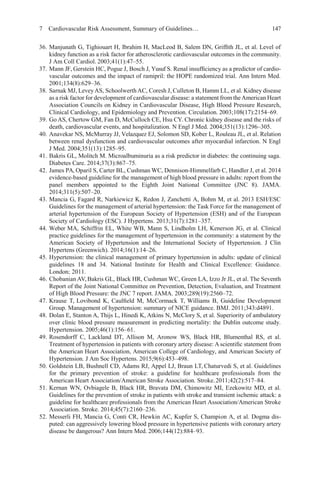
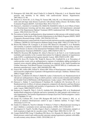
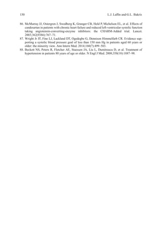
![Chapter 8
Xử trí THA sau đột quỵ
J. Dedrick Jordan and William J. Powers
Tăng huyết áp (HA) hay gặp kèm với cơn đột quỵ thiếu máu cục bộ cấp tính; Tuy
nhiên, việc xử trí huyết áp là vấn đề khó giải quyết. Trong khi có những lợi ích rõ ràng
cho việc giảm HA lâu dài để phòng ngừa đột quỵ thứ phát, tranh cãi vẫn tồn tại trong
giai đoạn ngay sau đột quỵ thiếu máu cục bộ. Mối quan tâm chính ở đây là hạ huyết áp
làm giảm tưới máu não xung quanh khu vực nhồi máu. Những giữ huyết áp cao sẽ làm
tăng nguy cơ dẫn đến xuất huyết và các biến chứng toàn thân. Chương này sẽ xem xét
các bằng chứng hiện tại về xử trí huyết áp trong trường hợp đột quỵ thiếu máu cục bộ
cấp tính và ảnh hưởng đến thần kinh.
Tăng huyết áp có đột quỵ thiếu máu cục bộ cấp tính
Tỷ lệ mắc
Nhiều bệnh nhân bị đột quỵ thiếu máu cục bộ cấp tính vào cấp cứu kèm theo có huyết
áp cao; Tuy nhiên, vẫn chưa rõ đây là dấu hiệu bù trừ do giảm tưới máu não hay có
liên quan đến nguyên nhân đáp ứng hệ thống [1, 2]. Một số nghiên cứu đã giúp làm
sáng tỏ những thay đổi huyết áp sau đột quỵ thiếu máu cục bộ cấp tính.
J.D. Jordan, M.D., Ph.D. (*) • W.J. Powers, M.D.
Departments of Neurology and Neurosurgery, University of North Carolina School
of Medicine, 170 Manning Drive, POB 2118, CB #7025, Chapel Hill,
NC 27599-7025, USA
e-mail: dedrick@unc.edu; powersw@neurology.unc.edu
© Springer International Publishing Switzerland 2016
V.Aiyagari, P.B. Gorelick (eds.), Hypertension and Stroke, Clinical
Hypertension and Vascular Diseases, DOI 10.1007/978-3-319-29152-9_8
151](https://image.slidesharecdn.com/hypertensionandstroke-170712211353/85/Hypertension-and-stroke-142-320.jpg)
![152 J.D. Jordan and W.J. Powers
Tỷ lệ đột quỵ kèm huyết áp cao gặp ở 76,5% trong một phân tích hồi cứu lớn của
276.734 bệnh nhân đến khám tại phòng cấp cứu [3]. Điều này tiếp tục được hỗ trợ
bởi hai thử nghiệm lớn về đột quỵ thiếu máu cục bộ. nghiên cứu CAST với 21.106
bệnh nhân và nghiên cứu đột quỵ quốc tế (IST) với 19.435 bệnh nhân, được ghi nhận
là huyết áp tâm thu >140 mmHg trong 75 % và 80% bệnh nhân. Hơn nữa, THA
nghiêm trọng, như định nghĩa là huyết áp tâm thu> 180 mmHg, đã được báo cáo ở
25% bệnh nhân trong CAST và 28% bệnh nhân trong IST [4, 5].
Thông thường quá trình THA này sẽ giảm trong vài ngày tới [1, 2, 6, 7]. Một nghiên
cứu đột quỵ lớn cho thấy HA tâm thu giảm từ 20-30% kèm theo quá trình phục hồi
thần kinh [8].
Ảnh hưởng của tăng huyết áp với hậu quae quả sau đột quỵ thiếu máu cục bộ cấp tính
Các mục tiêu tối ưu cho huyết áp sau đột quỵ thiếu máu cục bộ cấp tính vẫn chưa rõ
ràng, như một số nghiên cứu đã chứng minh sự tương quan hình chữ U giữa huyết áp
và tiên lượng xấu. huyết áp rất cao hoặc rất thấp khi nhập viện đều đi kèm với tiên
lượng xấu.
Nguy cơ tương đối của tỉ lệ tử vong ở 1 tháng và 1 năm tăng với mỗi thay đổi 10
mmHg huyết áp tâm thu trên hoặc dưới 130 mmHg [10]. Dữ liệu từ IST chứng minh
rằng cả BP cao và BP thấp là chỉ số tiên lượng độc lập cho tiên lượng xấu. HA tâm
thu 140- 179 mmHg thường tiên lượng không xấu[11].
Các nghiên cứu khác đã chứng minh mối liên quan giữa huyết áp tâm thu cao hay
thấp và kết quả bệnh nhân. Một nghiên cứu như vậy cho thấy những bệnh nhân có
huyết áp tâm thu dưới 155 mmHg có nhiều khả năng sẽ chết trong vòng 90 ngày so
với những bệnh nhân có huyết áp tâm thu giữa 155 và 220 mmHg [12]. Nghiên cứu
về dùng nimodipine tĩnh mạch ở bn đột quỵ Châu Âu cho thấy huyết áp tâm thu lớn
hơn 160 mmHg, là một yếu tố dự báo tử vong hoặc tàn tật so với những bệnh nhân
lúc vào huyết áp bình thường (HA tâm thu 120-160 mmHg và huyết áp tâm trương
BP 60- 90 mmHg) [13]. phân tích VISTA xét mối quan hệ giữa các biện pháp huyết
động, biến đổi huyết áp, và sự thay đổi huyết áp trong vòng 24 giờ đầu tiên sau khi
đột quỵ thiếu máu cục bộ cấp tính. Nghiên cứu này đã chứng minh rằng huyết áp tâm
thu liên tục tăng trong 24h đầu liên quan đáng kể với tăng rối loạn thần kinh và tiên
lượng xấu. Ngoài ra, thay đổi huyết áp trong 24 h đầu tiên này có liên quan đáng kể
đến tiên lượng xấu khi giảm lớn (> 75 mmHg) hoặc tăng (> 25 mmHg) huyết áp [14
]. Phân tích đột quỵ Fukuoka đã chứng minh rằng huyết áp tâm thu (trung bình hơn
48 giờ đầu tiên) khoảng 144-153 mmHg làm giảm khả năng phục hồi thần kinh. HA
tâm thu cao cũng làm tăng nguy cơ suy giảm thần kinh và tiên lượng xấu [15].](https://image.slidesharecdn.com/hypertensionandstroke-170712211353/85/Hypertension-and-stroke-143-320.jpg)
![8 Acute Blood Pressure Management After Ischemic Stroke 153
Sinh lý bệnh mạch máu não và đột quỵ thiếu máu cục bộ
Trong điều kiện sinh lý bình thường, lưu lượng máu não (CBF) được tự điều bất
chấp sự khác biệt về áp lực khu vực não được tưới máu (CPP) [17]. CPP khu vực
bằng áp lực trung bình động mạch (MAP) trừ đi áp lực nội sọ tại chỗ (ICP). Trong
trường hợp không có tắc động mạch hoặc hẹp hoặc tăng ICP, CPP tương đương
MAP. Các mạch máu não hoặc sẽ co hoặc giãn duy trì ổn định CBF trong một khu
vực có nghĩa là phạm vi của CPP 50-150 mmHg [18, 19]. Khi CPP khu vực giảm
xuống dưới giới hạn dưới của tự điều chỉnh, CBF khu vực bị giảm dẫn đến thiếu
máu cục bộ não. Ngược lại, khi CPP trên giới hạn trên của quá trình tự điều chỉnh
CBF khu vực, có thể dẫn đến phù nề hoặc xuất huyết não. Việc tự điều chỉnh của
CBF có thể bị ảnh hưởng bởi tăng huyết áp hệ thống mãn tính. Sự thay đổi này có
thể dẫn đến giảm tưới máu não ngay cả khi MAP trong phạm vi sinh lý bình
thường 50-150 mmHg. Trước khi ra đời chụp cắt lớp não hiện đại như CT, PET,
MRI, các nghiên cứu CBF ở người sử dụng kỹ thuật tiêm chất đánh dấu phóng xạ
vào động mạch cảnh để phát hiện các tinh thể lấp lánh trên da đầu cho thấy bất
thường trong những ngày đầu tiên sau đột quỵ thiếu máu cục bộ.; Tuy nhiên, nó
không thể xác định những thay đổi ở mô nhồi máu, vùng ven vùng nhồi máu hoặc
mô không thiếu máu cục bộ [20-22]. Những nghiên cứu này đã dẫn đến quan điểm
rằng áp lực tưới máu não CBF đáp ứng với những thay đổi trong huyết áp hệ
thống bị suy giảm trong đột quỵ thiếu máu cục bộ cấp tính. Nhiều dữ liệu gần đây
sử dụng kỹ thuật tạo ảnh để đo CBF có độ phân giải không gian tốt hơn đã cho ra
kết quả khác nhau từ các nghiên cứu trước đó. Trong ba nghiên cứu sử dụng các
thuốc truyền tĩnh mạch để giảm nhanh chóng sau đó ổn định huyết áp, không có
quá trình tự điều chỉnh vùng ven nhồi máu để giảm MAP ở những bệnh nhân được
nghiên cứu trong vòng 6 giờ hoặc 1-11 ngày sau khi khởi phát đột quỵ [23-25].
Hai nghiên cứu bổ sung sử dụng thuốc uống để giảm huyết áp hơn 6-8 h cũng
không chứng minh được quá trình tự điều chỉnh tưới máu não sau 2-8 ngày kể từ
khi khởi phát đột quỵ. Những nghiên cứu này đã không giải quyết được vấn đề
nhồi máu phù nề lớn gây tăng áp lực nội sọ hoặc những người có tắc động mạch
lớn kéo dài gây giảm tưới máu MAP. Trong những tình huống, khi CPP khu vực
thấp hơn MAP hệ thống, giảm MAP hệ thống trong phạm vi 50-150 mmHg có thể
gây ra giảm CPP khu vực dưới giới hạn tự điều chỉnh và giảm CBF dù quá trình tự
điều chỉnh của mạch máu não là bình thường.](https://image.slidesharecdn.com/hypertensionandstroke-170712211353/85/Hypertension-and-stroke-144-320.jpg)
![154 J.D. Jordan and W.J. Powers
Xử trí cấp tính huyết áp sau đột quỵ thiếu máu cục bộ
Table 8.1 Studies evaluating the effect of blood pressure control after acute ischemic stroke
Trial Design Study arms/cohort
Number
of patients
Time from
onset to
presentation
(h) Results
CHHIPS [29] Randomized, double-blinded,
prospective
Labetalol vs. lisinopril vs.
placebo
179 <36 No difference in death or dependency at
2 weeks
Reduced 3 month mortality in treatment
arm
SCAST [30] Randomized, double-blinded,
prospective
Candesartan vs. placebo 2004 <30 Trend toward increased risk of poor
functional outcome at 6 months in
treatment group
CATIS [31] Randomized, single-blind,
blinded endpoint, prospective
Antihypertensive treatment
vs. discontinue all
antihypertensive medications
4071 <48 No difference in death or major disability at
hospital discharge or at 2 weeks or 3 months
COSSACS [32] Open, blinded-endpoint,
prospective
Continue vs. discontinue
preexisting antihypertensive
drug
763 <48 No difference in death or dependency at
2 weeks
ENOS [33] Randomized, single-blinded,
blinded-outcome, prospective
Glyceryl trinitrate vs. no
treatment; subset of patients
taking antihypertensive agents
on admission randomized to
continue or stop these
medications
4011 <48 No difference in modified Rankin Score at
90 days in either treatment comparison
Patients continued on antihypertensive
agents had increased risk of hospital death
or have been discharged to an institution,
and be dead or disabled
VENTURE [34] Randomized, blinded-end-
point, open-label, prospective
Valsartan vs. no treatment 393 <24 No reduction in death or dependency and
major vascular events at 90 days
Increased risk of early neurological
deterioration
8AcuteBloodPressureManagementAfterIschemicStroke155](https://image.slidesharecdn.com/hypertensionandstroke-170712211353/85/Hypertension-and-stroke-145-320.jpg)
![156 J.D. Jordan and W.J. Powers
thuốc hạ áp với mục tiêu để giảm huyết áp tâm thu 10-25% trong vòng 24 h và
huyết áp dưới 140/90 mmHg trong vòng 7 ngày. Tiêu chí chính là giảm tỷ lệ tử
vong và tàn tật sau 14 ngày hoặc khi xuất viện.
vẫn có tranh luận có nên tiếp tục hay ngừng thuốc hạ huyết áp mà bệnh nhân đã
dùng trước khi nhập viện vì cơn đột quỵ thiếu máu cục bộ cấp tính. Nghiên cứu
(COSSACS) đánh giá hiệu quả và tính an toàn của việc tiếp tục hay dừng thuốc hạ
áp ở những bệnh nhân có đột quỵ cấp. Bệnh nhân được chọn ngẫu nhiên để tiếp tục
(n = 379; 67% đột quỵ thiếu máu cục bộ) hoặc dừng (n = 384; 58% đột quỵ thiếu
máu cục bộ) thuốc chống tăng huyết áp dùng từ trước của họ trong vòng 48 giờ sau
khi khởi phát. BP trong nhóm thuốc tiếp tục dùng thấp hơn đáng kể sau 2 tuần giảm
13/8 mmHg; Tuy nhiên không có sự khác biệt trong kết quả ban đầu về tỷ lệ tử
vong trong 2 tuần. Cuộc thử nghiệm này cung cấp dữ liệu chỉ ra rằng tiếp tục dùng
thuốc hạ áp trước đó là an toàn;
Tính hiệu quả của Nitric Oxide trong thử nghiệm đột quỵ (Enos) ngẫu nhiên ở bệnh
nhân đột quỵ cấp tính, khoảng 85% bệnh nhân thiếu máu cục bộ và 15% xuất huyết
có tăng huyết áp tâm thu dùng miếng dán thẩm thấu qua da glyceryl trinitrate hoặc
không dùng trinitrate glyceryl, vẫn dùng hạ áp trước khi vào viện trong vòng 48 giờ
sau khởi phát đột quỵ. Trong khi có một sự giảm đáng kể huyết áp trong 24 h ở
những người dùng glyceryl trinitrate cũng như những người tiếp tục dùng thuốc hạ
áp trước đây của họ. Người ta thấy với bệnh nhân tiếp tục dùng thuốc hạ huyết áp
trước vào viện có nguy cơ tử vong cao hơn (trong vòng 90 ngày) so với những
người ngừng thuốc huyết áp đang dùng khi nhập viện. Hơn nữa, về tri giác những
người tiếp tục dùng thuốc giảm hơn và tăng nguy cơ mắc bệnh viêm phổi [33].).
Trong thử nghiệm ngẫu nhiên, với 393 bệnh nhân bị đột quỵ thiếu máu cục bộ cấp
tính và tăng huyết áp hoặc dùng valsartan hoặc không dùng. Trong khi HA tâm thu
không khác biệt giữa hai nhóm, HA tâm trương (huyết áp tâm trương) thấp hơn
đáng kể trong thời gian 7 ngày khoảng 2 mmHg ở nhóm điều trị valsartan. Nhóm
dùng valsartan không giảm nguy cơ tử vong hoặc giảm các biến cố mạch máu lớn
trong 90 ngày, tuy nhiên làm tăng suy giảm thần kinh sớm [34].](https://image.slidesharecdn.com/hypertensionandstroke-170712211353/85/Hypertension-and-stroke-146-320.jpg)
![8 Acute Blood Pressure Management After Ischemic Stroke 157
chúng tôi tiếp tục dùng thuốc hạ huyết áp sau khi nhập viện hoặc bằng miệng hoặc
qua sonde dạ dày. Chúng tôi ban đầu thường giảm liều một loại thuốc duy nhất hoặc
giảm số lượng thuốc chống làm tụt nhanh huyết áp trong trường hợp bệnh nhân
không tuân thủ điều trị ngoại trú. Sau 3 hoặc 4 ngày, chúng tôi sẽ bắt đầu thêm thuốc
trở lại. Đối với bệnh nhân tăng huyết áp mới được chẩn đoán hoặc tăng huyết áp
được biết đến trước đó nhưng không được điều trị, chúng tôi bắt đầu một loại thuốc
hạ huyết áp duy nhất sau 3-4 ngày theo JNC 8 [35]. chúng tôi muốn tránh việc sử
dụng các thuốc hạ huyết áp tĩnh mạch để kiểm soát huyết áp, trừ khi có biểu hiện cấp
tính như suy tim hoặc thiếu máu cục bộ cơ tim.
Xử trí huyết áp ở bệnh nhân hội đủ điều kiện dùng liệu pháp tiêu huyết khối
Trong khi xử trí THA ở những bệnh nhân bị đột quỵ thiếu máu cục bộ cấp tính đã
được thảo luận, hạ áp cho bệnh nhân có huyết áp> 185/110 mmHg được khuyến cáo
cho bệnh nhân hội đủ điều kiện để điều trị tiêu huyết khối.
Một nghiên cứu đánh giá các yếu tố liên quan đến xuất huyết nội sọ (ICH) sau khi sử
dụng liệu pháp tan huyết khối phát hiện ra rằng nguy cơ ICH kết hợp với tăng huyết
áp tâm trương [36]. Do đó, cần kiểm soát HA <185/110 mmHg và duy trì trong 24 h
với mục tiêu BP <180/105 mmHg [37 ].
Tiên lượng tốt nhất ở những bệnh nhân có giá trị huyết áp tâm thu giữa 141 và 150
mmHg đến 24 h sau liệu pháp tiêu huyết khối [40].](https://image.slidesharecdn.com/hypertensionandstroke-170712211353/85/Hypertension-and-stroke-147-320.jpg)
![158 J.D. Jordan and W.J. Powers
Hướng dẫn xử trí huyết áp ở bệnh nhân đột quỵ thiếu máu cục bộ
(AHA) / (ASA) hướng dẫn xử trí thận trọng huyết áp ở bệnh nhân sau đột quỵ
thiếu máu cục bộ cấp tính. Đối với những bệnh nhân dùng thuốc làm tan huyết
khối, BP nên giảm xuống dưới 185/110 mmHg trước khi bắt đầu điều trị và nên
được duy trì ở mức dưới 180/105 mmHg trong 24 giờ sau khi điều trị. Việc sử
dụng labetalol truyền tĩnh mạch và nicardipin được khuyến cáo như là thuốc đầu
tiên mặc dù có rất ít thông tin để hỗ trợ khuyến cáo này. Đối với bệnh nhân không
dùng tiêu huyết khối hướng dẫn chưa nên dùng thuốc gì trừ khi huyết áp tâm thu
lớn hơn 220 mmHg hoặc huyết áp tâm trương lớn hơn 120 mmHg. Khuyến nghị
này là một ý kiến thống nhất tuy nhiên không dựa trên các nghiên cứu ngẫu nhiên
[26].
Tổ chức đột quỵ châu Âu đề nghị hướng dẫn tương tự cho những người điều trị
với tiêu huyết khối. Đối với các bệnh nhân không dùng tiêu huyết khối, họ khuyên
không nên dùng thuốc trừ khí huyết áp trên 220/120 mmHg hoặc có bằng chứng
rối loạn cơ quan đích [41]. (AHA) / (ASA) 2013 đề nghị không điều trị hạ huyết
trong tuần đầu tiên sau khi đột quỵ cấp tính "bất kể mức huyết áp, mặc dù đánh
giá lâm sàng nên được sử dụng khi đối mặt với các giá trị huyết áp tâm thu rất cao
"[42].
lựa chọn thuốc hạ huyết áp trong đột quỵ thiếu máu cục bộ
Hai nghiên cứu so sánh đánh giá đáp ứng điều trị và khả năng dung nạp của
labetalol và nicardipin sau đột quỵ cấp [43, 44]. Cả hai thử nghiệm được đánh giá
theo hướng dẫn của AHA và ASA liên quan đến điều trị BP sau đột quỵ thiếu máu
cục bộ hoặc xuất huyết cấp tính và bệnh nhân được đánh giá trong 24 giờ đầu tiên.
Nghiên cứu đầu tiên là một nghiên cứu hồi cứu không ngẫu nhiên đánh giá giảm
BP giữa labetalol và nicardipin. Bệnh nhân dùng nicardipin đạt mục tiêu HA sau
1h tốt hơn so với dùng labetalol. Hơn nữa, bệnh nhân được điều trị với nicardipin
cần điều chỉnh liều ít hơn hoặc phải phối hợp thuốc hạ áp khác so với khi dùng
labetalol.
Một nghiên cứu hồi cứu 54 bệnh nhân đột quỵ thiếu máu cục bộ hoặc xuất
huyết cấp với cao huyết áp. Các bệnh nhân được dùng labetalol hoặc nicardipin
trong 24 giờ đầu tiên sau khi nhập viện. Bệnh nhân được điều trị với nicardipin đạt
mục tiêu huyết áp trong vòng 60 phút khi bắt đầu thuốc, duy trì huyết áp tốt
hơn.Không có bệnh nhân nào cần phối hợp thêm thuốc khi dùng nicardipin trong
khi 72,7% số người được chọn ngẫu nhiên để dùng labetalol cần một thuốc khác
để đạt mục tiêu BP [44]. Bảng 8.2 tóm tắt các thuốc hạ áp ưa thích dùng trong
điều trị tăng huyết áp – đột quỵ thiếu máu cục bộ cấp tính.](https://image.slidesharecdn.com/hypertensionandstroke-170712211353/85/Hypertension-and-stroke-148-320.jpg)
![8 Acute Blood Pressure Management After Ischemic Stroke 159
Table 8.2 Rapidly acting antihypertensive agents for the treatment of hypertension immediately
after acute ischemic stroke
Thuốc Cơ chế Liều tĩnh mạch
Time
to
onset
(min) Cân nhắc
Nicardipine Dihydropyridine
calcium channel blocker
Infusion at
5–15 mg/h
5–10 Liều chuẩn
Không cần theo dõi
HA xâm nhập
Thuốc đầu tay
Labetalol Mixed α- and β-receptor
antagonist
10–40 mg bolus
every 15 min;
max of 300 mg
5–10 Có thể nhịp chậm
Không tác động
mạch não
Infusion at
0.5–2 mg/min
Thuốc lựa chọn thứ 2
Hydralazine Vasodilator 5–10 mg bolus 5–10 Có thể tăng áp nội
sọ
Enalaprilat ACE inhibitor 0.625–1.25 mg
bolus every 6 h
15 Chỉnh liều theo chức
năng thận
Thời gian tác
dụng 4–6 h
Data from [26, 40, 42]
ACE angiotensin converting enzyme
.
References
1. Wallace JD, Levy LL. Blood pressure after stroke. JAMA. 1981;246:2177–80.
2. Britton M, Carlsson A, de Faire U. Blood pressure course in patients with acute stroke and
matched controls. Stroke. 1986;17:861–4.
3. Qureshi AI, Ezzeddine MA, Nasar A, Suri MFK, Kirmani JF, Hussein HM, Divani AA, Reddi
AS. Prevalence of elevated blood pressure in 563,704 adult patients with stroke presenting to
the ED in the United States. Am J Emerg Med. 2007;25:32–8.
4. CAST: randomised placebo-controlled trial of early aspirin use in 20,000 patients with acute
ischaemic stroke. CAST (Chinese Acute Stroke Trial) Collaborative Group. Lancet
1997;349:1641–49.
5. The International Stroke Trial (IST): a randomised trial of aspirin, subcutaneous heparin, both,
or neither among 19435 patients with acute ischaemic stroke. International Stroke Trial
Collaborative Group. Lancet 1997;349:1569–81.](https://image.slidesharecdn.com/hypertensionandstroke-170712211353/85/Hypertension-and-stroke-149-320.jpg)


Tài liệu này trình bày về định nghĩa, chẩn đoán và điều trị tăng huyết áp, nhấn mạnh mối liên hệ giữa huyết áp và nguy cơ đột quỵ. Các nghiên cứu chỉ ra rằng huyết áp cao (≥140/90 mmHg) có thể làm tăng nguy cơ tử vong và kéo dài thời gian sống. Hướng dẫn chẩn đoán khuyến nghị việc đo huyết áp nhiều lần để xác nhận tình trạng tăng huyết áp và xác định các yếu tố nguy cơ.