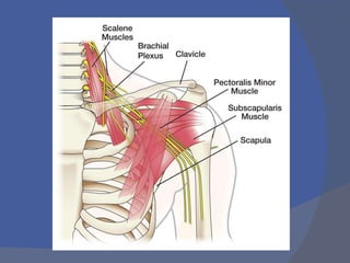
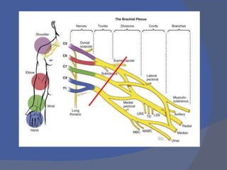
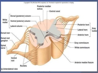
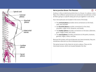
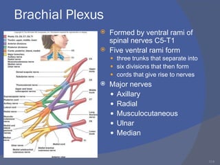
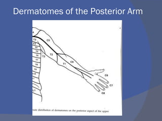
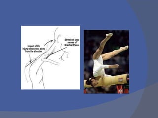
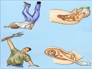
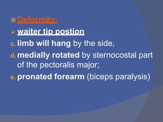
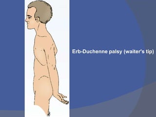
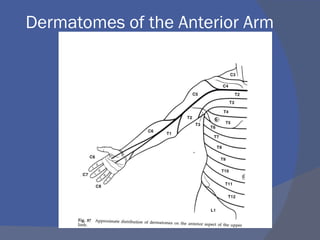
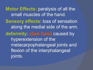
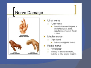
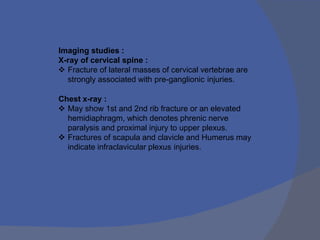
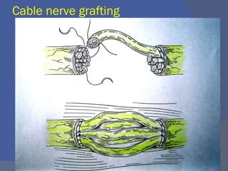
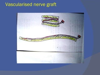
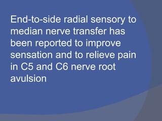
Dr P. ROHIT RAJ
MBBS MS ORTHO
ASSISTANT PROFESSOR
ORTHOPAEDIC DEPARTMENT
VISHWABHARATHI MEDICAL COLLEGE
ANATOMY
INJURY- Classification
- Etiology
- diagnosis
- Management
12-2
Spinal Nerves
 Spinal nerves attach to
the spinal cord via roots
 Dorsal root



Has only sensory neurons
Attached to cord via rootlets
Dorsal root ganglion
○

Bulge formed by cell bodies
of unipolar sensory neurons
Ventral root


Has only motor neurons
No ganglion - all cell bodies
of motor neurons found in
gray matter of spinal cord
12-3
Spinal Nerves
 31 pair
 each contains thousands of nerve fibers
 All are mixed nerves have both sensory and motor
neurons)
 Connect to the spinal cord
 Named for point of issue from the spinal cord
 8 pairs of cervical nerves (C1-C8)
 12 pairs of thoracic nerves (T1-T12)
 5 pairs of lumbar nerves (L1-L5)
 5 pairs of sacral nerves (S1-S5)
 1 pair of coccygeal nerves (Co1)
12-4
Formation of Rami



Rami are lateral branches of a
spinal nerve
Rami contain both sensory
and motor neurons
Two major groups
 Dorsal ramus
○ Neurons innervate the
dorsal regions of the body
 Ventral ramus
○ Larger
○ Neurons innervate the
ventral regions of the
body
○ Braid together to form
plexuses (plexi)
12-8
Brachial Plexus


Formed by ventral rami of
spinal nerves C5-T1
Five ventral rami form



three trunks that separate into
six divisions that then form
cords that give rise to nerves
 Major nerves
 Axillary
 Radial
 Musculocutaneous
 Ulnar
 Median
12-9
Brachial Plexus: Axillary Nerve
 Motor neurons stimulate
 Deltoid, teres minor
○ Abducts arm- deltoid
○ Laterally rotate arm-teres
minor
 Sensory neurons
 Skin: inferior lateral
shoulder
12-10
Brachial Plexus: Radial Nerve
 Motor components stimulate
 Posterior muscles of arm, forearm, and
hand
○ Triceps, supinator, brachioradialis,
extensors
○ Cause extension movements at elbow
and wrist, thumb movements
 Sensory components
 Skin on posterior surface of arm and
forearm, hand
12-11
Brachial Plexus:
Musculocutaneous Nerve


Motor components stimulate
 Flexors in anterior upper arm:
(biceps brachii, brachialis)
○ Cause flexion movements at
shoulder and elbow
Sensory: Skin along lateral
surface of forearm
12-12
Brachial Plexus: Ulnar Nerve
 Motor components
stimulate
 Flexor muscles in anterior
forearm (FCU, FDP, most
intrinsic muscles of hand)
 Results in wrist and finger
flexion
 Sensory: Skin on medial
part of hand
12-13
Brachial Plexus: Median Nerve
 Motor components
stimulate

 All but one of the flexors of the
wrist and fingers, and thenar
muscles at base of thumb
(Palmaris longus, FCR, FDS,
FPL, pronator)
Causes flexion of the wrist
and fingers and thumb
 Sensory components
 Stimulate skin on lateral part
of hand
Dermatomes of the Posterior Arm
Dermatomes of the Anterior Arm
Etiology
 traffic accidents
 birth injuries
 brachial plexus neuritis
 stab and bullet wounds
Mechanisms of Injury to the Brachial
Plexus
A.Traction: direct blow to the
shoulder with the neck laterally
flexed toward the unaffected
shoulder (gymnast falls on beam)
B.Direct trauma: direct blow to the
supraclavicular fossa over Erb’s point
C.Compression: Occurs when the
neck is flexed laterally toward the
patient’s affected shoulder,
compressing / irritating the nerves,
resulting in point tenderness over
involved vertebrae of affected
nerve(s)
(Troub, 2001)
Brachialplexus
Injury
Paediatric/Obstetric
Upper/Erb’s
(C5C6,C7)
Lower/Klumpke’s
(C8 T1)
Adult
Brachial Plexus Injuries
 Upper Lesions of the Brachial
Plexus (Erb’s Palsy): resulting from
excessive displacement of the head to
opposite side and depression of shoulder
on the same side.
 This causes excessive traction or
even tearing of C5 and 6 roots of
the plexus. It occurs in infants
during a difficult delivery or in
adults after a blow to or fall on
shoulder.
Dermatomes of the Posterior Arm
Dermatomes of the Anterior Arm
 Effects:
Motor: paralysis of
 the supraspinatus,
 infraspinatus,
 subclavius,
 biceps brachii,
 part of brachialis,
 coracobrachialis;
 deltoid
 teres minor.
Sensroy: sensory loss on the lateral side
of the arm.
 Deformity:
 waiter tip postion
c. limb will hang by the side,
d. medially rotated by sternocostal part
of the pectoralis major;
e. pronated forearm (biceps paralysis)
Erb-Duchenne palsy (waiter's tip)
Lower Lesions of the Brachial Plexus
(Klumpke Palsy)

 traction injuries by excessive abduction of
the arm
i.e. occurs if person falling from a height
clutching at an object to save himself or
herself.
Can be caused by cervical rib.
 T1 is usually torn (ulnar and median
nerves)
Dermatomes of the Posterior Arm
Dermatomes of the Anterior Arm
Motor Effects: paralysis of all the
small muscles of the hand.
Sensory effects: loss of sensation
along the medial side of the arm.
deformity: claw hand caused by
hyperextension of the
metacarpophalangeal joints and
flexion of the interphalangeal
joints.
Symptoms:
• a limp or paralyzed arm
• severe pain, especially in
the neck and shoulders,
but also in the arm
• numbness in the arm or
hand
• diminished arterial pulses
in the arm
INVESTIGATIONS
• Imaging Studies
• Electromyogram
• Nerve Conduction Velocity
• Intraoperative Nerve Action Potential
• Myelography
• CT scan for any tumours
• MRI
Imaging studies :
X-ray of cervical spine :
 Fracture of lateral masses of cervical vertebrae are
strongly associated with pre-ganglionic injuries.
Chest x-ray :
 May show 1st and 2nd rib fracture or an elevated
hemidiaphragm, which denotes phrenic nerve
paralysis and proximal injury to upper plexus.
 Fractures of scapula and clavicle and Humerus may
indicate infraclavicular plexus injuries.
INVESTIGATIONS
EMG :
 Most important use of EMG studies is for serial evaluation
of injury to search for signs of re innervation.
 A decreased in number of fibrillation potentials and positive
sharp potentials  typically seen in dennervated muscles
 regenerating axons have reached the motor end plates.
 The appearance of prolonged, polyphasic and low-amp
indicated  re-innervation.
 Seen several weeks before the onset of voluntary muscle
contraction and signify that a further period of
observation is in order.
CT Myelography :
 If plexus injury is strongly suspected a myelogram and
subsequent CT scan should be obtained 2-3 months
after injury.
It may be inaccurate early after the injury because clotted
blood may occlude the opening into the pseudomeningocele.
A delay of 6-12 weeks is recommended before myelogram is
advised.
Advantages:
-detect partial root avulsion
-excellent visualization of bony structures
-no CSF flow artifacts and
-multiplanar reconstruction.
Disadvantages:
- high radiation dose
-poor visualization of lower brachial plexus
due to bony artifacts.
CT myelogram showing a normal brachial plexus (left) and injured brachial plexus
(right)
MRI
Nerve root avulsion. Axial T2-weighted (A) and
coronal MIP 3D STIR SPACE (B) images show the
avulsed left T1 nerve root (large arrows) and C8
nerve root (small arrow) with
pseudomeningocele formation
management
2 clinical types brachial plexus injuries
-OBSTETRIC OR NEONATAL
-ADULT
OBSTETRIC OR NEONATAL
Erbs palsy
OBSTETRIC OR NEONATAL
Consevative
• The majority of patients with brachial plexus palsy
at birth will recover from neurologic deficit.
• 1-2 week rest of affected limb
• No traction of affected arm, no pressure under
axila.
• Those who do not recover during 3-6month
period will Require surgical intervention.
Treatment
Physiotherapy
Avoid contractures
Avoid deformities
Surgical intervension to correct
contactures and deformities.
-muscle releases
-osteotomies
-arthrodesis
Treatment Surgical options
d. nerve transfers
e. nerve grafting
f. muscle transfers
g. free muscle transfers
h. neurolysis of scar around the brachial
plexus in incomplete lesions.
Advances in nerve injury Rx
 Carlstedt obtained promising initial
results with the repair of preganglionic
lesions by replanting nerve rootlets
directly into the spinal cord.
 This is a dramatic advance because
preganglionic lesions were previously
thought to be irreparable
Types of Nerve repairs
Epineural repair
Group fascicular repair
Perineural repair
Epineural repair
Epi perineural repair
Cable nerve grafting
Vascularised nerve graft
End-to-side radial sensory to
median nerve transfer has
been reported to improve
sensation and to relieve pain
in C5 and C6 nerve root
avulsion
MNEmonic
s
Thank
you

Brachialplexusinjuries

  • 1.
    Dr P. ROHITRAJ MBBS MS ORTHO ASSISTANT PROFESSOR ORTHOPAEDIC DEPARTMENT VISHWABHARATHI MEDICAL COLLEGE
  • 2.
  • 6.
    12-2 Spinal Nerves  Spinalnerves attach to the spinal cord via roots  Dorsal root    Has only sensory neurons Attached to cord via rootlets Dorsal root ganglion ○  Bulge formed by cell bodies of unipolar sensory neurons Ventral root   Has only motor neurons No ganglion - all cell bodies of motor neurons found in gray matter of spinal cord
  • 7.
    12-3 Spinal Nerves  31pair  each contains thousands of nerve fibers  All are mixed nerves have both sensory and motor neurons)  Connect to the spinal cord  Named for point of issue from the spinal cord  8 pairs of cervical nerves (C1-C8)  12 pairs of thoracic nerves (T1-T12)  5 pairs of lumbar nerves (L1-L5)  5 pairs of sacral nerves (S1-S5)  1 pair of coccygeal nerves (Co1)
  • 8.
    12-4 Formation of Rami    Ramiare lateral branches of a spinal nerve Rami contain both sensory and motor neurons Two major groups  Dorsal ramus ○ Neurons innervate the dorsal regions of the body  Ventral ramus ○ Larger ○ Neurons innervate the ventral regions of the body ○ Braid together to form plexuses (plexi)
  • 11.
    12-8 Brachial Plexus   Formed byventral rami of spinal nerves C5-T1 Five ventral rami form    three trunks that separate into six divisions that then form cords that give rise to nerves  Major nerves  Axillary  Radial  Musculocutaneous  Ulnar  Median
  • 12.
    12-9 Brachial Plexus: AxillaryNerve  Motor neurons stimulate  Deltoid, teres minor ○ Abducts arm- deltoid ○ Laterally rotate arm-teres minor  Sensory neurons  Skin: inferior lateral shoulder
  • 13.
    12-10 Brachial Plexus: RadialNerve  Motor components stimulate  Posterior muscles of arm, forearm, and hand ○ Triceps, supinator, brachioradialis, extensors ○ Cause extension movements at elbow and wrist, thumb movements  Sensory components  Skin on posterior surface of arm and forearm, hand
  • 14.
    12-11 Brachial Plexus: Musculocutaneous Nerve   Motorcomponents stimulate  Flexors in anterior upper arm: (biceps brachii, brachialis) ○ Cause flexion movements at shoulder and elbow Sensory: Skin along lateral surface of forearm
  • 15.
    12-12 Brachial Plexus: UlnarNerve  Motor components stimulate  Flexor muscles in anterior forearm (FCU, FDP, most intrinsic muscles of hand)  Results in wrist and finger flexion  Sensory: Skin on medial part of hand
  • 16.
    12-13 Brachial Plexus: MedianNerve  Motor components stimulate   All but one of the flexors of the wrist and fingers, and thenar muscles at base of thumb (Palmaris longus, FCR, FDS, FPL, pronator) Causes flexion of the wrist and fingers and thumb  Sensory components  Stimulate skin on lateral part of hand
  • 17.
    Dermatomes of thePosterior Arm
  • 18.
    Dermatomes of theAnterior Arm
  • 22.
    Etiology  traffic accidents birth injuries  brachial plexus neuritis  stab and bullet wounds
  • 23.
    Mechanisms of Injuryto the Brachial Plexus A.Traction: direct blow to the shoulder with the neck laterally flexed toward the unaffected shoulder (gymnast falls on beam) B.Direct trauma: direct blow to the supraclavicular fossa over Erb’s point C.Compression: Occurs when the neck is flexed laterally toward the patient’s affected shoulder, compressing / irritating the nerves, resulting in point tenderness over involved vertebrae of affected nerve(s) (Troub, 2001)
  • 26.
  • 28.
    Brachial Plexus Injuries Upper Lesions of the Brachial Plexus (Erb’s Palsy): resulting from excessive displacement of the head to opposite side and depression of shoulder on the same side.
  • 29.
     This causesexcessive traction or even tearing of C5 and 6 roots of the plexus. It occurs in infants during a difficult delivery or in adults after a blow to or fall on shoulder.
  • 30.
    Dermatomes of thePosterior Arm
  • 31.
    Dermatomes of theAnterior Arm
  • 32.
     Effects: Motor: paralysisof  the supraspinatus,  infraspinatus,  subclavius,  biceps brachii,  part of brachialis,  coracobrachialis;  deltoid  teres minor. Sensroy: sensory loss on the lateral side of the arm.
  • 33.
     Deformity:  waitertip postion c. limb will hang by the side, d. medially rotated by sternocostal part of the pectoralis major; e. pronated forearm (biceps paralysis)
  • 34.
  • 36.
    Lower Lesions ofthe Brachial Plexus (Klumpke Palsy)   traction injuries by excessive abduction of the arm i.e. occurs if person falling from a height clutching at an object to save himself or herself. Can be caused by cervical rib.  T1 is usually torn (ulnar and median nerves)
  • 38.
    Dermatomes of thePosterior Arm
  • 39.
    Dermatomes of theAnterior Arm
  • 40.
    Motor Effects: paralysisof all the small muscles of the hand. Sensory effects: loss of sensation along the medial side of the arm. deformity: claw hand caused by hyperextension of the metacarpophalangeal joints and flexion of the interphalangeal joints.
  • 44.
    Symptoms: • a limpor paralyzed arm • severe pain, especially in the neck and shoulders, but also in the arm • numbness in the arm or hand • diminished arterial pulses in the arm
  • 45.
    INVESTIGATIONS • Imaging Studies •Electromyogram • Nerve Conduction Velocity • Intraoperative Nerve Action Potential • Myelography • CT scan for any tumours • MRI
  • 46.
    Imaging studies : X-rayof cervical spine :  Fracture of lateral masses of cervical vertebrae are strongly associated with pre-ganglionic injuries. Chest x-ray :  May show 1st and 2nd rib fracture or an elevated hemidiaphragm, which denotes phrenic nerve paralysis and proximal injury to upper plexus.  Fractures of scapula and clavicle and Humerus may indicate infraclavicular plexus injuries.
  • 47.
    INVESTIGATIONS EMG :  Mostimportant use of EMG studies is for serial evaluation of injury to search for signs of re innervation.  A decreased in number of fibrillation potentials and positive sharp potentials  typically seen in dennervated muscles  regenerating axons have reached the motor end plates.  The appearance of prolonged, polyphasic and low-amp indicated  re-innervation.  Seen several weeks before the onset of voluntary muscle contraction and signify that a further period of observation is in order.
  • 50.
    CT Myelography : If plexus injury is strongly suspected a myelogram and subsequent CT scan should be obtained 2-3 months after injury. It may be inaccurate early after the injury because clotted blood may occlude the opening into the pseudomeningocele. A delay of 6-12 weeks is recommended before myelogram is advised. Advantages: -detect partial root avulsion -excellent visualization of bony structures -no CSF flow artifacts and -multiplanar reconstruction. Disadvantages: - high radiation dose -poor visualization of lower brachial plexus due to bony artifacts.
  • 52.
    CT myelogram showinga normal brachial plexus (left) and injured brachial plexus (right)
  • 53.
  • 54.
    Nerve root avulsion.Axial T2-weighted (A) and coronal MIP 3D STIR SPACE (B) images show the avulsed left T1 nerve root (large arrows) and C8 nerve root (small arrow) with pseudomeningocele formation
  • 55.
    management 2 clinical typesbrachial plexus injuries -OBSTETRIC OR NEONATAL -ADULT
  • 56.
  • 57.
  • 58.
    OBSTETRIC OR NEONATAL Consevative •The majority of patients with brachial plexus palsy at birth will recover from neurologic deficit. • 1-2 week rest of affected limb • No traction of affected arm, no pressure under axila. • Those who do not recover during 3-6month period will Require surgical intervention.
  • 59.
    Treatment Physiotherapy Avoid contractures Avoid deformities Surgicalintervension to correct contactures and deformities. -muscle releases -osteotomies -arthrodesis
  • 60.
    Treatment Surgical options d.nerve transfers e. nerve grafting f. muscle transfers g. free muscle transfers h. neurolysis of scar around the brachial plexus in incomplete lesions.
  • 61.
    Advances in nerveinjury Rx  Carlstedt obtained promising initial results with the repair of preganglionic lesions by replanting nerve rootlets directly into the spinal cord.  This is a dramatic advance because preganglionic lesions were previously thought to be irreparable
  • 63.
    Types of Nerverepairs Epineural repair Group fascicular repair Perineural repair
  • 64.
  • 65.
  • 66.
  • 67.
  • 68.
    End-to-side radial sensoryto median nerve transfer has been reported to improve sensation and to relieve pain in C5 and C6 nerve root avulsion
  • 69.
  • 70.