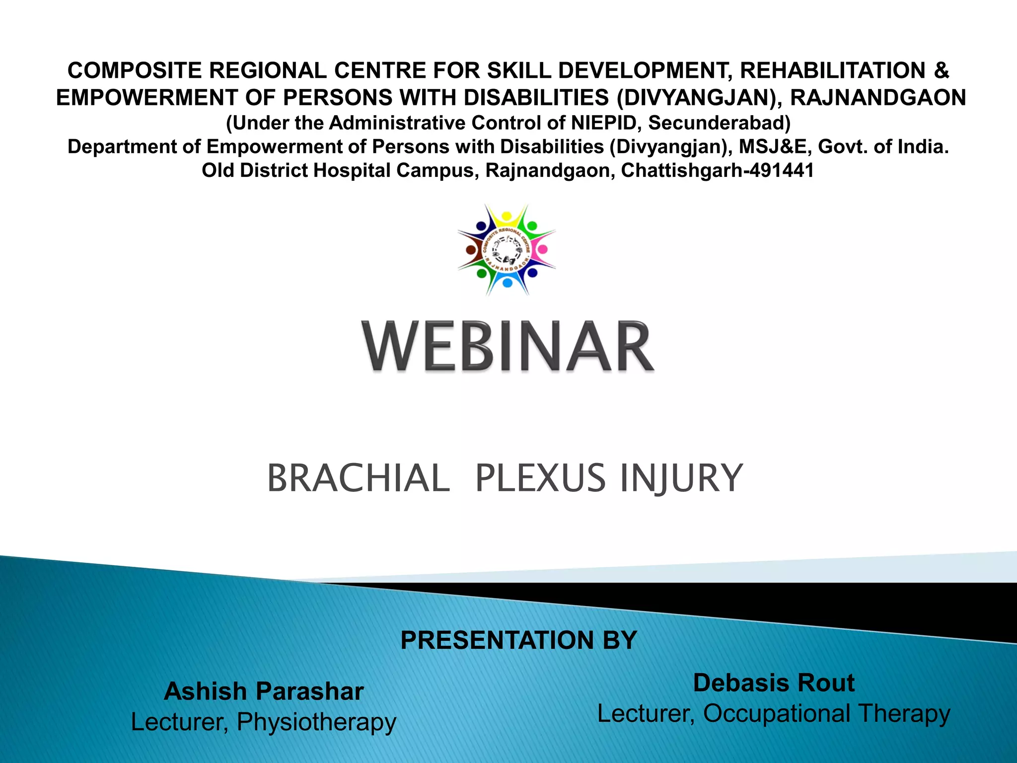
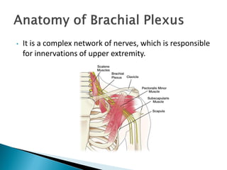
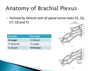
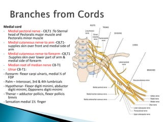
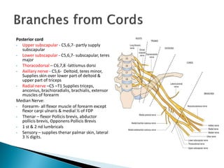
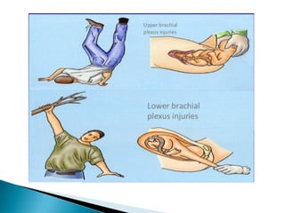
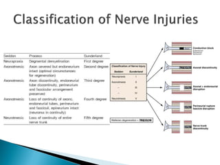
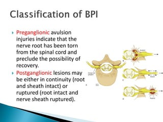
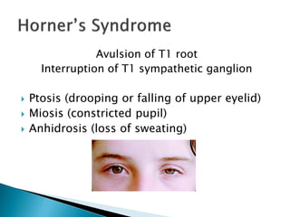
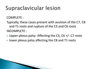
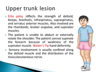
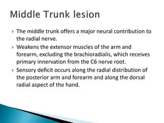
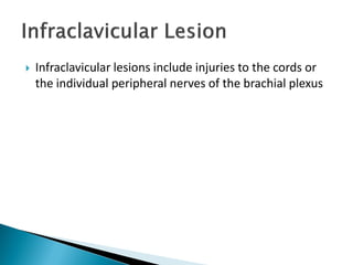
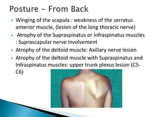
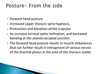
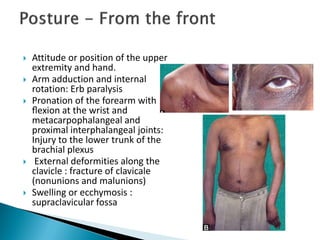
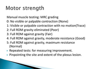
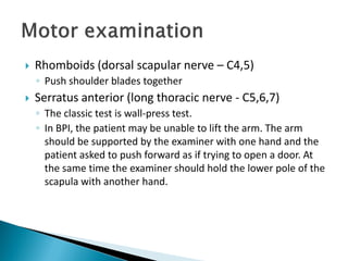
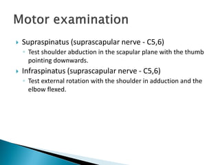
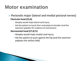
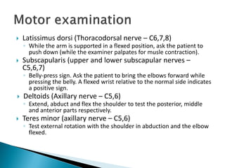
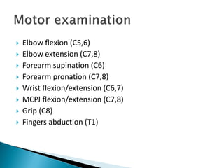
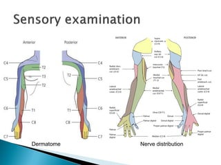
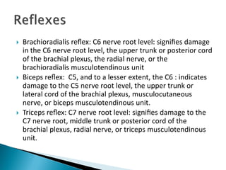
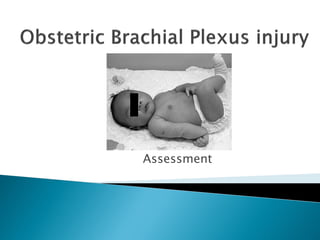
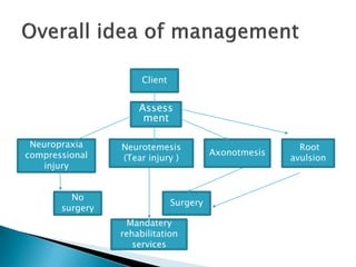
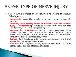
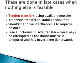
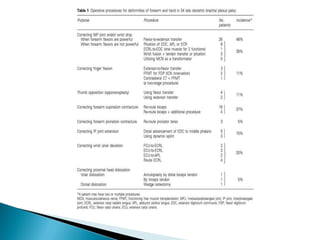
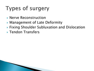
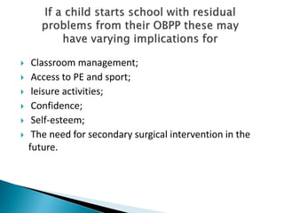
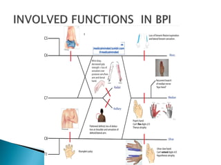
The document provides an in-depth overview of the brachial plexus anatomy, including its formation from spinal nerve roots and the associated major nerves. It discusses various causes of brachial plexus injuries, such as trauma and obstetric palsy, detailing different types and presentations, including Erb's and Klumpke's palsies. Assessment techniques and potential motor and sensory deficits, as well as the implications of treatment, are also addressed, highlighting the complexity and significance of brachial plexus injuries.


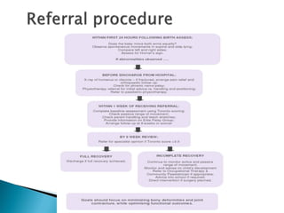
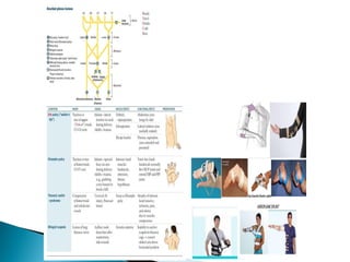
![ Early diagnosis and follow-up, if possible, within two to
three weeks after the child’s birth [21]
Conservative treatment should involve a
multidisciplinary team, composed of physiatrists,
clinical neurophysiologist, neurosurgeons, occupational
therapists, and physiotherapists](https://image.slidesharecdn.com/bpi-200731092820/85/BRACHIAL-PLEXUS-INJURY-EVALUATION-AND-MANAGEMENT-71-320.jpg)
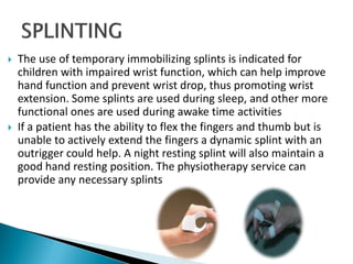
![ Electrical stimulation/electrostimulation is a
complementary means or technique used in conservative
therapies for the rehabilitation of brachial plexus palsy
[4,19], that promotes gaining muscle tone/strength on the
affected muscles, and significant improvements in the
mobility of the injured limb.
These therapies aim to ensure the conditions needed for
the functional recovery of the limb following nerve
regeneration, which implies the prevention of muscle
shrinkage, sagging, joint deformities, and muscle
contractures [20,21]
Most studies reveal that conservative treatment performed
by therapists significantly reduces injuries, removing the
need for surgical intervention [28,29].](https://image.slidesharecdn.com/bpi-200731092820/85/BRACHIAL-PLEXUS-INJURY-EVALUATION-AND-MANAGEMENT-72-320.jpg)
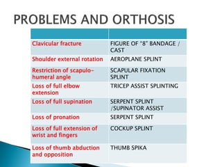
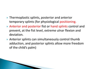
![ There are different tools that are used as means and/or
complementary techniques to the
conservative/surgical treatment of neonatal brachial
plexus palsy, such as electrostimulation, botulinum
toxin injection, thermoplastic splints, posterior and
anterior temporary splints (for physiological
positioning, facilitating functional motor function, and
preventing vicious postures. and constraint induced
movement therapy [20,21].](https://image.slidesharecdn.com/bpi-200731092820/85/BRACHIAL-PLEXUS-INJURY-EVALUATION-AND-MANAGEMENT-73-320.jpg)
![ Constraint induced movement therapy demonstrates
that performing activities at home for one hour a day
can improve mobility, functional capacity, speed, range
of motion, and hand manipulation ability [23].](https://image.slidesharecdn.com/bpi-200731092820/85/BRACHIAL-PLEXUS-INJURY-EVALUATION-AND-MANAGEMENT-74-320.jpg)
![ Surgical nerve reconstruction may be necessary for
rehabilitating patients with neonatal brachial plexus palsy,
especially children who do not show spontaneous recovery
during the first months of life [17].
When surgical intervention is required, both primary and
secondary microsurgeries are available. Primary
microsurgery techniques include recession and
reconstruction of the neuroma, neurolysis, and nerve
transfer [16,20,22]. Studies reveal that, as a primary
surgery for neonatal brachial plexus palsy, neurolysis
combined with nerve transfer produces good results [34].](https://image.slidesharecdn.com/bpi-200731092820/85/BRACHIAL-PLEXUS-INJURY-EVALUATION-AND-MANAGEMENT-75-320.jpg)
![ In situations where the lesion affects the suprascapular
nerve, shoulder function is impaired (abduction and
external rotation). Grafts extracted from the proximal
C5 root stump or the accessory nerve are often used to
reconstruct the suprascapular nerve. The use of the
phrenic nerve has also been shown to provide a similar
level of recovery to the use of the median nerve,
increasing the number of graft options available to
recover suprascapular nerve function [24].](https://image.slidesharecdn.com/bpi-200731092820/85/BRACHIAL-PLEXUS-INJURY-EVALUATION-AND-MANAGEMENT-76-320.jpg)
![ In Erb’s palsy, affecting shoulder abduction and
external rotation, elbow flexion, and forearm
supination, and when there is no evidence of
spontaneous recovery, surgery is a valid treatment
option
The Oberlin’s procedure involves the transfer of the
ulnar nerve to the cutaneous nerve and is an effective
way of recovering the elbow function, improving elbow
flexion and leading to increased functional use of the
affected limb [25].](https://image.slidesharecdn.com/bpi-200731092820/85/BRACHIAL-PLEXUS-INJURY-EVALUATION-AND-MANAGEMENT-77-320.jpg)


![ONFH[AVN HIP] -TRIPLE REGIME -A NOVAL SURGICAL CONCEPT .pptx](https://cdn.slidesharecdn.com/ss_thumbnails/onfhavnhip2026koaconcalicutdrgokuldevdrmashraf-260210064517-213ec005-thumbnail.jpg?width=640&height=640&fit=bounds)


![CTEV [ clubfoot] DR ARUN LAL ,DR MOHAMED ASHRAF travancore medical college k...](https://cdn.slidesharecdn.com/ss_thumbnails/ctevclubfootdrarunlaldrmohamedashraftravancoremedicalcollegekollamkeralaindia-260208063247-18fc466c-thumbnail.jpg?width=640&height=640&fit=bounds)


