SPINA BIFIDA
Definition
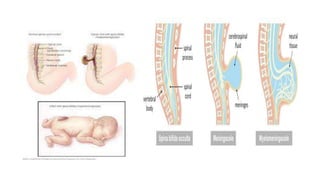
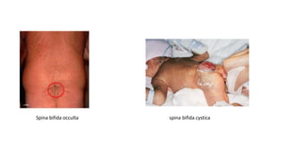
Spina bifida is a neural tube defect where there is an incomplete closure of the
vertebrae and neural tube(hollow structure from which brain and spinal cord develop
in embryo).
Incidence
Spina bifida is the most common developmental defect of central nervous system,
occurring in about 1 or 2 per 1000 live births.
Etiology
• Genetic: A child of parents with spina bifida carries 15 times more probability of
inheriting the defect.
• Matemal Age: The disorder is twice more common in pregnant mothers over 35
years of age or below 20 years of age.
• Environmental: Radiations increase the risk of defective neural tube development.
Regional differences are also seen. The incidence of spina bifida is more in Great
Britain. It is three times more than in USA.
• Diet: Folic acid deficiency in the pregnancy increases the risk of spina bifida
• Maternal obesity
• Maternal hypertension
• Intake of anticonvulant medications.
• Maternal diabetes
• Maternal epileptic seizures
• Maternal infection or fever in the first trimester
Types
1) Spina bifida occulta: It is a defect which results from failure of formation of
bony arch around the spinal cord, but spinal cord and meninges are
normal. It is not visible externally and is asymptomatic. It presents as a tuft
of hair or a dimple in the malformed vertebra
2) Spina bifida cystica: Spina bifida cystica is a defect in the closure of
posterior vertebral arch with protrusion of spinal cord and meninges
through the defect. It is divided into
• Meningocele: It is a sac-like herniation through the bony malformation,
containing meninges and cerebrospinal fluid. The covering of the sac may be
thin and translucent or membranous.
• Myelomeningocele : It is a sac like protrusion of spinal nerves,spinal cord,
CSF and meninges through spinal cleft
The defect most commonly occur in lumbosacral region as this
is the last part of the neural tube to fuse
Spina bifida occulta spina bifida cystica
Clinical features
Spina Bifida Occulta
• Most of the patients with spina bifida occulta are asymptomatic. The only features
seen are:
• A dimple in the skin or growth of hair over malformed vertebra
• As the child grows, he may develop foot weakness or disturbances of bladder and
bowel sphincter.
Meningocele
• An external cystic defect can be seen at the back. The sac is composed only of
meninges and is filled with CSF.
Myelomeningocele
• Manifestations of myelomeningocele depend on the location and extent of defect.
The higher the deformity the more neurological deficits will be present.
A round, raised, poorly epithelialized, herniated mass is present over the vertebral
column, mainly in the lumbosacral region.
• Approximately, 90% of infants with severe spina bifida develop hydrocephalus
due to associated Arnold-chiari syndrome(brain tissues extend into spinal
canal).
• Loss of motor control and sensation occurs, below the level of lesion.
• A low thoracic lesion may cause total flaccid paralysis.
• A Sacral lesion leads to weakness of the lower limbs.
• Bowel and bladder may or may not be affected; there may be fecal and urinary
incontinence.
• There may be renal impairment due to faulty renal innervation. Urinary tract
infections are common complication associated with the defect.
• Congenital skeletal anomalies like club foot, developmental dysplasia of hip,
kyphosis or scoliosis - due to denervation of muscles
• The skeletal and muscular deformities may also lead to back pain, respiratory
distress, recurrent skin breakdown and difficulty in movement.
• Developmental delays are commonly seen in speech, mobility etc.
Diagnostic Evaluation
• Prenatal diagnosis of neural tube defects
Ultrasound
Fetal MRI
Amniocentesis : It may reveal an increase in alpha-fetoprotein, a fetal specific
gamma-globulin in the amniotic fluid that indicates presence of
meningomyelocele.
• Diagnosis after birth
On neonatal examination, a sac may be seen on the back of the baby
CT Scan and MRI of spinal cord and brain
Laboratory tests like urine test for presence of infection, Renal function test,
WBC counts, ESR
Other tests include neurological assessment for motor response and sensory
reactions
Developmental assessment to detect any delay in milestones etc.
Management
• Usually no intervention is required for spina bifida occulta.
• For spina bifida cystica ,surgery is required-Laminectomy and closure of the
defect or removal of the sac is done within 24-48 hours of birth. T-Closure of skin graft is
done.
• A multidisciplinary team approach is required to maximize the physical and intellectual
potential of each affected child.
• A team of neurologist, neurosurgeon, orthopedic surgeon, urologist, primary care provider,
nurse, speech therapist and physiotherapist need to work together for managing the child.
Nursing Management
The main aims of pre and post-operative nursing care are:
• Prevention of infection
• Preventing injury to the sac
• Prevention of skin breakdown
• Preventing urinary tract infection
• Preventing leg and hip deformities
• Provision of adequate nutrition
Pre-operative Care
• Risk of impaired skin integrity related to altered motor and sensory functions.
Avoid positioning the baby on back to prevent pressure on the sac.
Change the position of the baby frequently.
Provide meticulous skin and back care to keep the skin clean and dry.
Skin may be massaged periodically to stimulate blood circulation, with special
attention to bony prominences.
• Risk of injury and infection to the sac.
Do not position the baby on the back, as pressure over the sac may lead to rupture
of sac.
Clean the sac aseptically using normal saline.
Cover the sac with sterile gauze pieces dipped in normal saline. This prevents the sac
from
dryness and rupture.
Change the wet saline gauze dressings every 2-4 hours, in order to keep the sac
moist.
Prevent contamination of the sac with urine and feces. So, do not put diaper, as
passing urine or stool inside the diaper keeps care giver unaware of soiling.
Frequently check the sac for any CSF leakage.
Prophylactic antibiotics must be administered to the infant.
Infant must be handled carefully while feeding or giving care, to prevent injury to the sac
While the infant is placed in side lying position, keep a pillow behind the back to prevent sudden
rolling of infant on the back.
• Impaired bladder and bowel function related to neurological deficit.
Children with spina bifida may have the problem of retention of urine so intermittent
catheterization is done and also taught to parents.
Since bladder is an abdominal organ in infancy, pressure is applied gently on the lower abdomen to
manually express urine.
Monitor for sign and symptoms of urinary tract infection like cloudy or foul-smelling urine, fever etc.
Administer prophylactic antibiotics to decrease the risk of urinary tract infection among patients
who are catheterized.
Administer increased amount of fluids to the child as it helps in flushing out the bladder and urinary
tract.
If the child has urine incontinence, then also catheterization is essential.
In case of bowel incontinence, keep the child clean and dry
 Provide, frequent buttock care and perianal care to infants with fecal
incontinence, to prevent excoriation of perianal skin.
Provide soft bland food to the child having fecal incontinence.
In case, the child has constipation administer laxatives as prescribed.
• Inadequate cerebral tissue perfusion related to complications like
hydrocephalus.
Monitor for signs of hydrocephalus like irritability, feeding difficulty, reduced
alertness, tense or bulging fontanelle etc.
Administer diuretics like Mannitol and Acetazolamide as prescribed, to reduce
the intra cranial pressure.
 Avoid excess stimulation to the child
Elevate the head end of the child to reduce CSF pressure
Post-operative Care
• Ineffective thermoregulation related to surgery.
Monitor the vital signs frequently after surgery.
Keep the infant warm, using radiant warmer or incubator.
 Avoid exposing the child unnecessarily.
Maintain thermoneutral environment.
• Risk of development of infection related to surgery.
Keep the operated site clean and dry.
 Observe for any drainage from the operated site.
Keep the baby in prone position, to avoid pressure on the operated site.
Dressing of the site should be done using aseptic techniques.
Administer the prescribed antibiotics.
• Altered nutrition less than body requirement related to disease process.
Administer intravenous fluids as ordered.
Maintain intake and output chart.
Begin oral feeds as soon as infant starts tolerating the feeds.
Feed the infant in side lying position.
The baby can be held gently for feeding as soon as the surgical area is sufficiently healed.
 Burp the baby frequently between feeds
Check the weight of the baby daily .
• Risk of development of complications related to the disease process.
In order to prevent development of hip or joint deformity, the nurse must pay attention to
the position in which the infant is placed.
An infant with associated hip dysplasia is placed in prone position with legs slightly to
moderately abducted. A pillow is placed between the knees to counteract hip subluxation.
In order to reduce chances of CSF leakage from the incision site, the infant may be placed
in low trendelenburg's position as this helps in reducing CSF pressure at the site.
Monitor for development of hydrocephalus.
If hydrocephalus is suspected, elevate the head end of bed to reduce the intracranial
pressure.
Provide passive range of motion exercises to prevent development of contractures.
Educate the parents regarding surgeries that may be needed for correction of
deformities or contractures .Such surgeries must be performed before the infant
begins to walk.
Clubfoot may be corrected by repeated application of plaster cast.
 Administer oxygen if saturation falls post operatively.
• Altered bladder and bowel function related to neurological deficit.
Provide bowel training to the child to compensate for reduced sensation.
Medications such as stool softeners, suppositories and enema may be used in case of
constipation
Encourage fluid intake.
In case of urine incontinence, catheterization may be done and taught to parents.
 Urinary diversions can be considered later
• Knowledge deficit in parents related to care of the infant after discharge from
the hospital.
Teach the parents about special techniques required for holding, positioning
and feeding the baby.
Encourage the parents to provide tender loving care to the baby by making
close contact
Teach the parents about passive range of motion exercises that need to be
done at home.
Advise the parents that children with paralysis or difficulty in movements are
at risk of obesity due to inactivity, so they should be given low fat diet.
Timely immunization is essential to protect the infant from killer diseases.
Teach the parents about monitoring growth and development of the infant.
Tell parents about the need and importance of follow up care.
Tell the parents to treat the child as other normal children.
Complications
• Frequent urinary tract infections
• Hydrocephalus
• Loss of bladder and bowel control
• Meningitis
• Permanent weakness or paralysis of legs
• joint contractures
Prevention
• Genetic counseling of at risk couple may be recommended.
• If severe defect is detected early in pregnancy, a therapeutic abortion may be
considered.
• The incidence of spina bifida can be decreased by upto 70% if daily folic acid
supplements are taken by women prior to conception.
• A pregnant women needs 1mg folic acid per day.
HYDROCEPHALUS
Definition
Hydrocephalus is defined as an imbalance between the production and absorption of
cerebrospinal fluid. It is characterized by abnormal increase in the volume of
cerebrospinal fluid within the intracranial cavity resulting in enlargement of the
infant's head.
Incidence
It occurs in approximately 3-4 per 1000 live births
Classification
Depending upon the pathology, hydrocephalus is of two types:
a. Congenital
b. Acquired
Another classification based on obstruction
a. Non-communicating or obstructive hydrocephalus
b. Communicating or non obstructive hydrocephalus
CSF Pathway
Non- communicating or obstructive hydrocephalus
• In this type, there is obstruction in the flow of cerebrospinal fluid, within the
ventricles and subarachnoid space, resulting in interference with the circulation
and absorption of CSF. This type is more common inchildren.
• In Non-Communicating or obstructive hydrocephalus, the accumulated CSF
distends the ventricles. This leads to gradual thinning of brain substance, which
is compressed between the distended ventricles and the skull.
Congenital causes
• Stenosis of aqueduct of sylvius
• Meningomyelocele
• Dandy-walker syndrome (obstruction of foramen of luschka or magendie)
• Arnold-chiari malformation (a brain defect resulting in herniation of
cerebellum, medulla, pons and fourth ventricle into the spinal cord)
Acquired causes
• Infections caused by rubella, cytomegalovirus and toxoplasmosis
• Spontaneous intracranial hemorrhage.
• Intracranial tumors like medulloblastoma, craniopharyngioma or astrocytoma
• Head injury
Communicating or non-obstructive hydrocephalus
• In communicating hydrocephalus there is normal flow of CSF, within the ventricular system but
there is interference outside the ventricles causing decreased absorption of CSF in the subarachnoid
villi. The CSF that accumulates in the subarachnoid space compresses the brain and distends the
cranial cavity
• This excessive CSF around the brain leads to flattening and atrophy of convolutions of brain.
Causes
• Subarachnoid hemorrhage
• Meningitis
• Toxoplasmosis or Cytomegalovirus infection in which there is obliteration of sub arachnoid space
due to inflammatory reaction.
• Diseases of connective tissue like hurler's syndrome and achondroplasia (a bone disorder . The
stunted bone growth at the base of the skull and the spine can cause the spinal cord and brain stem
to become compressed.)
Clinical Features
• In Infants:
There is accumulation of CSF in ventricles leading to enlargement of the skull
Sutures become widely separated
Delayed closure of anterior fontanelle
Tense bulging fontanelle
A hollow or "cracked pot" sound is heard on percussion of skull (Macewen's sign)
Scalp veins are prominent and scalp appears shiny
The eyes may have a wide bridge between them and visible sclera above the iris (sun setting
sign)
Neurologically, the infant may be fussy, restless, irritable, apathetic
or have an altered or diminished level of consciousness
Sluggish pupillary response to light,
 Abnormal posturing and spasticity of lower limbs.
In addition, there may be feeding difficulty and high pitch cry due to increased
intracranial pressure.
The infant may have physical or mental developmental retardation
• In older children
Headache on awakening in the morning
Nausea and vomiting
Irritability and high pitch cry
Lethargy
Apathy
Confusion
Impaired judgement and reasoning skills
Affected motor abilities (ataxia, spasticity)
Papilledema, Strabismus or decreased visual acuity secondary to compression of
optic nerve
Diagnostic Evaluation
• Rapid head enlargement and other clinical features suggestive of increased
Intra Cranial Pressure.
• CT scan and MRI - reveal ventricular enlargement or dilatation as well as any
structural defect, if present.
• ultrasound or echoencephalographyin
• X-ray of skull may show a large skull with wide cranial sutures.
Management
Management of hydrocephalus is directed towards:
• Relief of hydrocephalus or reducing the intracranial pressure
• Prevention and management of complications
• Managing problems caused by the pathology
• The treatment of hydrocephalus depends on the cause.
• Medical management includes the use of osmotic diuretic-Acetazolamide and
Frusemide to reduce the rate of CSF production. These medicines
provide temporary relief
• The main management is surgery for removal of any space-occupying lesion
and insertion of a shunt (VP shunt).
• A shunt is made up of radio-opaque plastic and has a ventricular catheter, a
unidirectional pressure valve, pumping chamber and a distal catheter that
directs the flow of CSF from the ventricles to other areas of body from where it
is absorbed. Thus the shunt helps in removing excessive CSF from the
ventricles, thereby reducing the increased intracranial pressure.
• Four types of shunts are available:
1. Ventriculoperitoneal shunt( from ventricle to peritoneal cavity)
2. Ventriculoatrial shunt (from ventricles to left atrium)
3. Ventriculopleural shunt (from ventricles to the pleural cavity)
4. Ventriculoureteric shunt (from ventricles to the ureter)
• The complications that may arise with shunt include kinking, separation or
plugging of shunt tubing.
• The most serious complication is infection that mostly occurs within two
months of surgery
• Infection of shunt may lead to ventriculitis and septicemia. Conservative
treatment for shunt infection is administration of antibiotics.
Nursing Management
Pre-operative Nursing Care
Nursing care in the preoperative period focuses on recognition of signs of
increased intracranial pressure, providing supportive care and preventing
complications.
• Measure the head circumference of the child daily.
• Palpate the fontanelle for evidence of increased intracranial pressure. The
anterior fontanelle is bulging and tense.
• Assess the pupillary response and level of consciousness.
• Monitor vital signs regularly. Any changes in vital signs should be promptly reported.
• As the infant is irritable and restless, provide calm and quiet environment so that the
infant may take adequate rest.
• Prevent the child from nosocomial infections by using barrier nursing and strict
aseptic techniques while providing care.
• Position the body with neck adequately supported.
• As the scalp becomes thin, there is an increased chance for breakdown of scalp so, a
water pillow or lamb's wool may be used to keep head over it.
• Change the infant's position frequently.
• Support the infant's head and neck while handling because the head may be too large
and neck muscles may be too weak to support the head.
• The infant is prone to vomiting, so provide small, frequent feeding with intermittent
burping.
• Keep the infant clean and dry.
Post-operative Care
• Post operatively, place the infant in flat position to prevent rapid CSF drainage and
on unoperated side to avoid pressure on the valve of shunt. If CSF is drained too
rapidly, there is a risk of subdural hematoma caused by tearing of the vessels
secondary to the cerebral cortex pulling away from the duramater.
• Check vital signs every 15-30 minutes in immediate post-operative period.
• Assess the neurological status and level of consciousness frequently.
• Assess the head circumference regularly.
• Monitor the intake and output, as fluids may be restricted during first 24 hours post
operatively.
• Oral rehydration must be started after the bowel sounds reappear
• Check dressings for any drainage.
• Avoid constipation as straining can increase ICP
• Meticulous skin care should be provided.
• The child must be observed for signs of infection such as fever, increased heart and
respiratory rate, poor feeding or vomiting, altered mental status and local symptoms
such as redness or CSF leakage at the surgical site.
• If the fontanelle becomes sunken, notify the physician immediately. Also immediately
lower the head end of the bed to decrease the outflow of CSF through the shunt. This
will help in reducing the risk of subdural hematoma.
• Prophylactic antibiotics are administered as prescribed to prevent infections.
• Teach parents about care of the child at home, after discharge from hospital. Tell
them about
Handling the baby while feeding and positioning the child.
Recognizing the signs of increased intracranial pressure and malfunctioning or
blockage of shunt.
Pumping of the shunt in case of increased intracranial pressure.
Preventing constipation if the child has Ventriculoperitoneal shunt, as straining during
defecation may increase the intracranial pressure.
Importance of follow up care.
CEREBRAL PALSY
Definition
Causes
Spastic (Pyramidal) Cerebral Palsy
• characterized by persistent primitive reflexes, positive babinski reflex,
• deep tendon reflex exaggerated
• Clasp knife spasticity: rigidity of the extensor muscles of a joint that gives resistance
to passive flexion initially but gives away abruptly to allow easy flexion
• ankle clonus- series of clonic jerky movements when we stretch the limb across the
joint( due to hyperexcitability of the muscles)
• exaggerated stretch reflex and eventual development of contractures.
• Type of spastic cerebral palsy:
Hemiplegia : One side of the body is affected
Diplegia: lower extremities more effected than upper. ‘Scissors gait’ normal
Tetraplegia (quadriplegia): all four extremities involved.
Triplegia: involving three extremities.
Monoplegia: involving only one extremities.
Extrapyramidal
• Hypotonic muscle tone
• deep tendon reflexes are depressed
• Babinski reflex –ve
• Athetoid: chorea (involuntary, irregular, jerking movements),
characterized by slow, wormlike, writhing movements that usually
involve the extremities, trunk, neck, facial muscle and tongue. Abnormal
posture common
Involvement of the pharyngeal and oral muscle causing drooling
and dysarthria (slurred and slow speech)
• Dystonic: repetitive muscle contractions result in twisting and repetitive movements or
abnormal fixed postures.
• Ataxic : lesion in the cerebellum. Mainly associated with inco-orination and balance
disturbance. Wide based gait
Mixed cerebral palsy
• Include both the features of pyramidal and extrapyramidal cerebral palsy
Signs indicative of CP
• poor head control after 3 months of age
• stiff or rigid arms or legs and toe walking
• arching back
• floppy or limp body posture
• cannot sit up without support by 8 months
• uses only one side of the body, or only the arms to crawl
• clenched hands after 3 months( cortical thumb sign)
• a leg scissoring
• Seizures
• sensory impairment (hearing, vision)
• persistent tongue thrusting after 6 months of age,
• Feeding difficulty
• Extreme irritability or crying
Nursing diagnosis
• Risk for injury related to spasms, uncontrolled movements, and seizures.
• Impaired physical mobility related to spasms and muscle weakness.
• Changes in growth and development related to neuromuscular disorders.
• Impaired verbal communication related to difficulty in articulation.
• Risk for aspiration related to neuromuscular disorders.
• Disturbed thought processes related to cerebral injury, learning disabilities.
• Self-care deficit related to muscle spasms, increased activity, cognitive
changes.
• Deficient knowledge related to home care and therapeutic needs
Nursing interventions
• Ensure therapeutic communication. To ease the change of environment,
the nurse needs to communicate with the family to learn as much as possible about
the child’s activities at home.
• Enhance self-esteem. Assist the patient to increase his/her personal judgment of
self-worth.
• Provide emotional support. Provide of reassurance, acceptance, and
encouragement during times of stress.
• Strengthen family support. Utilize the family’s strengths to influence patient’s
health in a positive direction.
• Prevent injury. Prevent physical injury by providing the child with a safe
environment, appropriate toys, and protective gear (helmet, kneepads) if needed.
• Prevent deformity. Prevent physical deformity by ensuring the correct use of
prescribed braces and other devices and by performing ROM exercises.
• Encourage mobility. Promote mobility by encouraging the child to perform age-and
condition-appropriate motor activities.
• Increase oral fluid intake. Promote adequate fluid and nutritional intake.
• Manage sleep and rest periods. Foster relaxation and general health by
providing rest periods.
• Enhance self-care. Encourage self-care by urging the child to participate in
activities of daily living (ADLs) (e.g. using utensils and implements that are
appropriate for the child’s age and condition).
• Facilitate communication. Talk to the child deliberately and slowly, using
pictures to reinforce speech when needed; encourage early speech therapy to
prevent poor or maladaptive communication habits; and provide means of
articulate speech such as sign language or a picture board.
• Enforce therapeutic measures. Assist in multidisciplinary therapeutic
measures designed to establish locomotion, communication, and self-help,
gain optimal appearance and integration of motor functions.

Gnm cns congenital defects

  • 1.
    SPINA BIFIDA Definition Spina bifidais a neural tube defect where there is an incomplete closure of the vertebrae and neural tube(hollow structure from which brain and spinal cord develop in embryo). Incidence Spina bifida is the most common developmental defect of central nervous system, occurring in about 1 or 2 per 1000 live births. Etiology • Genetic: A child of parents with spina bifida carries 15 times more probability of inheriting the defect. • Matemal Age: The disorder is twice more common in pregnant mothers over 35 years of age or below 20 years of age. • Environmental: Radiations increase the risk of defective neural tube development. Regional differences are also seen. The incidence of spina bifida is more in Great Britain. It is three times more than in USA. • Diet: Folic acid deficiency in the pregnancy increases the risk of spina bifida
  • 2.
    • Maternal obesity •Maternal hypertension • Intake of anticonvulant medications. • Maternal diabetes • Maternal epileptic seizures • Maternal infection or fever in the first trimester
  • 4.
    Types 1) Spina bifidaocculta: It is a defect which results from failure of formation of bony arch around the spinal cord, but spinal cord and meninges are normal. It is not visible externally and is asymptomatic. It presents as a tuft of hair or a dimple in the malformed vertebra 2) Spina bifida cystica: Spina bifida cystica is a defect in the closure of posterior vertebral arch with protrusion of spinal cord and meninges through the defect. It is divided into • Meningocele: It is a sac-like herniation through the bony malformation, containing meninges and cerebrospinal fluid. The covering of the sac may be thin and translucent or membranous. • Myelomeningocele : It is a sac like protrusion of spinal nerves,spinal cord, CSF and meninges through spinal cleft The defect most commonly occur in lumbosacral region as this is the last part of the neural tube to fuse
  • 6.
    Spina bifida occultaspina bifida cystica
  • 7.
    Clinical features Spina BifidaOcculta • Most of the patients with spina bifida occulta are asymptomatic. The only features seen are: • A dimple in the skin or growth of hair over malformed vertebra • As the child grows, he may develop foot weakness or disturbances of bladder and bowel sphincter. Meningocele • An external cystic defect can be seen at the back. The sac is composed only of meninges and is filled with CSF. Myelomeningocele • Manifestations of myelomeningocele depend on the location and extent of defect. The higher the deformity the more neurological deficits will be present. A round, raised, poorly epithelialized, herniated mass is present over the vertebral column, mainly in the lumbosacral region.
  • 8.
    • Approximately, 90%of infants with severe spina bifida develop hydrocephalus due to associated Arnold-chiari syndrome(brain tissues extend into spinal canal). • Loss of motor control and sensation occurs, below the level of lesion. • A low thoracic lesion may cause total flaccid paralysis. • A Sacral lesion leads to weakness of the lower limbs. • Bowel and bladder may or may not be affected; there may be fecal and urinary incontinence. • There may be renal impairment due to faulty renal innervation. Urinary tract infections are common complication associated with the defect. • Congenital skeletal anomalies like club foot, developmental dysplasia of hip, kyphosis or scoliosis - due to denervation of muscles • The skeletal and muscular deformities may also lead to back pain, respiratory distress, recurrent skin breakdown and difficulty in movement. • Developmental delays are commonly seen in speech, mobility etc.
  • 9.
    Diagnostic Evaluation • Prenataldiagnosis of neural tube defects Ultrasound Fetal MRI Amniocentesis : It may reveal an increase in alpha-fetoprotein, a fetal specific gamma-globulin in the amniotic fluid that indicates presence of meningomyelocele. • Diagnosis after birth On neonatal examination, a sac may be seen on the back of the baby CT Scan and MRI of spinal cord and brain Laboratory tests like urine test for presence of infection, Renal function test, WBC counts, ESR Other tests include neurological assessment for motor response and sensory reactions Developmental assessment to detect any delay in milestones etc.
  • 10.
    Management • Usually nointervention is required for spina bifida occulta. • For spina bifida cystica ,surgery is required-Laminectomy and closure of the defect or removal of the sac is done within 24-48 hours of birth. T-Closure of skin graft is done. • A multidisciplinary team approach is required to maximize the physical and intellectual potential of each affected child. • A team of neurologist, neurosurgeon, orthopedic surgeon, urologist, primary care provider, nurse, speech therapist and physiotherapist need to work together for managing the child. Nursing Management The main aims of pre and post-operative nursing care are: • Prevention of infection • Preventing injury to the sac • Prevention of skin breakdown • Preventing urinary tract infection • Preventing leg and hip deformities • Provision of adequate nutrition
  • 11.
    Pre-operative Care • Riskof impaired skin integrity related to altered motor and sensory functions. Avoid positioning the baby on back to prevent pressure on the sac. Change the position of the baby frequently. Provide meticulous skin and back care to keep the skin clean and dry. Skin may be massaged periodically to stimulate blood circulation, with special attention to bony prominences. • Risk of injury and infection to the sac. Do not position the baby on the back, as pressure over the sac may lead to rupture of sac. Clean the sac aseptically using normal saline. Cover the sac with sterile gauze pieces dipped in normal saline. This prevents the sac from dryness and rupture. Change the wet saline gauze dressings every 2-4 hours, in order to keep the sac moist. Prevent contamination of the sac with urine and feces. So, do not put diaper, as passing urine or stool inside the diaper keeps care giver unaware of soiling.
  • 12.
    Frequently check thesac for any CSF leakage. Prophylactic antibiotics must be administered to the infant. Infant must be handled carefully while feeding or giving care, to prevent injury to the sac While the infant is placed in side lying position, keep a pillow behind the back to prevent sudden rolling of infant on the back. • Impaired bladder and bowel function related to neurological deficit. Children with spina bifida may have the problem of retention of urine so intermittent catheterization is done and also taught to parents. Since bladder is an abdominal organ in infancy, pressure is applied gently on the lower abdomen to manually express urine. Monitor for sign and symptoms of urinary tract infection like cloudy or foul-smelling urine, fever etc. Administer prophylactic antibiotics to decrease the risk of urinary tract infection among patients who are catheterized. Administer increased amount of fluids to the child as it helps in flushing out the bladder and urinary tract. If the child has urine incontinence, then also catheterization is essential. In case of bowel incontinence, keep the child clean and dry
  • 13.
     Provide, frequentbuttock care and perianal care to infants with fecal incontinence, to prevent excoriation of perianal skin. Provide soft bland food to the child having fecal incontinence. In case, the child has constipation administer laxatives as prescribed. • Inadequate cerebral tissue perfusion related to complications like hydrocephalus. Monitor for signs of hydrocephalus like irritability, feeding difficulty, reduced alertness, tense or bulging fontanelle etc. Administer diuretics like Mannitol and Acetazolamide as prescribed, to reduce the intra cranial pressure.  Avoid excess stimulation to the child Elevate the head end of the child to reduce CSF pressure
  • 14.
    Post-operative Care • Ineffectivethermoregulation related to surgery. Monitor the vital signs frequently after surgery. Keep the infant warm, using radiant warmer or incubator.  Avoid exposing the child unnecessarily. Maintain thermoneutral environment. • Risk of development of infection related to surgery. Keep the operated site clean and dry.  Observe for any drainage from the operated site. Keep the baby in prone position, to avoid pressure on the operated site. Dressing of the site should be done using aseptic techniques. Administer the prescribed antibiotics.
  • 15.
    • Altered nutritionless than body requirement related to disease process. Administer intravenous fluids as ordered. Maintain intake and output chart. Begin oral feeds as soon as infant starts tolerating the feeds. Feed the infant in side lying position. The baby can be held gently for feeding as soon as the surgical area is sufficiently healed.  Burp the baby frequently between feeds Check the weight of the baby daily . • Risk of development of complications related to the disease process. In order to prevent development of hip or joint deformity, the nurse must pay attention to the position in which the infant is placed. An infant with associated hip dysplasia is placed in prone position with legs slightly to moderately abducted. A pillow is placed between the knees to counteract hip subluxation. In order to reduce chances of CSF leakage from the incision site, the infant may be placed in low trendelenburg's position as this helps in reducing CSF pressure at the site. Monitor for development of hydrocephalus.
  • 16.
    If hydrocephalus issuspected, elevate the head end of bed to reduce the intracranial pressure. Provide passive range of motion exercises to prevent development of contractures. Educate the parents regarding surgeries that may be needed for correction of deformities or contractures .Such surgeries must be performed before the infant begins to walk. Clubfoot may be corrected by repeated application of plaster cast.  Administer oxygen if saturation falls post operatively. • Altered bladder and bowel function related to neurological deficit. Provide bowel training to the child to compensate for reduced sensation. Medications such as stool softeners, suppositories and enema may be used in case of constipation Encourage fluid intake. In case of urine incontinence, catheterization may be done and taught to parents.  Urinary diversions can be considered later
  • 17.
    • Knowledge deficitin parents related to care of the infant after discharge from the hospital. Teach the parents about special techniques required for holding, positioning and feeding the baby. Encourage the parents to provide tender loving care to the baby by making close contact Teach the parents about passive range of motion exercises that need to be done at home. Advise the parents that children with paralysis or difficulty in movements are at risk of obesity due to inactivity, so they should be given low fat diet. Timely immunization is essential to protect the infant from killer diseases. Teach the parents about monitoring growth and development of the infant. Tell parents about the need and importance of follow up care. Tell the parents to treat the child as other normal children.
  • 18.
    Complications • Frequent urinarytract infections • Hydrocephalus • Loss of bladder and bowel control • Meningitis • Permanent weakness or paralysis of legs • joint contractures Prevention • Genetic counseling of at risk couple may be recommended. • If severe defect is detected early in pregnancy, a therapeutic abortion may be considered. • The incidence of spina bifida can be decreased by upto 70% if daily folic acid supplements are taken by women prior to conception. • A pregnant women needs 1mg folic acid per day.
  • 19.
    HYDROCEPHALUS Definition Hydrocephalus is definedas an imbalance between the production and absorption of cerebrospinal fluid. It is characterized by abnormal increase in the volume of cerebrospinal fluid within the intracranial cavity resulting in enlargement of the infant's head. Incidence It occurs in approximately 3-4 per 1000 live births Classification Depending upon the pathology, hydrocephalus is of two types: a. Congenital b. Acquired Another classification based on obstruction a. Non-communicating or obstructive hydrocephalus b. Communicating or non obstructive hydrocephalus
  • 20.
  • 21.
    Non- communicating orobstructive hydrocephalus • In this type, there is obstruction in the flow of cerebrospinal fluid, within the ventricles and subarachnoid space, resulting in interference with the circulation and absorption of CSF. This type is more common inchildren. • In Non-Communicating or obstructive hydrocephalus, the accumulated CSF distends the ventricles. This leads to gradual thinning of brain substance, which is compressed between the distended ventricles and the skull. Congenital causes • Stenosis of aqueduct of sylvius • Meningomyelocele • Dandy-walker syndrome (obstruction of foramen of luschka or magendie) • Arnold-chiari malformation (a brain defect resulting in herniation of cerebellum, medulla, pons and fourth ventricle into the spinal cord)
  • 22.
    Acquired causes • Infectionscaused by rubella, cytomegalovirus and toxoplasmosis • Spontaneous intracranial hemorrhage. • Intracranial tumors like medulloblastoma, craniopharyngioma or astrocytoma • Head injury Communicating or non-obstructive hydrocephalus • In communicating hydrocephalus there is normal flow of CSF, within the ventricular system but there is interference outside the ventricles causing decreased absorption of CSF in the subarachnoid villi. The CSF that accumulates in the subarachnoid space compresses the brain and distends the cranial cavity • This excessive CSF around the brain leads to flattening and atrophy of convolutions of brain. Causes • Subarachnoid hemorrhage • Meningitis • Toxoplasmosis or Cytomegalovirus infection in which there is obliteration of sub arachnoid space due to inflammatory reaction. • Diseases of connective tissue like hurler's syndrome and achondroplasia (a bone disorder . The stunted bone growth at the base of the skull and the spine can cause the spinal cord and brain stem to become compressed.)
  • 23.
    Clinical Features • InInfants: There is accumulation of CSF in ventricles leading to enlargement of the skull Sutures become widely separated Delayed closure of anterior fontanelle Tense bulging fontanelle A hollow or "cracked pot" sound is heard on percussion of skull (Macewen's sign) Scalp veins are prominent and scalp appears shiny The eyes may have a wide bridge between them and visible sclera above the iris (sun setting sign) Neurologically, the infant may be fussy, restless, irritable, apathetic or have an altered or diminished level of consciousness
  • 24.
    Sluggish pupillary responseto light,  Abnormal posturing and spasticity of lower limbs. In addition, there may be feeding difficulty and high pitch cry due to increased intracranial pressure. The infant may have physical or mental developmental retardation • In older children Headache on awakening in the morning Nausea and vomiting Irritability and high pitch cry Lethargy Apathy Confusion Impaired judgement and reasoning skills Affected motor abilities (ataxia, spasticity) Papilledema, Strabismus or decreased visual acuity secondary to compression of optic nerve
  • 25.
    Diagnostic Evaluation • Rapidhead enlargement and other clinical features suggestive of increased Intra Cranial Pressure. • CT scan and MRI - reveal ventricular enlargement or dilatation as well as any structural defect, if present. • ultrasound or echoencephalographyin • X-ray of skull may show a large skull with wide cranial sutures. Management Management of hydrocephalus is directed towards: • Relief of hydrocephalus or reducing the intracranial pressure • Prevention and management of complications • Managing problems caused by the pathology
  • 26.
    • The treatmentof hydrocephalus depends on the cause. • Medical management includes the use of osmotic diuretic-Acetazolamide and Frusemide to reduce the rate of CSF production. These medicines provide temporary relief • The main management is surgery for removal of any space-occupying lesion and insertion of a shunt (VP shunt). • A shunt is made up of radio-opaque plastic and has a ventricular catheter, a unidirectional pressure valve, pumping chamber and a distal catheter that directs the flow of CSF from the ventricles to other areas of body from where it is absorbed. Thus the shunt helps in removing excessive CSF from the ventricles, thereby reducing the increased intracranial pressure. • Four types of shunts are available: 1. Ventriculoperitoneal shunt( from ventricle to peritoneal cavity) 2. Ventriculoatrial shunt (from ventricles to left atrium) 3. Ventriculopleural shunt (from ventricles to the pleural cavity) 4. Ventriculoureteric shunt (from ventricles to the ureter)
  • 28.
    • The complicationsthat may arise with shunt include kinking, separation or plugging of shunt tubing. • The most serious complication is infection that mostly occurs within two months of surgery • Infection of shunt may lead to ventriculitis and septicemia. Conservative treatment for shunt infection is administration of antibiotics. Nursing Management Pre-operative Nursing Care Nursing care in the preoperative period focuses on recognition of signs of increased intracranial pressure, providing supportive care and preventing complications. • Measure the head circumference of the child daily. • Palpate the fontanelle for evidence of increased intracranial pressure. The anterior fontanelle is bulging and tense.
  • 29.
    • Assess thepupillary response and level of consciousness. • Monitor vital signs regularly. Any changes in vital signs should be promptly reported. • As the infant is irritable and restless, provide calm and quiet environment so that the infant may take adequate rest. • Prevent the child from nosocomial infections by using barrier nursing and strict aseptic techniques while providing care. • Position the body with neck adequately supported. • As the scalp becomes thin, there is an increased chance for breakdown of scalp so, a water pillow or lamb's wool may be used to keep head over it. • Change the infant's position frequently. • Support the infant's head and neck while handling because the head may be too large and neck muscles may be too weak to support the head. • The infant is prone to vomiting, so provide small, frequent feeding with intermittent burping. • Keep the infant clean and dry.
  • 30.
    Post-operative Care • Postoperatively, place the infant in flat position to prevent rapid CSF drainage and on unoperated side to avoid pressure on the valve of shunt. If CSF is drained too rapidly, there is a risk of subdural hematoma caused by tearing of the vessels secondary to the cerebral cortex pulling away from the duramater. • Check vital signs every 15-30 minutes in immediate post-operative period. • Assess the neurological status and level of consciousness frequently. • Assess the head circumference regularly. • Monitor the intake and output, as fluids may be restricted during first 24 hours post operatively. • Oral rehydration must be started after the bowel sounds reappear • Check dressings for any drainage. • Avoid constipation as straining can increase ICP • Meticulous skin care should be provided.
  • 31.
    • The childmust be observed for signs of infection such as fever, increased heart and respiratory rate, poor feeding or vomiting, altered mental status and local symptoms such as redness or CSF leakage at the surgical site. • If the fontanelle becomes sunken, notify the physician immediately. Also immediately lower the head end of the bed to decrease the outflow of CSF through the shunt. This will help in reducing the risk of subdural hematoma. • Prophylactic antibiotics are administered as prescribed to prevent infections. • Teach parents about care of the child at home, after discharge from hospital. Tell them about Handling the baby while feeding and positioning the child. Recognizing the signs of increased intracranial pressure and malfunctioning or blockage of shunt. Pumping of the shunt in case of increased intracranial pressure. Preventing constipation if the child has Ventriculoperitoneal shunt, as straining during defecation may increase the intracranial pressure. Importance of follow up care.
  • 32.
  • 33.
  • 34.
  • 42.
    Spastic (Pyramidal) CerebralPalsy • characterized by persistent primitive reflexes, positive babinski reflex, • deep tendon reflex exaggerated • Clasp knife spasticity: rigidity of the extensor muscles of a joint that gives resistance to passive flexion initially but gives away abruptly to allow easy flexion • ankle clonus- series of clonic jerky movements when we stretch the limb across the joint( due to hyperexcitability of the muscles) • exaggerated stretch reflex and eventual development of contractures. • Type of spastic cerebral palsy: Hemiplegia : One side of the body is affected Diplegia: lower extremities more effected than upper. ‘Scissors gait’ normal Tetraplegia (quadriplegia): all four extremities involved. Triplegia: involving three extremities. Monoplegia: involving only one extremities.
  • 43.
    Extrapyramidal • Hypotonic muscletone • deep tendon reflexes are depressed • Babinski reflex –ve • Athetoid: chorea (involuntary, irregular, jerking movements), characterized by slow, wormlike, writhing movements that usually involve the extremities, trunk, neck, facial muscle and tongue. Abnormal posture common Involvement of the pharyngeal and oral muscle causing drooling and dysarthria (slurred and slow speech)
  • 44.
    • Dystonic: repetitivemuscle contractions result in twisting and repetitive movements or abnormal fixed postures. • Ataxic : lesion in the cerebellum. Mainly associated with inco-orination and balance disturbance. Wide based gait Mixed cerebral palsy • Include both the features of pyramidal and extrapyramidal cerebral palsy
  • 46.
    Signs indicative ofCP • poor head control after 3 months of age • stiff or rigid arms or legs and toe walking • arching back • floppy or limp body posture • cannot sit up without support by 8 months • uses only one side of the body, or only the arms to crawl • clenched hands after 3 months( cortical thumb sign) • a leg scissoring • Seizures • sensory impairment (hearing, vision) • persistent tongue thrusting after 6 months of age, • Feeding difficulty • Extreme irritability or crying
  • 56.
    Nursing diagnosis • Riskfor injury related to spasms, uncontrolled movements, and seizures. • Impaired physical mobility related to spasms and muscle weakness. • Changes in growth and development related to neuromuscular disorders. • Impaired verbal communication related to difficulty in articulation. • Risk for aspiration related to neuromuscular disorders. • Disturbed thought processes related to cerebral injury, learning disabilities. • Self-care deficit related to muscle spasms, increased activity, cognitive changes. • Deficient knowledge related to home care and therapeutic needs
  • 57.
    Nursing interventions • Ensuretherapeutic communication. To ease the change of environment, the nurse needs to communicate with the family to learn as much as possible about the child’s activities at home. • Enhance self-esteem. Assist the patient to increase his/her personal judgment of self-worth. • Provide emotional support. Provide of reassurance, acceptance, and encouragement during times of stress. • Strengthen family support. Utilize the family’s strengths to influence patient’s health in a positive direction. • Prevent injury. Prevent physical injury by providing the child with a safe environment, appropriate toys, and protective gear (helmet, kneepads) if needed. • Prevent deformity. Prevent physical deformity by ensuring the correct use of prescribed braces and other devices and by performing ROM exercises. • Encourage mobility. Promote mobility by encouraging the child to perform age-and condition-appropriate motor activities.
  • 58.
    • Increase oralfluid intake. Promote adequate fluid and nutritional intake. • Manage sleep and rest periods. Foster relaxation and general health by providing rest periods. • Enhance self-care. Encourage self-care by urging the child to participate in activities of daily living (ADLs) (e.g. using utensils and implements that are appropriate for the child’s age and condition). • Facilitate communication. Talk to the child deliberately and slowly, using pictures to reinforce speech when needed; encourage early speech therapy to prevent poor or maladaptive communication habits; and provide means of articulate speech such as sign language or a picture board. • Enforce therapeutic measures. Assist in multidisciplinary therapeutic measures designed to establish locomotion, communication, and self-help, gain optimal appearance and integration of motor functions.