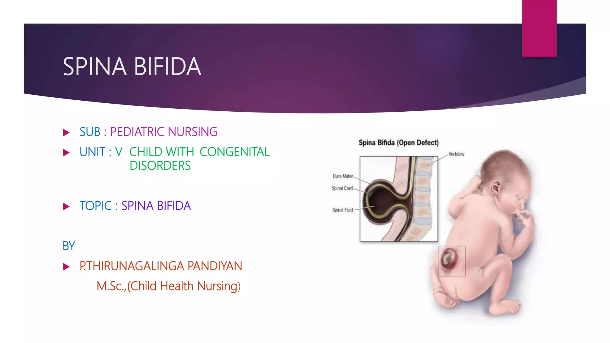
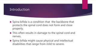
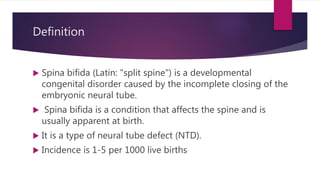
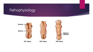
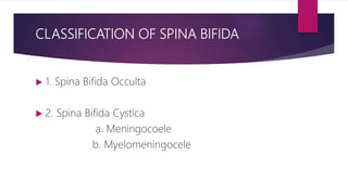
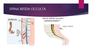
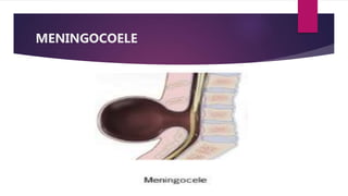
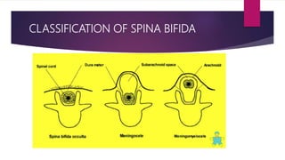
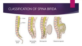
SPINA BIFIDA
 SUB : PEDIATRIC NURSING
 UNIT : V CHILD WITH CONGENITAL
DISORDERS
 TOPIC : SPINA BIFIDA
BY
 P.THIRUNAGALINGA PANDIYAN
M.Sc.,(Child Health Nursing)
Introduction
 Spina bifida is a condition that the backbone that
protects the spinal cord does not form and close
properly.
 This often results in damage to the spinal cord and
nerves.
 Spina bifida might cause physical and intellectual
disabilities that range from mild to severe.
Definition
 Spina bifida (Latin: "split spine") is a developmental
congenital disorder caused by the incomplete closing of the
embryonic neural tube.
 Spina bifida is a condition that affects the spine and is
usually apparent at birth.
 It is a type of neural tube defect (NTD).
 Incidence is 1-5 per 1000 live births
Causes
 The exact cause is unknown
Risk factors are
 Consanguinity marriage
 Drugs (valproate)
 Folic acid deficiency
 Exposure to chemicals and radiation during
antenatal period
Pathophysiology
 The neural plate folds along its central axis to form a neural
groove lined on each side by a neural fold.
 The two neural folds fuse together and pinch off to become
the neural tube.
 Fusion of the neural folds begins in the middle of the
embryo and moves cranially and caudally and form neural
tube closure during 3 -5 weeks of gestation
 Risk factors like folic acid deficiency ,drugs, radiation causes
genetic mutation which may results in abnormal
development and end in neural tube defects
Pathophysiology
CLASSIFICATION OF SPINA BIFIDA
 1. Spina Bifida Occulta
 2. Spina Bifida Cystica
a. Meningocoele
b. Myelomeningocele
CLASSIFICATION OF SPINA BIFIDA
 SPINA BIFIDA OCCULTA
Spina bifida occulta is the mildest type of spina bifida.
It is sometimes called “hidden” spina bifida. With it, there
is a small gap in the spine, but no opening or sac on the
back.
The spinal cord and the nerves usually are normal.
SPINA BIFIDA OCCULTA
CLASSIFICATION OF SPINA BIFIDA
Spina Bifida Occulta
 Isolated laminar defects are seen
 The defects is not visible externally
 It occurs most frequently in the lumbosacral area
 The spinal cord is usually normal
CLASSIFICATION OF SPINA BIFIDA
 SPINA BIFIDA CYSTICA
Spina bifida cystica classified into two
a . Meningocele
b. Meningomyelocele
CLASSIFICATION OF SPINA BIFIDA
A.MENINGOCOELE
 It is condition that the meninges herniates through
the gap in the spine.
 This creates a sac filled with fluid (called a
Meningocoele) on the baby’s back.
 There’s usually little or no nerve damage
 The sac containing meninges and CSF
MENINGOCOELE
CLASSIFICATION OF SPINA BIFIDA
 B.MYELOMENINGOCELE
 It is condition that the meninges herniates
through the gap in the spine with nerves.
 There’s usually with nerve damage
 The sac containing meninges and CSF and
nerves
MYELOMENINGOCELE
CLASSIFICATION OF SPINA BIFIDA
CLINICAL MANIFESTATIONS
Spina Bifida Occulta
 Frequently no observable manifestations
 May be associated with one or more cutaneous
manifestations
 Skin depression, port wine angiomatous nevi,dark tufts
of hair ,soft subcutaneous lyphoma
 Foot weakness
 Bowel and bladder sphincter disturbances
CLINICAL MANIFESTATIONS
 Spina Bifida Cystica
 Sensory disturbances
 Flaccid (weakness)
 Partial paralysis of lower extremities
 Overflow incontinence with constant dribbling of
urine
 Hydrocephalus, lack of bowel control
Diagnosis
 Maternal serum alpha-fetoprotein (MSAFP) test.
a sample of the mother's blood is drawn and tested for alpha-
fetoprotein abnormally high levels of AFP suggest that the baby has a
neural tube defect.
 Ultrasound
Fetal ultrasound is the most accurate method to diagnose spina bifida
 Investigations such as X-ray, MRI, or CT, to get a clearer view of the
baby’s spine and the bones in the back.
MANAGEMENT
 Surgical correction
The spinal cord and its nerve roots are put back
inside the spine and covered with meninges.
• Management is complicated and should involve a
multidisciplinary team
• Team should include pediatrician, orthopaedic
surgeon, neurologist, physiotherapist etc
MANAGEMENT
PREVENTION
 Folic acid supplementation
Dietary supplementation with folic acid has been
shown to be helpful in preventing spina bifida .
Sources of folic acid include whole grains, fortified
breakfast cereals, dried beans, leaf vegetables and
fruits.
NURSING MANAGEMENT
 PREOPERATIVE CARE
 Kept flat on his abdomen with a single layer of sterile gauze.
 The genitalia and buttocks must be kept clean.
 The ankles to be supported with foam rubber pads
 Antibiotics must be given as order if infection is suspected.
NURSING MANAGEMENT
 Emptying the infant’s bladder every 2 hours during the day
and once at night
 If evidence of urinary infection occurs culture should be done
to determine the antibiotics.
 The infant to be feeding properly .
 Records the activity of the legs and the degree of continence
 All the vital signs should be taken and recorded.
NURSING MANAGEMENT
POSTOPERATIVE CARE
 The nurse is responsible for observing Temperature,
Pulse, Respiration, symptoms of shock, abdominal
distention.
 Head circumference of the infant must be measured
frequently.
 Surgical dressing should be kept clean.
 Fluid and electrolyte management
NURSING DIAGNOSES
 Impaired physical mobility related to neuromuscular impairment
 Bowel incontinence related to neuromuscular impairment
 Impaired urinary elimination related to neuromuscular impairment
 Body image disturbances related to biophysical factors of child
 Altered family process related to situational crisis of long term condition
of child
 Impaired skin integrity related to skeletal prominence
Spina bifida ppt

Spina bifida ppt

  • 1.
    SPINA BIFIDA  SUB: PEDIATRIC NURSING  UNIT : V CHILD WITH CONGENITAL DISORDERS  TOPIC : SPINA BIFIDA BY  P.THIRUNAGALINGA PANDIYAN M.Sc.,(Child Health Nursing)
  • 2.
    Introduction  Spina bifidais a condition that the backbone that protects the spinal cord does not form and close properly.  This often results in damage to the spinal cord and nerves.  Spina bifida might cause physical and intellectual disabilities that range from mild to severe.
  • 3.
    Definition  Spina bifida(Latin: "split spine") is a developmental congenital disorder caused by the incomplete closing of the embryonic neural tube.  Spina bifida is a condition that affects the spine and is usually apparent at birth.  It is a type of neural tube defect (NTD).  Incidence is 1-5 per 1000 live births
  • 4.
    Causes  The exactcause is unknown Risk factors are  Consanguinity marriage  Drugs (valproate)  Folic acid deficiency  Exposure to chemicals and radiation during antenatal period
  • 5.
    Pathophysiology  The neuralplate folds along its central axis to form a neural groove lined on each side by a neural fold.  The two neural folds fuse together and pinch off to become the neural tube.  Fusion of the neural folds begins in the middle of the embryo and moves cranially and caudally and form neural tube closure during 3 -5 weeks of gestation  Risk factors like folic acid deficiency ,drugs, radiation causes genetic mutation which may results in abnormal development and end in neural tube defects
  • 6.
  • 7.
    CLASSIFICATION OF SPINABIFIDA  1. Spina Bifida Occulta  2. Spina Bifida Cystica a. Meningocoele b. Myelomeningocele
  • 8.
    CLASSIFICATION OF SPINABIFIDA  SPINA BIFIDA OCCULTA Spina bifida occulta is the mildest type of spina bifida. It is sometimes called “hidden” spina bifida. With it, there is a small gap in the spine, but no opening or sac on the back. The spinal cord and the nerves usually are normal.
  • 9.
  • 10.
    CLASSIFICATION OF SPINABIFIDA Spina Bifida Occulta  Isolated laminar defects are seen  The defects is not visible externally  It occurs most frequently in the lumbosacral area  The spinal cord is usually normal
  • 11.
    CLASSIFICATION OF SPINABIFIDA  SPINA BIFIDA CYSTICA Spina bifida cystica classified into two a . Meningocele b. Meningomyelocele
  • 12.
    CLASSIFICATION OF SPINABIFIDA A.MENINGOCOELE  It is condition that the meninges herniates through the gap in the spine.  This creates a sac filled with fluid (called a Meningocoele) on the baby’s back.  There’s usually little or no nerve damage  The sac containing meninges and CSF
  • 13.
  • 14.
    CLASSIFICATION OF SPINABIFIDA  B.MYELOMENINGOCELE  It is condition that the meninges herniates through the gap in the spine with nerves.  There’s usually with nerve damage  The sac containing meninges and CSF and nerves
  • 15.
  • 16.
  • 17.
  • 18.
    CLINICAL MANIFESTATIONS Spina BifidaOcculta  Frequently no observable manifestations  May be associated with one or more cutaneous manifestations  Skin depression, port wine angiomatous nevi,dark tufts of hair ,soft subcutaneous lyphoma  Foot weakness  Bowel and bladder sphincter disturbances
  • 19.
    CLINICAL MANIFESTATIONS  SpinaBifida Cystica  Sensory disturbances  Flaccid (weakness)  Partial paralysis of lower extremities  Overflow incontinence with constant dribbling of urine  Hydrocephalus, lack of bowel control
  • 20.
    Diagnosis  Maternal serumalpha-fetoprotein (MSAFP) test. a sample of the mother's blood is drawn and tested for alpha- fetoprotein abnormally high levels of AFP suggest that the baby has a neural tube defect.  Ultrasound Fetal ultrasound is the most accurate method to diagnose spina bifida  Investigations such as X-ray, MRI, or CT, to get a clearer view of the baby’s spine and the bones in the back.
  • 21.
    MANAGEMENT  Surgical correction Thespinal cord and its nerve roots are put back inside the spine and covered with meninges. • Management is complicated and should involve a multidisciplinary team • Team should include pediatrician, orthopaedic surgeon, neurologist, physiotherapist etc
  • 22.
  • 23.
    PREVENTION  Folic acidsupplementation Dietary supplementation with folic acid has been shown to be helpful in preventing spina bifida . Sources of folic acid include whole grains, fortified breakfast cereals, dried beans, leaf vegetables and fruits.
  • 24.
    NURSING MANAGEMENT  PREOPERATIVECARE  Kept flat on his abdomen with a single layer of sterile gauze.  The genitalia and buttocks must be kept clean.  The ankles to be supported with foam rubber pads  Antibiotics must be given as order if infection is suspected.
  • 25.
    NURSING MANAGEMENT  Emptyingthe infant’s bladder every 2 hours during the day and once at night  If evidence of urinary infection occurs culture should be done to determine the antibiotics.  The infant to be feeding properly .  Records the activity of the legs and the degree of continence  All the vital signs should be taken and recorded.
  • 26.
    NURSING MANAGEMENT POSTOPERATIVE CARE The nurse is responsible for observing Temperature, Pulse, Respiration, symptoms of shock, abdominal distention.  Head circumference of the infant must be measured frequently.  Surgical dressing should be kept clean.  Fluid and electrolyte management
  • 27.
    NURSING DIAGNOSES  Impairedphysical mobility related to neuromuscular impairment  Bowel incontinence related to neuromuscular impairment  Impaired urinary elimination related to neuromuscular impairment  Body image disturbances related to biophysical factors of child  Altered family process related to situational crisis of long term condition of child  Impaired skin integrity related to skeletal prominence