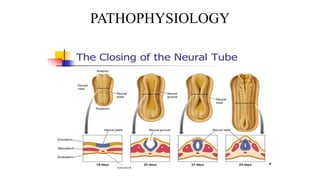
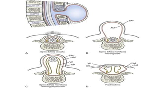
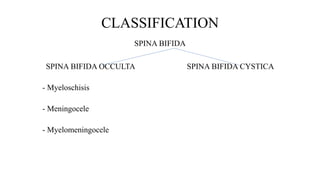
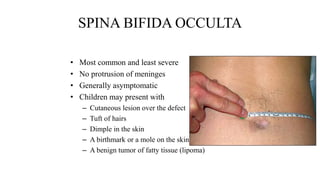
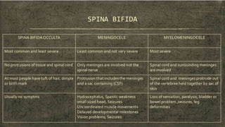
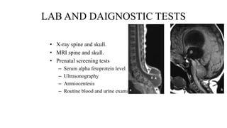
SPINA BIFIDA
Presented by: Dr.Bipul Borthakur
Professor, Dept of Orthopaedics,SMCH
INTRODUCTION
• It is a type of neural tube defect.
• A birth defect where there is incomplete closing of the neural arches and meningeal
layers around the spinal cord.
• It is a developmental congenital anomaly.
PATHOPHYSIOLOGY
Ectoderm (outermost layer) of fertilized egg develop a ridge, that eventually become
neural tube.
This neural tube forms spinal cord, brain and meninges.
Spina bifida occurs when a portion of neural tube fail to close properly
PATHOPHYSIOLOGY
CLASSIFICATION
SPINA BIFIDA
SPINA BIFIDA OCCULTA SPINA BIFIDA CYSTICA
- Myeloschisis
- Meningocele
- Myelomeningocele
ETIOLOGY
• Specific cause is unknown
• Various risk factors
– Folic acid deficiency
– Uncontrolled diabetes
– Obesity
– Hyperthermia during pregnancy
– Medications that interfere with folate metabolism
SPINA BIFIDA OCCULTA
• Most common and least severe
• No protrusion of meninges
• Generally asymptomatic
• Children may present with
– Cutaneous lesion over the defect
– Tuft of hairs
– Dimple in the skin
– A birthmark or a mole on the skin (naevus)
– A benign tumor of fatty tissue (lipoma)
SPINA BIFIDA OCCULTA
• After 6-8 years of age, children may present with
– Progressive deformity of foot
– Change in micturition pattern
– Alteration in gait
– Trophic ulcers over the foot and toes
SPINA BIFIDA OCCULTA
• Diagnosis: Myelogram, CT scan and MRI.
• Treatment:
– Progressive disorder needs surgical correction.
– Laminectomy and excision of intra-spinal lesion.
MENINGOCELE
• It is a protrusion of that includes the meninges and a sac containing
cerebrospinal fluid (CSF).
• It is covered by normal skin.
• Spinal cord is never involved.
• Usually seen in lumbosacral
region, but may also be seen
thoracic region and skull.
MENINGOCELE
• Symptoms include:
– Hydrocephalus – macrocephaly, headache, vomiting, urinary
incontinence OR an abnormally small sized head.
– Spastic weakness of all four limbs.
– Uncoordinated muscle movements.
– Delayed developmental milestones.
– Vision problems.
– Seizures.
MENINGOCELE
• Diagnosis: X ray spine, CT scan brain and MRI spine and brain.
• Treatment:
– Head circumference should be measured daily.
– Sac should be protected – prone position and sterile dressing over the
sac; risk of infection due to CSF leak.
– Surgical closure of the sac as early as
possible.
MYELOMENINGOCELE
• It consists of a cystic sac of meninges with spinal tissue and CSF,
which herniate through a defect in the posterior vertebral arch.
• There are two tpes:
– Open type – myelocele
– Closed type - myelomeningocele
MYELOMENINGOCELE
• The child may present with
– Flaccid paralysis
– Absence of sensation
– Drop reflex
– Postural abnormalities eg. Clubfoot
– Hydrocephalus is usually associated
– Musculoskeletal deformity
– Contracture of joints, scoliosis and kyphosis
– Rupture of sac and infection of CNS
MYELOMENINGOCELE
• Chiari malformation type II – it is a common brain abnormality in
children with the myelomeningocele form of spina bifida.
• The brainstem or lowest part of the above the spinal cord is
elongated and positioned lower than usual.
• Can cause problems with breathing and swallowing.
• Compression of brainstem occurs rarely, requiring decompression
surgery.
MYELOMENINGOCELE
• Diagnosis: Primarily by clinical manifestations
– X-ray spine, CT brain and MRI spine, complete neurological
assessment.
– Routine blood examination and urine routine.
– Prenatal diagnosis – amniocentesis and estimation of Alpha
fetoprotein.
– Monitor head circumference daily.
MYELOMENINGOCELE
• Treatment: Surgical correction of the defect
– Correction of the musculoskeletal deformities.
– Regulation of bowel and bladder function.
– Adequate nutrition.
– Prevention of rupture of sac by proper positioning of child and sterile
dressing of the sac.
LAB AND DAIGNOSTIC TESTS
• X-ray spine and skull.
• MRI spine and skull.
• Prenatal screening tests
– Serum alpha fetoprotein level
– Ultrasonography
– Amniocentesis
– Routine blood and urine examination
ASSESSMENT OF CHILD
• Musculoskeletal and neurological assessment
• Parents interactions with their infant and ability to cope with their child’s
condition.
• Extent of motor and sensory involvement, and presence of reflexes
• Signs and symptoms of dehydration or fluid overload
• Parents for preoperative and postoperative information and support
• Wound drainage and signs of infection
• Increased ICP
• Parents’ and child’s ability to manage home treatment regimen
• Parents’ and child’s needs for community services
NON-SURGICAL INTERVENTIONS
• Preoperatively:
– Motivation of parents regarding expression of grief over loss of perfect
child. Eg, - guilt, self-blame, anger etc.
– Emotional support to parents
– Monitoring of infant’s vital signs and neurologic status
– Promote optimal hydration and nutritional status
– Maintain integrity of defect, prevent further injury
– Prepare parents and infants for surgery
NON-SURGICAL INTERVENTIONS
• Post-operatively:
– Maintenance of nutrition and fluid intake
– Look for signs and symptoms of infection
– Promote healing of surgical site; sterile dressings to be enforced
– Monitor vital signs and neurologic status
– Emotional support to parents
NON-SURGICAL INTERVENTIONS
• Home care:
– Long term management of bowel and bladder training
– Provide info about techniques of facilitate mobility and independence
– Info about skin care and injury prevention
– Educate parents about normal growth and development and also
deviations from normal.
– Instruct parents and child to avoid contact with latex and natural rubber
THANK YOU
Chapter 4, Verse 8
“paritranaya sadhunaam vinaashaaya
chadushkritaam,dharma samsthaapanaarthaya
sambhavaami yuge yuge…
“for the protection of the good, for the destruction
of the evil, and for the establishment of dharma,
Iam bone from age to age..”

Spina bifida

  • 1.
    SPINA BIFIDA Presented by:Dr.Bipul Borthakur Professor, Dept of Orthopaedics,SMCH
  • 2.
    INTRODUCTION • It isa type of neural tube defect. • A birth defect where there is incomplete closing of the neural arches and meningeal layers around the spinal cord. • It is a developmental congenital anomaly.
  • 3.
    PATHOPHYSIOLOGY Ectoderm (outermost layer)of fertilized egg develop a ridge, that eventually become neural tube. This neural tube forms spinal cord, brain and meninges. Spina bifida occurs when a portion of neural tube fail to close properly
  • 4.
  • 6.
    CLASSIFICATION SPINA BIFIDA SPINA BIFIDAOCCULTA SPINA BIFIDA CYSTICA - Myeloschisis - Meningocele - Myelomeningocele
  • 7.
    ETIOLOGY • Specific causeis unknown • Various risk factors – Folic acid deficiency – Uncontrolled diabetes – Obesity – Hyperthermia during pregnancy – Medications that interfere with folate metabolism
  • 8.
    SPINA BIFIDA OCCULTA •Most common and least severe • No protrusion of meninges • Generally asymptomatic • Children may present with – Cutaneous lesion over the defect – Tuft of hairs – Dimple in the skin – A birthmark or a mole on the skin (naevus) – A benign tumor of fatty tissue (lipoma)
  • 9.
    SPINA BIFIDA OCCULTA •After 6-8 years of age, children may present with – Progressive deformity of foot – Change in micturition pattern – Alteration in gait – Trophic ulcers over the foot and toes
  • 10.
    SPINA BIFIDA OCCULTA •Diagnosis: Myelogram, CT scan and MRI. • Treatment: – Progressive disorder needs surgical correction. – Laminectomy and excision of intra-spinal lesion.
  • 11.
    MENINGOCELE • It isa protrusion of that includes the meninges and a sac containing cerebrospinal fluid (CSF). • It is covered by normal skin. • Spinal cord is never involved. • Usually seen in lumbosacral region, but may also be seen thoracic region and skull.
  • 12.
    MENINGOCELE • Symptoms include: –Hydrocephalus – macrocephaly, headache, vomiting, urinary incontinence OR an abnormally small sized head. – Spastic weakness of all four limbs. – Uncoordinated muscle movements. – Delayed developmental milestones. – Vision problems. – Seizures.
  • 13.
    MENINGOCELE • Diagnosis: Xray spine, CT scan brain and MRI spine and brain. • Treatment: – Head circumference should be measured daily. – Sac should be protected – prone position and sterile dressing over the sac; risk of infection due to CSF leak. – Surgical closure of the sac as early as possible.
  • 14.
    MYELOMENINGOCELE • It consistsof a cystic sac of meninges with spinal tissue and CSF, which herniate through a defect in the posterior vertebral arch. • There are two tpes: – Open type – myelocele – Closed type - myelomeningocele
  • 15.
    MYELOMENINGOCELE • The childmay present with – Flaccid paralysis – Absence of sensation – Drop reflex – Postural abnormalities eg. Clubfoot – Hydrocephalus is usually associated – Musculoskeletal deformity – Contracture of joints, scoliosis and kyphosis – Rupture of sac and infection of CNS
  • 16.
    MYELOMENINGOCELE • Chiari malformationtype II – it is a common brain abnormality in children with the myelomeningocele form of spina bifida. • The brainstem or lowest part of the above the spinal cord is elongated and positioned lower than usual. • Can cause problems with breathing and swallowing. • Compression of brainstem occurs rarely, requiring decompression surgery.
  • 17.
    MYELOMENINGOCELE • Diagnosis: Primarilyby clinical manifestations – X-ray spine, CT brain and MRI spine, complete neurological assessment. – Routine blood examination and urine routine. – Prenatal diagnosis – amniocentesis and estimation of Alpha fetoprotein. – Monitor head circumference daily.
  • 18.
    MYELOMENINGOCELE • Treatment: Surgicalcorrection of the defect – Correction of the musculoskeletal deformities. – Regulation of bowel and bladder function. – Adequate nutrition. – Prevention of rupture of sac by proper positioning of child and sterile dressing of the sac.
  • 20.
    LAB AND DAIGNOSTICTESTS • X-ray spine and skull. • MRI spine and skull. • Prenatal screening tests – Serum alpha fetoprotein level – Ultrasonography – Amniocentesis – Routine blood and urine examination
  • 21.
    ASSESSMENT OF CHILD •Musculoskeletal and neurological assessment • Parents interactions with their infant and ability to cope with their child’s condition. • Extent of motor and sensory involvement, and presence of reflexes • Signs and symptoms of dehydration or fluid overload • Parents for preoperative and postoperative information and support • Wound drainage and signs of infection • Increased ICP • Parents’ and child’s ability to manage home treatment regimen • Parents’ and child’s needs for community services
  • 22.
    NON-SURGICAL INTERVENTIONS • Preoperatively: –Motivation of parents regarding expression of grief over loss of perfect child. Eg, - guilt, self-blame, anger etc. – Emotional support to parents – Monitoring of infant’s vital signs and neurologic status – Promote optimal hydration and nutritional status – Maintain integrity of defect, prevent further injury – Prepare parents and infants for surgery
  • 23.
    NON-SURGICAL INTERVENTIONS • Post-operatively: –Maintenance of nutrition and fluid intake – Look for signs and symptoms of infection – Promote healing of surgical site; sterile dressings to be enforced – Monitor vital signs and neurologic status – Emotional support to parents
  • 24.
    NON-SURGICAL INTERVENTIONS • Homecare: – Long term management of bowel and bladder training – Provide info about techniques of facilitate mobility and independence – Info about skin care and injury prevention – Educate parents about normal growth and development and also deviations from normal. – Instruct parents and child to avoid contact with latex and natural rubber
  • 25.
    THANK YOU Chapter 4,Verse 8 “paritranaya sadhunaam vinaashaaya chadushkritaam,dharma samsthaapanaarthaya sambhavaami yuge yuge… “for the protection of the good, for the destruction of the evil, and for the establishment of dharma, Iam bone from age to age..”