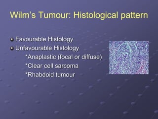
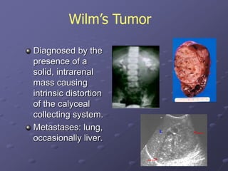
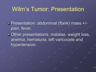
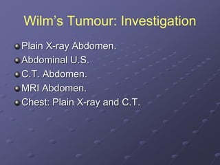
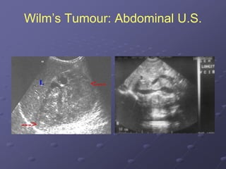
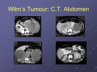
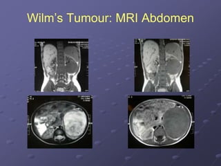
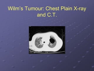
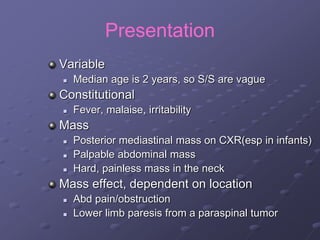
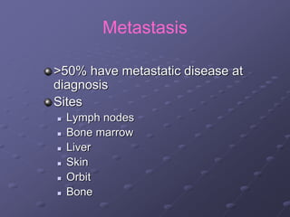
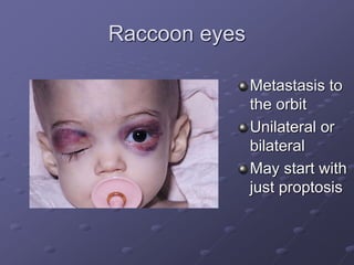
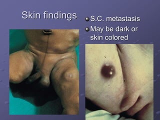
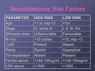
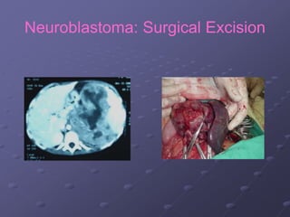
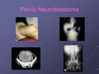
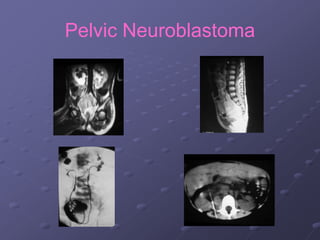
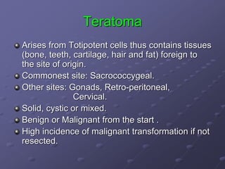
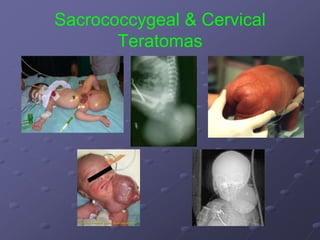
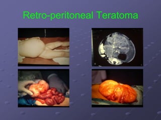
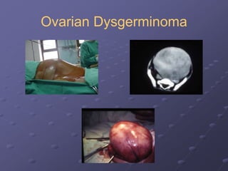
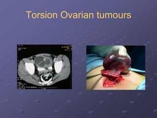
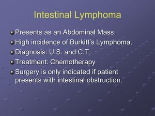
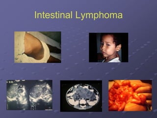
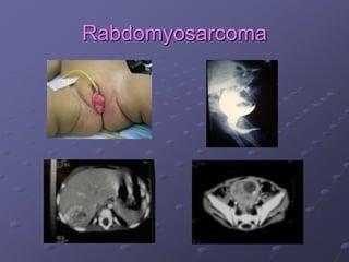
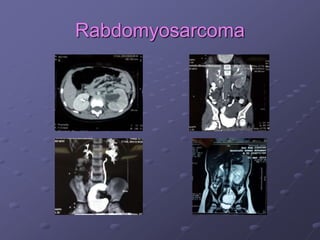
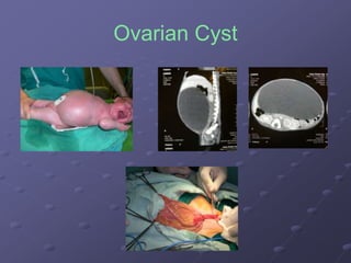
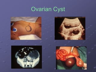
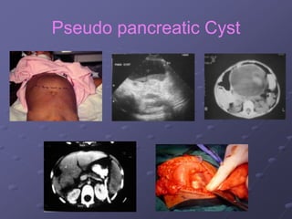
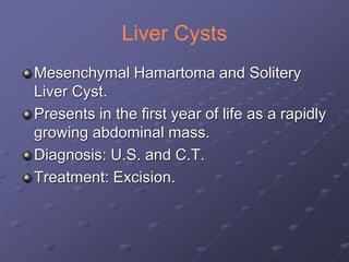
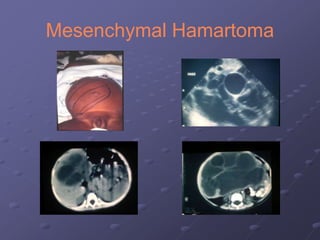
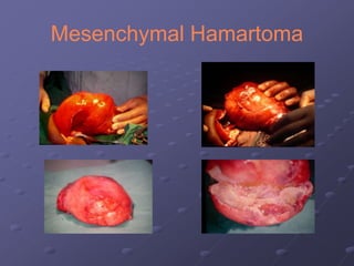
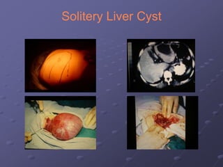
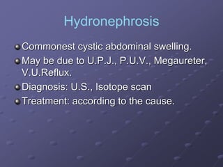
Solid And Cystic Pediatric
Abdominal Tumors
Tarek A. Hassan
FRCS,MD
Prof. of Pediatric Surgery
Ain Shams University
Classification
Solid Tumours:
*Nephroblastoma (Wilm’s Tumour ).
*Neuroblastoma.
*Hepatoblastoma.
*Teratoma.
*Intestinal Lymphoma.
*Rabdomyosarcoma.
Classification
Cystic Swellings:
*Hydronephrosis
*Mesenteric and Duplication Cysts
*Liver cysts (Simple, Mesenchymal
hamartoma)
*Pseudo pancreatic Cyst
*Ovarian cysts
*Teratoma
Wilm’s Tumor (Nephroblastoma)
The second most common solid intra-
abdominal malignant tumor in children.
Commonest renal mass in children
Peak incidence: 3-5 years of age.
Wilm’s Tumour: Pathology
Derived from primitive metanephric
blastema cells.
Histopathology: Triphasic (three cell types)
stromal, epithelial, and blastemal. Thus
may contain tissue not found in the normal
kidney eg. Skeletal, muscles, cartilage and
squamous epithelium.
Wilm’s Tumour: Histological pattern
Favourable Histology
Unfavourable Histology
*Anaplastic (focal or diffuse)
*Clear cell sarcoma
*Rhabdoid tumour
Diagnosed by the
presence of a
solid, intrarenal
mass causing
intrinsic distortion
of the calyceal
collecting system.
Metastases: lung,
occasionally liver.
Wilm’s Tumor
Wilm’s Tumor: Presentation
Presentation: abdominal (flank) mass +/-
pain, fever.
Other presentations: malaise, weight loss,
anemia, hematuria, left varicocele and
hypertension.
Wilm’s Tumour: Investigation
Plain X-ray Abdomen.
Abdominal U.S.
C.T. Abdomen.
MRI Abdomen.
Chest: Plain X-ray and C.T.
Wilm’s Tumour: Abdominal U.S.
Wilm’s Tumour: C.T. Abdomen
Wilm’s Tumour: MRI Abdomen
Wilm’s Tumour: Chest Plain X-ray
and C.T.
Staging by National Wilm’s Tumor
Study Group:
Stage I: Tumor limited to kidney and
completely resected.
Stage II: Tumor extends beyond the
kidney but completely excised.
Stage III: Residual non-hematogenous
tumor confined to the abdomen.
Stage IV: Hematogenous metastasis.
Stage V: Bilateral tumors.
Wilm’s Tumour: Treatment
Surgical Excision followed by Chemotherapy +/-
Radiotherapy according to the stage and histology
(favorable or unfavorable).
Aim of surgery: Radical nephrectomy without
spillage + accurate staging (spread of disease).
Chemotherapy commonly used: Actinomycin D
(A), Vincristine (V), Doxorubicin (D), Cytoxan (C).
Wilm’s Tumour: NWTS-4 treatment
STAGE HISTOLOGY NEPHRE
CTOMY
CHEMO
THERAPY
RADIO
THERAPY
I FV, UFV + A, V NO
II FV + A, V NO
III, IV FV + A, V, D YES
II, III, IV UFV + A, V, D +/-
C
YES
Wilm’s Tumour: Surgical Excision
Wilm’s Tumor : Prognosis
Disease-free survival is 95% for Stage I
and approximately 77% for all patients.
Good prognosis:
-Stage I & II
-Para-aortic LN: -ve
-Favorable histology
-No tumor ruptured during surgery
Congenital Mesoblastic Nephroma
presents in infants < 30 days of age
(< 6 months), is commonly benign
and invasive locally.
Operative removal is curative.
Ruptured of tumor increases
recurrences.
Chemo and radiotherapy are not
indicated.
Mesoblastic Nephroma
Neuroblastoma
most common extracranial solid tumor
of childhood.
8-10% of all pediatric malignancies
Median age at diagnosis is 2 years.
Arises from the adrenals or sympathetic
chain
Higher frequency seen in certain
syndromes (Myoclonus-opsoclonus
syndrome ,Watery diarrhea )
Pathology
“Small round blue cell” tumors
 Originates from neural crest cells in the sympathetic
nervous system
Arises from the adrenals or sympathetic chain
3 subgroups
 Neuroblastoma- most primitive
 Ganglioneuroma- benign and mature
 Ganglioneuroblastoma- in between
Secrete neurogenically derived substance
 Urinary catecholamines in 75-90%
*VMA & HVA
 Others
*Neuron specific enolase (NSE)
*Ferritin
Pathology : Pseudorosettes
Shimada Classification:
favorable histology&
unfavorable histology
NBL metastasized to the
bone marrow
Clumps of tumor cells
Cytogenetics
Chromosomal abnormalities in most
cases of NBL
N-myc amplification (normally 2 copies)
 >10 copies/cell
 30% of NBL
 Poor prognosis
Ploidy
 Hyperdiploid = DNA index > 1.0
 Good prognosis, at least in those < 2 years
Presentation
Variable
 Median age is 2 years, so S/S are vague
Constitutional
 Fever, malaise, irritability
Mass
 Posterior mediastinal mass on CXR(esp in infants)
 Palpable abdominal mass
 Hard, painless mass in the neck
Mass effect, dependent on location
 Abd pain/obstruction
 Lower limb paresis from a paraspinal tumor
Metastasis
>50% have metastatic disease at
diagnosis
Sites
 Lymph nodes
 Bone marrow
 Liver
 Skin
 Orbit
 Bone
Raccoon eyes
Metastasis to
the orbit
Unilateral or
bilateral
May start with
just proptosis
Skin findings S.C. metastasis
May be dark or
skin colored
Neuroblastoma: Work-up
Blood
 CBC
 Ferritin
 NSE
 LDH
Urine
 Catecholamines
VMA / HVA
Tumor tissues
 Shimada class.
 N-myc
 1p deletion
Radiology
 CXR
 Abdominal U.S.
 C.T. and MRI
 Skeletal survey
 Bone scan
 MIBG
Bone marrow
 Aspirate and biopsy
from both post iliac
crests
Neuroblastoma: C.T.
Neuroblastoma: MIBG scan
International Neuroblastoma
Staging System (INSS)
Treatment
Surgery
 Localized tumors that don’t encase vasculature
 Complete resection = excellent chance for cure
 Palliative treatment for tumors compressing vital
organs
Chemotherapy
 The mainstay of therapy since most are metastatic
 May be very intensive requiring stem cell rescue
Radiation
 Used for localized but unresectable tumor (stage III)
 Control of tumor that is unresponsive to chemo
Neuroblastoma: Treatment
Stage I: Complete surgical resection.
Stage II: Incomplete surgical resection 
Chemotherapy  second look surgical
resection.
Stage III: Incomplete surgical resection 
Chemotherapy +/- Radioth.  second look
surgical resection.
Stage IV: Chemotherapy  Surgical
resection if possible.
Neuroblastoma: Risk Factors
PARAMETER HIGH RISK LOW RISK
Age >1 yr, esp >2 <1yr
Stage IV, some III I, II, III, IVs
Shimada class. Unfavourable Favourable
N-myc >10 copies <10, esp <3
1p36 Present Absent
Ploidy Diploid Hyperploid
Trk-expression Absent Present
Ferritin serum >145-150ng/ml <145-150ng/ml
LDH serum >1500 <1500
Neuroblastoma: Surgical Excision
Pelvic Neuroblastoma
Liver Tumors
Hepatoblastoma and hepatocellular carcinoma
2% of all malignancies in childhood and 15% of
malignant abdominal masses
Hepatoblastoma
Hepatoblastoma is the
most common primary
malignant neoplasm of
the liver in children
Males, < four year of age
Hepatoblastoma: Investigations
Plain X-ray, U.S., C.T., MRI: predict
resectability and tumor extension.
Tumour markers: alpha-fetoprotein and
gamma-glutamyl transferase II
Hepatoblastoma: Investigations
Hepatoblastoma: Treatment
Surgery + Chemotherapy +/- Radiotherapy
Surgery: Optimum treatment is complete
resection.
Chemotherapy:
*Adjuvant following resection.
*Pre-operative for non-resectable.
*Palliative for metastatic disease.
Radiotherapy: for non-resectable tumor to
reduce its size.
Hepatoblastoma: Treatment
Surgical Excision and Chemotherapy.
Hepatoblastoma: Preoperative
chemotherapy
Teratoma
Arises from Totipotent cells thus contains tissues
(bone, teeth, cartilage, hair and fat) foreign to
the site of origin.
Commonest site: Sacrococcygeal.
Other sites: Gonads, Retro-peritoneal,
Cervical.
Solid, cystic or mixed.
Benign or Malignant from the start .
High incidence of malignant transformation if not
resected.
Teratoma: Investigations
Plain X-ray, U.S., C.T.
High Alpha Feto-protien.
Sacrococcygeal & Cervical
Teratomas
Retro-peritoneal Teratoma
Teratoma: Treatment
Surgical Excision.
Chemotherapy if histopathology proves it
to be malignant.
Ovarian tumours
Uncommon childhood malignancies (1%)
Presents as an Abdominal Mass or Acute
Abdomen in cases of torsion.
Common histology is germ cell:
dysgerminoma, teratoma, and endodermal
sinus tumor.
Other: Epithelial (older adolescent), lipid-
cell, and gonadoblastoma.
Ovarian Tumour: Investigations
Plain X-ray, U.S., C.T., MRI
Alpha feto protein
Ovarian Teratoma
Ovarian Dysgerminoma
Torsion Ovarian tumours
Ovarian Tumours: Treatment
Surgical excision with staging.
Chemotherapy for malignant cases.
Intestinal Lymphoma
Presents as an Abdominal Mass.
High incidence of Burkitt’s Lymphoma.
Diagnosis: U.S. and C.T.
Treatment: Chemotherapy
Surgery is only indicated if patient
presents with intestinal obstruction.
Intestinal Lymphoma
Rabdomyosarcoma
Rabdomyosarcoma
Mesenteric Cysts
Presents by Abdominal mass.
Diagnosis: U.S. and C.T.
Treatment: Surgical Excision
Mesenteric Cysts
Ovarian Cyst
Mostly functional and presents in the
neonatal period.
Can be diagnosed antenatally.
Liable for torsion.
Diagnosis: U.S ,C.T
Treatment is surgical excision of the cyst if
its size exceeds 4 to 5 cm.
Ovarian Cyst
Ovarian Cyst
Pseudo pancreatic Cyst
Commonly follows abdominal trauma
50% resolves spontaneously
Intervention is indicated
if it persists >4-6 W ,its size >4-6 cm,
develops complications
Surgical internal drainage, percutaneous
U.S. or C.T. guided drainage , endoscopic
internal drainage
Pseudo pancreatic Cyst
Liver Cysts
Mesenchymal Hamartoma and Solitery
Liver Cyst.
Presents in the first year of life as a rapidly
growing abdominal mass.
Diagnosis: U.S. and C.T.
Treatment: Excision.
Mesenchymal Hamartoma
Solitery Liver Cyst
Hydronephrosis
Commonest cystic abdominal swelling.
May be due to U.P.J., P.U.V., Megaureter,
V.U.Reflux.
Diagnosis: U.S., Isotope scan
Treatment: according to the cause.
THANK YOU

Solid and cystic pediatric abdominal tumors