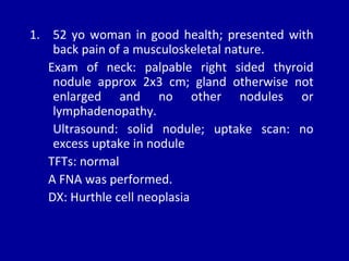
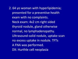
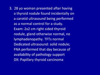
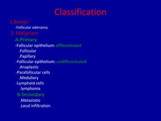
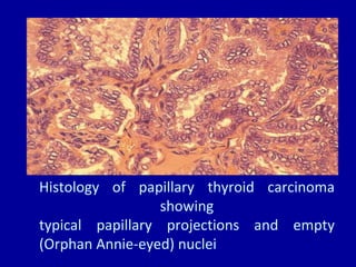
‫الرحيم‬ ‫الرحمن‬ ‫هللا‬ ‫بسم‬
Thyroid Cancer
Dr.MOSTAFA HEGAZY
1. 52 yo woman in good health; presented with
back pain of a musculoskeletal nature.
Exam of neck: palpable right sided thyroid
nodule approx 2x3 cm; gland otherwise not
enlarged and no other nodules or
lymphadenopathy.
Ultrasound: solid nodule; uptake scan: no
excess uptake in nodule
TFTs: normal
A FNA was performed.
DX: Hurthle cell neoplasia
2. 64 yo woman with hyperlipidemia;
presented for a preventive health
exam with no complaints.
Neck exam: 4x2 cm right sided
thyroid nodule, gland otherwise
normal, no lymphadenopathy.
Ultrasound-solid nodule, uptake scan
no excess uptake in nodule. TFTs
A FNA was performed.
DX: Hurthle cell neoplasia
3. 28 yo woman presented after having
a thyroid nodule found incidentally on
a carotid ultrasound being performed
as a normal control for a study.
Exam: 2x2 cm right sided thyroid
nodule, gland otherwise normal, no
lymphadenopathy. TFTs normal
Dedicated ultrasound: solid nodule;
FNA performed that day because of
availability of pathology support
DX: Papillary thyroid carcinoma
Epidemiology
• Thyroid nodules: very common
• Clinically detectable thyroid carcinoma:
rare: <1% of all cancers
• Female to male ratio- 2.5:1
• Median age at dx: 45-50
• Overall incidence is rising:
• In 1935: 1.3/100,000 women, .2/100,000
men
• By 1991: 5.8/100,000 women, 2.5/100,000
men
• Incidence has continued to rise in past 10
years: most rapid rate of increase in all
tracked cancers
Reason for rise?
• Neck irradiation: used between 1910 and
1960
• Better diagnosis?
BUT: only rise is in papillary type; if better
diagnosis was reason, would expect rise in
all types
Hegedus, L. N Engl J Med 2004;351:1764-1771
Algorithm for the Cost-Effective Evaluation and Treatment of a Clinically Detectable Solitary
Thyroid Nodule
Classification
1.Benign
-Follicular adenoma.
2. Malignant
A-Primary
-Follicular epithelium: differentiated
.Follicular
.Papillary
-Follicular epithelium: undifferentiated
.Anaplastic
-Parafollicular cells
.Medullary
-Lymphoid cells
.lymphoma
B-Secondary
.Metastatic
.Local infiltration
Benign Tumour
• Follicular adenoma :
- It is present clinically as solitary nodule.
-The differentiation between the follicular
carcinoma and adenoma can only made by
histological examination ,that in adenoma there is
no invasion of the capsule or of pericapsular
blood vessels .
-Treatment ___ wide excision ( preferably a
lobectomy ) .
Malignant tumours
60
17
13
6
4
0
10
20
30
40
50
60
papillary ca
follicular ca
anaplastic ca
medullary ca
malignant lymphoma
Thyroid cancer: epithelial types
Differentiated:
Papillary: 70-75 % of all thyroid cancers
Follicular: 15-25%
Undifferentiated:
Anaplastic: 2-5%
Thyroid cancer: non epithelial
Medullary thyroid cancer
• Sporadic
• Familial
• MEN-2A and B
Others:
lymphoma, mets from breast, colon,
renal and melanoma
Clinical characteristics that suggest
Malignancy
1.Gender ___ male gender(F:M__3:1 ??)
2.Age <15,>60 years
3.Hx of head & neck radiation
4.Family hx of thyroid cancer
5.Rapidly enlarging nodule
6.Hard single nodule & nodules fixed to
surrounding structures
7.Hx of thyroiditis
8.Hoarseness
9.Cervical lymphadenopathy
Isolated swelling in the upper
pole of the right thyroid lobe
Papillary carcinoma
- Slow growing ,60% multicentric .
- M:F--- 1:3 .
- Common 20-30 yrs old.
- Painless lump in the thyroid gland with enlarged
lymph gland.
- 80-90% of postradiation ca of the thyroid.
- Spread by lymphatics (50% have +ve node at
diagnosis) ,with good prognosis ,the presence of
nodes does not affect prognosis.
- Type of surgery __Lobectomy with
isthmusectomy .
• If the tumor is >3 cm ,male >40 , female
>50 , distant metastasis or angioinvasion
then _____ Total thyroidectomy is
indicated because of poor prognosis.
• 85% 10 yr survival.
• Occult carcinoma:
- Papillary carcinoma may present as an
enlarged lymph node in Jugular chain with no
palpable abnormality of the thyroid .The
primary tumour may be about few millimeter
in size & termed occult.
-The term occult is now applied to all papillary
carcinoma < 1.5 cm
- These tumor have an excellent prognosis.
Papillary thyroid carcinoma
Pathogenesis:
1. Activation of tyrosine kinase receptors by
rearrangement or gene amplification
• Results in a chimeric gene
• Occurs either by radiation or sporadic
2. Point mutations in BRAF gene
• 10X increased risk of thyroid cancer in relatives of
thyroid cancer patients: suggests a genetic link
PTC
Presentation:
• Solitary nodule most common
• Pathology: typically unencapsulated; may
be cystic
Papillae: 1 or 2 layers of tumor surrounding
fibrovascular core
Follicles and colloid are typically absent
PTC
• Psommoma bodies: scarred remnants of
tumor papillae that have infarcted
• Present in half of papillary thyroid
carcinomas
Histology of papillary thyroid carcinoma
showing
typical papillary projections and empty
(Orphan Annie-eyed) nuclei
PTC
Growth and behavior: minor to major
• Microcarcinoma: occult papillary
carcinoma, with tumor <1cm
• Found in up to 50% of glands at autopsy
(rarer in children)
• Incidental finding of no clinical importance
PTC
• Other end of spectrum: aggressive metastasis
through interthyroidal lymphatic channels to
form multifocal tumors
• Involves regional lymph nodes
• At diagnosis: clinically detectable nodes more
common in children (50%) than adults
• 2-10% distant mets at dx: 2/3 pulmonary, 1/4
skeletal; also brain, kidneys, liver, adrenals
PTC
Prognosis
• Most patients do not die of their disease
• 80-95% 10 year survival rates
• Patients between 20-45: best long term survival
• Patients older than 45 with lymph node
recurrences are most likely to die from PTC
PTC
• Prognosis is poorer in patients with large
tumors: one large series showed cancer
related mortality of 6%/2-3.9cm, 16%/4-
6.9cm and 50%7 cm and above
• Several variants have a worse prognosis:
tall cell variant=1% of PTC; more aggressive
and invasive
Magnetic resonance imaging scan of papillary cancer
with multiple node metastases.
Schlumberger, M. J. N Engl J Med 1998;338:297-306
Survival Rate among 1701 Patients with Papillary or Follicular Carcinoma and No Distant
Metastases at the Time of Diagnosis
Follicular carcinoma
• Unifocal , more aggressive.
• M:F___1:3
• 30_40 yrs old.
• Common presentation __ lump in the neck.
• If tumor spread beyond the thyroid__ pt may
complain of breathlessness , chest pain , pain
or swelling in the bone.
• Capsular & vascular invasion are prominent
feature.
• Blood-borne metastases.
• Metastasis to lung &bone
• Total thyroidectomy is indicated .
• 40% 10 yr survival.
Follicular thyroid carcinoma
• Characterized by follicular differentiation and
encapsulation
• Invasion of the capsule and blood vessels is the
main determinant between adenomas and
carcinoma
• 2 main forms: minimally invasive and widely
invasive
• Multicentricity and lymph node involvement are
less frequent than in PTC
Follicular neoplasm of the thyroid presenting
as an
isolated swelling.
Histology of follicular thyroid carcinoma showing
vascular (red arrow) and capsular (black arrow) invasion
Metastasis in the humerus from
a carcinoma of the thyroid
Metastasis in the left parietal
bone from a carcinoma of the
thyroid
Follicular carcinoma of the thyroid with skull secondaries
FTC
• Minimally invasive FTC behaves more like
PTC
• Widely invasive behaves more like
anaplastic thyroid carcinoma
• Hurthle Cell variant:more aggressive
• FTC is more likely than PTC to be
nonresponsive to I 131.
Anaplastic carcinoma
• Very aggressive tumor.
• It is the worst variety of thyroid tumor due its rapid
spread.
• Local infiltration is an early feature.
• The common complaint is a swelling in the neck
rather than lump ( because the tumor is diffuse &
infiltrating)
• A dull aching neck pain is quit common.
• Hoarseness or change in voice quality is a diagnostic
symptom because it implies infiltration of recurrent
laryngeal nerve.
• Many of these aggressive lesion present in an
advanced stage with tracheal obstruction
&required urgent tracheal decompression.
• Spread by lymphatic & blood stream.
• Chemotherapy & radiation may improve 5 %.
• Mean survival 2-4 months.( poor prognosis)
Anaplastic thyroid carcinoma
• Undifferentiated tumor of thyroid follicular
epithelium
• Very aggressive, with a disease specific mortality
approaching 100%
• 2/1,000,000 annual incidence
• Typical patient is older than differentiated
carcinoma, mean age 65
• <10% under 50
• 60-70% women
ATC
• 20% of ATC: history of differentiated
thyroid carcinoma, most papillary
• 10% of Hurthle cell carcinoma: has
anaplastic tumor within
• Up to 1/2 of ATC: history of multinodular
goiter
ATC
• Presentation:
• Nearly all present with a thyroid mass
• Regional or distant spread is present 90% of the
time at dx
• Lungs, bones, brain most common mets
• Rapidly enlarging tumor; often causes
compression symptoms like dyspnea, dysphagia,
hoarseness
• Constitutional symptoms like fatigue, anorexia,
wt loss
ATC
• 50% have palpable nodes at dx
• Dx: made by FNA, then CT neck and mediastinum,
CXR
• Prognostic factors: tumor size
<6 cm=25% 2 yr survival
>6cm=3-15% 2 yr survival
Others: older age, male sex, dyspnea at
presentation
• No effective treatment for advanced or metastatic
ATC: uniformly fatal, with median survival 3-7 mo
Treatment of differentiated
thyroid carcinoma
• Surgery: goal is to remove all tumor tissue
from neck
• Total or near total thyroidectomy because
of risk of multicentricity
• Removal of local nodes in PTC, only
palpable nodes in FTC because of lower
rate of lymph node involvement
Treatment
• I 131: given post op: destroys any remaining
normal thyroid tissue, and may destroy occult
microcarcinomas
• Increases sensitivity of subsequent 1 131 total
body scans
• 4-6 wks after surgery a total body scan off thyroid
replacement with low dose 1 131; if any uptake, a
treatment dose is given (2 mCi vs. 30-100 mCi)
• Radiation: only if surgical excision is impossible
and tissue doesn’t take up I 131
Followup
Goals of followup:
• Maintain adequate thyroxine treatment
• Detect persistent or recurrent cancer
• Recurrences usually occur early but may
occur later so follow up for life
• Thyroxine treatment goals: initial serum
thyrotropin level 0.1 or less, serum free T3
normal
• Check U/S of thyroid area and nodal areas
• Serum thyroglobulin levels: TG produced by
follicular cells-should not be detectable after total
ablation; presence signifies persistent or
recurrent disease
• 80% of patients with TG >40 have detectable foci
or I 131 uptake
• I 131 scanning: needs to be done after
withdrawal of thyroxine tx, with TSH >30
needed
• Scanning is done 3 days after I 131 given
• Low risk patients with no I 131 uptake after
1 year: TSH maintained at low but
detectable level (0.1-0.5)
• Local or regional mets: occur in 5-20%
• Excision/I 131 tx/ Radiation tx if no I 131
uptake
• Distant mets: If I 131 uptake, high dose I
131 given + RT
Complications of treatment:
• I 131: nausea, sialadenitis common but mild and
short duration
• Genetic defects: can’t be given to pregnant
women
• Increased risk of miscarriage in pregnancies
within 1 year of tx
• Overall relative risk of a second type of cancer
only if high cumulative dose of I 131 and/or
radiation
Medullary carcinoma
• Aggressive tumors.
• It is a neoplasm of parafollicular ( c ) cell.
• C-cell calcitonin producing tumor.
• High level of serum calcitonin >0.08 ng/ml
• Sporadic form is unifocal with worse
prognosis , familial form is multifocal with
a better prognosis.
• Involvement of lymph node occurs in
50_60%.
• May occur in combination with adrenal
phaeochromocytoma &
hyperparathyroidism in syndrome known
as multiple endocrine neoplasia type IIa
(MEN IIa)
-Blood-born metastases are common.
-Treatment__ Total thyroidectomy with
resection of involved lymph node.
Medullary thyroid cancer
• Much less common than epithelial thyroid
cancers
• Involves abnormalities of parafollicular C-
cells
• Most cases are sporadic
Hyperplasia of parafollicular C
cells in a child from a
family with medullary cancer
Histology of medullary carcinoma
showing characteristic
‘cell balls’ and amyloid
MTC
• MEN 2 A: autosomal dominant disorder
characterized by MTC, pheochromocytoma, and
primary parathyroid hyperplasia
• MEN 2 B: same inheritance; MTC +
pheochromocytoma. Occurs at a younger age;
more aggressive.
• Familial MTC: like MEN 2 A but no other
associated abnormalities
Total thyroidectomy specimen from a young girl undergoing
surgery following genetic screening, showing a small medullary
cancer in the right lobe.
MTC
• Female to male ratio=1:1
• MEN 2 A and familiar MTC: peak in index
cases in 3rd decade
• MEN 2 B: children and teens most common
age of presentation.
• Basal serum calcitonin: usually correlates
with tumor mass and is almost always high
with palpable tumor
MTC
• MTC in MEN 2 B: more aggressive
• Early onset
• Surgery often not curative
• Death from MTC: 50% of MEN 2 B,
10 % MEN 2 A
Lymphoma
• Usually affect female( may have hx of
hashimoto’s)
• There is a rapid enlargement.
• Compressive symptoms are common.
• Sensitive for chemotherapy & radiotherapy
• Surgery for diagnosis & compressive
symptom
• Good prognosis if there is no involvement
of cervical LN.
THANK
YOU
THANK YOU

Thyroid cancer hegazy

  • 1.
  • 2.
  • 3.
    1. 52 yowoman in good health; presented with back pain of a musculoskeletal nature. Exam of neck: palpable right sided thyroid nodule approx 2x3 cm; gland otherwise not enlarged and no other nodules or lymphadenopathy. Ultrasound: solid nodule; uptake scan: no excess uptake in nodule TFTs: normal A FNA was performed. DX: Hurthle cell neoplasia
  • 4.
    2. 64 yowoman with hyperlipidemia; presented for a preventive health exam with no complaints. Neck exam: 4x2 cm right sided thyroid nodule, gland otherwise normal, no lymphadenopathy. Ultrasound-solid nodule, uptake scan no excess uptake in nodule. TFTs A FNA was performed. DX: Hurthle cell neoplasia
  • 5.
    3. 28 yowoman presented after having a thyroid nodule found incidentally on a carotid ultrasound being performed as a normal control for a study. Exam: 2x2 cm right sided thyroid nodule, gland otherwise normal, no lymphadenopathy. TFTs normal Dedicated ultrasound: solid nodule; FNA performed that day because of availability of pathology support DX: Papillary thyroid carcinoma
  • 6.
    Epidemiology • Thyroid nodules:very common • Clinically detectable thyroid carcinoma: rare: <1% of all cancers • Female to male ratio- 2.5:1 • Median age at dx: 45-50
  • 7.
    • Overall incidenceis rising: • In 1935: 1.3/100,000 women, .2/100,000 men • By 1991: 5.8/100,000 women, 2.5/100,000 men • Incidence has continued to rise in past 10 years: most rapid rate of increase in all tracked cancers
  • 8.
    Reason for rise? •Neck irradiation: used between 1910 and 1960 • Better diagnosis? BUT: only rise is in papillary type; if better diagnosis was reason, would expect rise in all types
  • 9.
    Hegedus, L. NEngl J Med 2004;351:1764-1771 Algorithm for the Cost-Effective Evaluation and Treatment of a Clinically Detectable Solitary Thyroid Nodule
  • 10.
    Classification 1.Benign -Follicular adenoma. 2. Malignant A-Primary -Follicularepithelium: differentiated .Follicular .Papillary -Follicular epithelium: undifferentiated .Anaplastic -Parafollicular cells .Medullary -Lymphoid cells .lymphoma B-Secondary .Metastatic .Local infiltration
  • 11.
    Benign Tumour • Follicularadenoma : - It is present clinically as solitary nodule. -The differentiation between the follicular carcinoma and adenoma can only made by histological examination ,that in adenoma there is no invasion of the capsule or of pericapsular blood vessels . -Treatment ___ wide excision ( preferably a lobectomy ) .
  • 12.
  • 13.
    Thyroid cancer: epithelialtypes Differentiated: Papillary: 70-75 % of all thyroid cancers Follicular: 15-25% Undifferentiated: Anaplastic: 2-5%
  • 14.
    Thyroid cancer: nonepithelial Medullary thyroid cancer • Sporadic • Familial • MEN-2A and B Others: lymphoma, mets from breast, colon, renal and melanoma
  • 15.
    Clinical characteristics thatsuggest Malignancy 1.Gender ___ male gender(F:M__3:1 ??) 2.Age <15,>60 years 3.Hx of head & neck radiation 4.Family hx of thyroid cancer 5.Rapidly enlarging nodule 6.Hard single nodule & nodules fixed to surrounding structures 7.Hx of thyroiditis 8.Hoarseness 9.Cervical lymphadenopathy
  • 16.
    Isolated swelling inthe upper pole of the right thyroid lobe
  • 17.
    Papillary carcinoma - Slowgrowing ,60% multicentric . - M:F--- 1:3 . - Common 20-30 yrs old. - Painless lump in the thyroid gland with enlarged lymph gland. - 80-90% of postradiation ca of the thyroid. - Spread by lymphatics (50% have +ve node at diagnosis) ,with good prognosis ,the presence of nodes does not affect prognosis. - Type of surgery __Lobectomy with isthmusectomy .
  • 18.
    • If thetumor is >3 cm ,male >40 , female >50 , distant metastasis or angioinvasion then _____ Total thyroidectomy is indicated because of poor prognosis. • 85% 10 yr survival.
  • 19.
    • Occult carcinoma: -Papillary carcinoma may present as an enlarged lymph node in Jugular chain with no palpable abnormality of the thyroid .The primary tumour may be about few millimeter in size & termed occult. -The term occult is now applied to all papillary carcinoma < 1.5 cm - These tumor have an excellent prognosis.
  • 20.
    Papillary thyroid carcinoma Pathogenesis: 1.Activation of tyrosine kinase receptors by rearrangement or gene amplification • Results in a chimeric gene • Occurs either by radiation or sporadic 2. Point mutations in BRAF gene • 10X increased risk of thyroid cancer in relatives of thyroid cancer patients: suggests a genetic link
  • 21.
    PTC Presentation: • Solitary nodulemost common • Pathology: typically unencapsulated; may be cystic Papillae: 1 or 2 layers of tumor surrounding fibrovascular core Follicles and colloid are typically absent
  • 22.
    PTC • Psommoma bodies:scarred remnants of tumor papillae that have infarcted • Present in half of papillary thyroid carcinomas
  • 23.
    Histology of papillarythyroid carcinoma showing typical papillary projections and empty (Orphan Annie-eyed) nuclei
  • 24.
    PTC Growth and behavior:minor to major • Microcarcinoma: occult papillary carcinoma, with tumor <1cm • Found in up to 50% of glands at autopsy (rarer in children) • Incidental finding of no clinical importance
  • 25.
    PTC • Other endof spectrum: aggressive metastasis through interthyroidal lymphatic channels to form multifocal tumors • Involves regional lymph nodes • At diagnosis: clinically detectable nodes more common in children (50%) than adults • 2-10% distant mets at dx: 2/3 pulmonary, 1/4 skeletal; also brain, kidneys, liver, adrenals
  • 26.
    PTC Prognosis • Most patientsdo not die of their disease • 80-95% 10 year survival rates • Patients between 20-45: best long term survival • Patients older than 45 with lymph node recurrences are most likely to die from PTC
  • 27.
    PTC • Prognosis ispoorer in patients with large tumors: one large series showed cancer related mortality of 6%/2-3.9cm, 16%/4- 6.9cm and 50%7 cm and above • Several variants have a worse prognosis: tall cell variant=1% of PTC; more aggressive and invasive
  • 28.
    Magnetic resonance imagingscan of papillary cancer with multiple node metastases.
  • 29.
    Schlumberger, M. J.N Engl J Med 1998;338:297-306 Survival Rate among 1701 Patients with Papillary or Follicular Carcinoma and No Distant Metastases at the Time of Diagnosis
  • 30.
    Follicular carcinoma • Unifocal, more aggressive. • M:F___1:3 • 30_40 yrs old. • Common presentation __ lump in the neck. • If tumor spread beyond the thyroid__ pt may complain of breathlessness , chest pain , pain or swelling in the bone.
  • 31.
    • Capsular &vascular invasion are prominent feature. • Blood-borne metastases. • Metastasis to lung &bone • Total thyroidectomy is indicated . • 40% 10 yr survival.
  • 32.
    Follicular thyroid carcinoma •Characterized by follicular differentiation and encapsulation • Invasion of the capsule and blood vessels is the main determinant between adenomas and carcinoma • 2 main forms: minimally invasive and widely invasive • Multicentricity and lymph node involvement are less frequent than in PTC
  • 33.
    Follicular neoplasm ofthe thyroid presenting as an isolated swelling.
  • 34.
    Histology of follicularthyroid carcinoma showing vascular (red arrow) and capsular (black arrow) invasion
  • 35.
    Metastasis in thehumerus from a carcinoma of the thyroid Metastasis in the left parietal bone from a carcinoma of the thyroid
  • 36.
    Follicular carcinoma ofthe thyroid with skull secondaries
  • 37.
    FTC • Minimally invasiveFTC behaves more like PTC • Widely invasive behaves more like anaplastic thyroid carcinoma • Hurthle Cell variant:more aggressive • FTC is more likely than PTC to be nonresponsive to I 131.
  • 38.
    Anaplastic carcinoma • Veryaggressive tumor. • It is the worst variety of thyroid tumor due its rapid spread. • Local infiltration is an early feature. • The common complaint is a swelling in the neck rather than lump ( because the tumor is diffuse & infiltrating) • A dull aching neck pain is quit common. • Hoarseness or change in voice quality is a diagnostic symptom because it implies infiltration of recurrent laryngeal nerve.
  • 39.
    • Many ofthese aggressive lesion present in an advanced stage with tracheal obstruction &required urgent tracheal decompression. • Spread by lymphatic & blood stream. • Chemotherapy & radiation may improve 5 %. • Mean survival 2-4 months.( poor prognosis)
  • 40.
    Anaplastic thyroid carcinoma •Undifferentiated tumor of thyroid follicular epithelium • Very aggressive, with a disease specific mortality approaching 100% • 2/1,000,000 annual incidence • Typical patient is older than differentiated carcinoma, mean age 65 • <10% under 50 • 60-70% women
  • 41.
    ATC • 20% ofATC: history of differentiated thyroid carcinoma, most papillary • 10% of Hurthle cell carcinoma: has anaplastic tumor within • Up to 1/2 of ATC: history of multinodular goiter
  • 42.
    ATC • Presentation: • Nearlyall present with a thyroid mass • Regional or distant spread is present 90% of the time at dx • Lungs, bones, brain most common mets • Rapidly enlarging tumor; often causes compression symptoms like dyspnea, dysphagia, hoarseness • Constitutional symptoms like fatigue, anorexia, wt loss
  • 43.
    ATC • 50% havepalpable nodes at dx • Dx: made by FNA, then CT neck and mediastinum, CXR • Prognostic factors: tumor size <6 cm=25% 2 yr survival >6cm=3-15% 2 yr survival Others: older age, male sex, dyspnea at presentation • No effective treatment for advanced or metastatic ATC: uniformly fatal, with median survival 3-7 mo
  • 44.
    Treatment of differentiated thyroidcarcinoma • Surgery: goal is to remove all tumor tissue from neck • Total or near total thyroidectomy because of risk of multicentricity • Removal of local nodes in PTC, only palpable nodes in FTC because of lower rate of lymph node involvement
  • 45.
    Treatment • I 131:given post op: destroys any remaining normal thyroid tissue, and may destroy occult microcarcinomas • Increases sensitivity of subsequent 1 131 total body scans • 4-6 wks after surgery a total body scan off thyroid replacement with low dose 1 131; if any uptake, a treatment dose is given (2 mCi vs. 30-100 mCi) • Radiation: only if surgical excision is impossible and tissue doesn’t take up I 131
  • 46.
    Followup Goals of followup: •Maintain adequate thyroxine treatment • Detect persistent or recurrent cancer • Recurrences usually occur early but may occur later so follow up for life
  • 47.
    • Thyroxine treatmentgoals: initial serum thyrotropin level 0.1 or less, serum free T3 normal • Check U/S of thyroid area and nodal areas • Serum thyroglobulin levels: TG produced by follicular cells-should not be detectable after total ablation; presence signifies persistent or recurrent disease • 80% of patients with TG >40 have detectable foci or I 131 uptake
  • 48.
    • I 131scanning: needs to be done after withdrawal of thyroxine tx, with TSH >30 needed • Scanning is done 3 days after I 131 given • Low risk patients with no I 131 uptake after 1 year: TSH maintained at low but detectable level (0.1-0.5)
  • 49.
    • Local orregional mets: occur in 5-20% • Excision/I 131 tx/ Radiation tx if no I 131 uptake • Distant mets: If I 131 uptake, high dose I 131 given + RT
  • 50.
    Complications of treatment: •I 131: nausea, sialadenitis common but mild and short duration • Genetic defects: can’t be given to pregnant women • Increased risk of miscarriage in pregnancies within 1 year of tx • Overall relative risk of a second type of cancer only if high cumulative dose of I 131 and/or radiation
  • 51.
    Medullary carcinoma • Aggressivetumors. • It is a neoplasm of parafollicular ( c ) cell. • C-cell calcitonin producing tumor. • High level of serum calcitonin >0.08 ng/ml • Sporadic form is unifocal with worse prognosis , familial form is multifocal with a better prognosis. • Involvement of lymph node occurs in 50_60%.
  • 52.
    • May occurin combination with adrenal phaeochromocytoma & hyperparathyroidism in syndrome known as multiple endocrine neoplasia type IIa (MEN IIa) -Blood-born metastases are common. -Treatment__ Total thyroidectomy with resection of involved lymph node.
  • 53.
    Medullary thyroid cancer •Much less common than epithelial thyroid cancers • Involves abnormalities of parafollicular C- cells • Most cases are sporadic
  • 54.
    Hyperplasia of parafollicularC cells in a child from a family with medullary cancer Histology of medullary carcinoma showing characteristic ‘cell balls’ and amyloid
  • 55.
    MTC • MEN 2A: autosomal dominant disorder characterized by MTC, pheochromocytoma, and primary parathyroid hyperplasia • MEN 2 B: same inheritance; MTC + pheochromocytoma. Occurs at a younger age; more aggressive. • Familial MTC: like MEN 2 A but no other associated abnormalities
  • 56.
    Total thyroidectomy specimenfrom a young girl undergoing surgery following genetic screening, showing a small medullary cancer in the right lobe.
  • 57.
    MTC • Female tomale ratio=1:1 • MEN 2 A and familiar MTC: peak in index cases in 3rd decade • MEN 2 B: children and teens most common age of presentation. • Basal serum calcitonin: usually correlates with tumor mass and is almost always high with palpable tumor
  • 58.
    MTC • MTC inMEN 2 B: more aggressive • Early onset • Surgery often not curative • Death from MTC: 50% of MEN 2 B, 10 % MEN 2 A
  • 59.
    Lymphoma • Usually affectfemale( may have hx of hashimoto’s) • There is a rapid enlargement. • Compressive symptoms are common. • Sensitive for chemotherapy & radiotherapy • Surgery for diagnosis & compressive symptom • Good prognosis if there is no involvement of cervical LN.
  • 60.