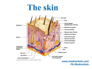
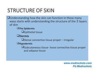
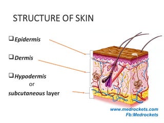
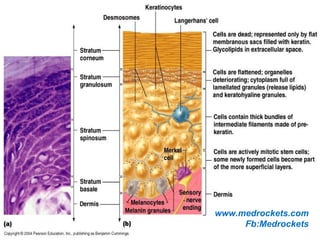
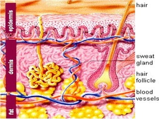
2. The skin (the interface between humans and their
environment) is the largest organ in the body. It weighs an
average of 4 kg and covers an area of 2 m2.
The skin has two layers:
1.The epidermis, outer epithelial layer.
2.The dermis, inner connective tissue.
Beneath the dermis is, the subcutis/hypodermis which usually
contains abundant fat.
The epidermis adheres to the dermis partly by the interlocking
of its downward projections (epidermal ridges or pegs) with
upward projections of the dermis (dermal papillae).
3.
4. The Integument
Is the largest system of the body
16% of body weight,
1.5 to 2m2
in area,
The integument is made up of two parts:
1. Cutaneous membrane
a. Epidermis– Superficial epithelium
b. Dermis – underlying CT with blood supply
2. Accessory structures
a. Hair
b. Nails
c. Exocrine Glands
5. Protection
First line of defense against
Bacteria
Viruses
Protects underlying structures from
Ultraviolet (UV) radiation
Dehydration
Vitamin D production
Needed for calcium absorption
Sensation
Sensory receptors
6. Body temperature regulation
If too hot
Dermal blood vessels dilate
Vessels carry more blood to surface so heat can escape
If too cold
Dermal blood vessels constrict
Prevents heat from escaping
Excretion
Small amounts of waste products are lost through
perspiration
www.medrockets.com
Fb:Medrockets
7. Understanding how the skin can function in these many
ways starts with understanding the structure of the 3 layers
of skin
The Epidermis
Epithelial tissue
Dermis
Dense connective tissue proper – irregular
Hypodermis
Subcutaneous tissue- loose connective tissue proper
and adipose tissue
www.medrockets.com
Fb:Medrockets
9. Epidermis:
The epidermis is formed from many layers of closely packed cells
called keratinocytes:
1.Basal layer (stratum germinativum(.
2.Prickle cell layer (stratum spinosum(.
3.Granular layer (stratum ranulosum(.
4.Horny layer (stratum corneum(
On the palms and soles a pale or pink layer, the stratum lucidum,
is noted just above the granular layer.
The epidermis varies in thickness from less than 0.1 mm on the
eyelids to nearly 1 mm on the palms and soles.
10. Structures of the Epidermis
The five strata of keratinocytes in thick skin
From basal lamina to free surface
1. Stratum basale
2. Stratum spinosum
3. Stratum granulosum
4. Stratum lucidum
5. Stratum corneum
www.medrockets.com
Fb:Medrockets
11. 11.The basal layer:
Is the deepest layer, rests on basement membrane,
which attaches it to the dermis.
This layer generate cells of the epidermis.
It is a single layer of columnar cells, whose basal
surfaces sprout many fine processes and
hemidesmosomes, anchoring them to the lamina densa
of the basement membrane.
www.medrockets.com
Fb:Medrockets
12. 22..The spinous or prickle cell layer:
Composed of differentiating cells, contain some tonofibrils
and kertohyalin granules, which synthesize keratins.
They are larger than basal cells.
Keratinocytes are firmly attached to each other by small
interlocking cytoplasmic processes, and by abundant
desmosomes.
Under the light microscope, the desmosomes look like
‘prickles’, they are specialized attachment plaques.
13. 3.Granular layer:
Consists of two or three layers of cells that are flatter than
those in the spinous layer, and have more tonofibrils.
As the name of the layer implies, these cells contain large
irregular basophilic granules of keratohyalin, which merge
with tonofibrils.
As keratinocytes migrate out through the outermost layers,
their keratohyalin granules break up and their contents are
dispersed throughout the cytoplasm, leading to keratinization
and the formation of a thick and tough peripheral protein
coating called the horny envelope.
14. Stratum Lucidum — the “clear layer”
Found only in thick skin
Covers stratum granulosum
Stratum Corneum — the “horn layer”
Exposed surface of skin
15 to 30 layers of keratinized cells
Water resistant
Shed and replaced every 2 weeks
www.medrockets.com
Fb:Medrockets
15. Other cells of the epidermis:
1. Melanocytes.
2. Langerhans cells.
3. Merckle cells
www.medrockets.com
Fb:Medrockets
16. Melanocytes are the only cells that can synthesize
melanin.
They migrate from the neural crest into the
basal layer of the ectoderm where, in human embryos,
they are seen as early as the eighth week of gestation.
They are also found in hair bulbs, the retina and pia
arachnoid.
Melanocytes
www.medrockets.com
Fb:Medrockets
17. Each dendritic melanocyte associates with a number
of keratinocytes, forming an ‘epidermal
melanin unit.
The dendritic processes of melanocytes wind between
the epidermal cells and end as discs in contact with
them.
Their cytoplasm contains discrete organelles, the
melanosomes, containing varying amounts of the
pigment melanin.
www.medrockets.com
Fb:Medrockets
18. The Langerhans cell is a dendritic cell like the
melanocyte.
It also lacks desmosomes and tonofibrils, but has a
lobulated nucleus.
The specific granules within the cell look like a tennis
racket when seen in two dimensions in an electron
micrograph. They are plate-like, with a rounded bleb
protruding from the surface.
Langerhans cells
www.medrockets.com
Fb:Medrockets
19. Langerhans cells come from precursors originating in the bone
marrow.
There are approximately 800 Langerhans cells per mm2.
Langerhans cells are alone among epidermal cells in
possessing surface receptors for C3b and the Fc portions of
IgG and IgE, and in bearing major histocompatibility complex
(MHC) Class II antigens (HLA-DR, -DP and -DQ).
They are best thought of as specialized macrophages.
20. Langerhans cells have a key role in many immune reactions.
They take up exogenous antigen, process
it and present it to T lymphocytes either in the skin
or in the local lymph nodes.
They probably play a part in immuno-surveillance for viral
and tumor antigens.
In this way, ultraviolet radiation can induce skin tumors both
by causing mutations in the epidermal cells, and by decreasing
the number of epidermal Langerhans cells.
Topical or systemic glucocorticoids also reduce the density of
epidermal Langerhans cells.
21. Merkel cells are found in normal epidermis.
Act as transducers for fine touch.
They are nondendritic cells.
Lying in or near the basal layer.
Are of the same size as keratinocytes.
They are concentrated in localized thickenings of the
epidermis near hair follicles (hair discs).
Contain membrane bound spherical granules, 80–100 nm in
diameter.
Sparse desmosomes connect these cells to neighbouring
keratinocytes.
Fine unmyelinated nerve endings are often associated with
Merkel cells.
Merkel cells
23. The basement membrane lies at the interface between the
epidermis and dermis.
With light microscopy it can be highlighted using a periodic
acid–Schiff (PAS) stain, because of its abundance of neutral
mucopolysaccharides..
The dermo-epidermal junction
24. Electron microscopy shows that the basement membrane has 4
componenets:
1.The plasma membrane of basal cells which has hemidesmosomes
(containing bullous pemphigoid antigens, collagen XVII and á6 â4
integrin).
2.Electron-lucent area, the lamina lucida which separate lamina densa
from the basal cells. The lamina lucida contains laminin-1, laminin-5 and
entactin.
3.Lamina densa (rich in type IV collagen).
4.Anchoring fibrils (of type VII collagen), dermal microfibril bundles
and single small collagen fibres (types I and III), extend from the
papillary dermis to the deep part of the lamina densa.
25.
26. The structures within the dermo-epidermal junction provide
mechanical support, encouraging the adhesion, growth,
differentiation and migration of the overlying basal cells, and
also act as a semipermeable filter that regulates the transfer of
nutrients and cells from dermis to epidermis.
www.medrockets.com
Fb:Medrockets
27. Dermis:
The dermis lies between the epidermis and the subcutaneous fat. It is tow
parts:
1.Upper papillary dermis.
2.Lower reticular dermis.
Its thickness varies, being greatest in the palms and soles and least in the
eyelids and penis.
In old age, the dermis thins and loses its elasticity.
The dermis interdigitates with the epidermis so that upward projections
of the dermis, the dermal papillae, interlock with downward ridges of the
epidermis, the rete pegs.
This interdigitation is responsible for the ridges seen most readily on the
fingertips (as fingerprints).
The dermis has three components:
cells, fibers and amorphous ground substance.
28. Cells of the dermis:
The main cells of the dermis are fibroblasts, but there
are also small numbers of resident and transitory mononuclear
phagocytes, lymphocytes, Langerhans cells and mast cells.
Other blood cells, e.g. polymorphs, are seen during
inflammation.
29. Fibres of the dermis:
The dermis is largely made up of interwoven fibresThe dermis is largely made up of interwoven fibres,,
principally of collagen, packed in bundlesprincipally of collagen, packed in bundles..
Those in the papillary dermis are finer than those in the deeperThose in the papillary dermis are finer than those in the deeper
reticular dermisreticular dermis..
When the skin is stretched, collagen, with its high tensileWhen the skin is stretched, collagen, with its high tensile
strength, prevents tearing, and the elastic fibres, intermingledstrength, prevents tearing, and the elastic fibres, intermingled
with the collagen, later return it to the unstretched statewith the collagen, later return it to the unstretched state..
www.medrockets.com
Fb:Medrockets
30. Collagen fibers:
Makes up to 70–80% of the dry weight of the dermis.
Its fibres are composed of thinner fibrils, which are in turn made up of
microfibrils built from individual collagen molecules. These molecules
consist of three polypeptide chains forming a triple helix with a non-
helical segment at both ends.
Collagen is an unusual protein as it contains a high proportion of proline
and hydroxyproline and many glycine residues.
Defects in the enzymes needed for collagen synthesis are responsible for
some skin diseases, including the Ehlers–Danlos syndrome and
osteogenesis imperfecta (fragility of bones(.
31. Elastic fibers:
Account for about 2% of the dry weight of adult dermis.
They have two distinct protein components:
an amorphous elastin core and a surrounding ‘elastic tissue microfibrillar
component’.
Abnormalities in the elastic tissue cause cutis laxa (sagging inelastic
skin( and pseudoxanthoma elasticum.
Reticulin fibres::
Are fine collagen fibers, seen in fetal skin and around the blood vessels
and appendages of adult skin.
32. The ground substance of the dermis consists largely of two
glycosaminoglycans (hyaluronic acid and dermatan sulphate( with
smaller amounts of heparan sulphate and chondroitin sulphate.
The ground substance has several important functions:
•it binds water, allowing nutrients, hormones and waste products to pass
through the dermis;
•it acts as a lubricant between the collagen and elastic fibre networks
during skin movement; and
•it provides bulk, allowing the dermis to act as a shock absorber.
Ground substance of the dermis:
33. Both smooth and striated muscle are found in the skin.
The smooth arrector pili muscles are used by animals to raise their fur
and so protect them from the cold.
They are vestigial in humans, but may help to express sebum. Smooth
muscle is also responsible for ‘goose pimples’ (bumps( from cold, nipple
erection, and the raising of the scrotum by the dartos muscle.
Striated fibres (e.g. the platysma( and some of the muscles of facial
expression, are also found in the dermis.
Muscles:
www.medrockets.com
Fb:Medrockets
34. Although the skin consumes little oxygen, its abundant blood supply
regulates body temperature.
The blood vessels lie in two main horizontal layers:
1.The deep plexus is just above the subcutaneous fat, and its arterioles
supply the sweat glands and hair papillae.
2.The superficial plexus is in the papillary dermis and arterioles from it
become capillary loops in the dermal papillae.
The blood vessels in the skin are important in thermoregulation.
Blood vessels:
35. :
Afferent lymphatics begin as blind-ended capillaries in the dermal
papilla and pass to a superficial lymphatic plexus in the papillary dermis.
There are also two deeper horizontal plexuses, and collecting lymphatics
from the deeper one run with the veins in the superficial fascia.
Cutaneous lymphatics
www.medrockets.com
Fb:Medrockets
36.
37. The skin is supplied with an estimated one million nerve fibres.
Most are found in the face and extremities.
Their cell bodies lie in the dorsal root ganglia.
Both myelinated and non-myelinated fibres exist, with the latter making
up an increasing proportion peripherally.
Most free sensory nerves end in the dermis; however, a few non-
myelinated nerve endings penetrate into the epidermis. Some of these are
associated with Merkel cells.
Nerves:
38. Free nerve endings detect stimuli of heat and pain (nocioceptors(, while
specialized end organs in the dermis, Pacinian and Meissner corpuscles,
detect pressure (mechanoreceptors(, vibration and touch.
Autonomic nerves supply the blood vessels, sweat glands and arrector
pili muscles.
Itching is an important feature of many skin diseases. It follows the
stimulation of fine free nerve endings lying close to the dermo-epidermal
junction.
Impulses from these free endings pass centrally in two ways:
quickly along myelinated A fibres, and more slowly along non-
myelinated C fibres.
In itchy skin diseases, pruritogenic chemicals such as histamine and
proteolytic enzymes are liberated close to the dermoepidermal junction.
39. The hard keratin of the nail plate is formed in the nail matrix,
which lies in an invagination of the epidermis (the nail fold(
on the back of the terminal phalanx of each digit.
The matrix runs from the proximal end of the floor of the nail
fold to the distal margin of the lunula.
From this area the nail plate grows forward over the nail bed,
ending in a free margin at the tip of the digit.
The nail:
40. The nail bed is capable of producing small amounts of keratin
which contribute to the nail and which are responsible for the
‘false nail’ formed when the nail matrix is obliterated by
surgery or injury.
The cuticle acts as a seal to protect the potential space of the
nail fold from chemicals and from infection.
The nails provide strength and protection for the terminal
phalanx.
Their presence helps with fine touch and with the handling of
small objects.
41. The rate at which nails grow varies from person to person:
fingernails average between 0.5 and 1.2 mm per week, while
toenails grow more slowly.
Nails grow faster in the summer, if they are bitten, and in
youth.
They change with ageing from the thin, occasionally spooned
nails of early childhood to the duller, paler and more opaque
nails of the very old.
42.
43.
44. Cosists of 2 parts:
1. Hair follicle, include: inner root sheath and outer rot sheath
2. Hair shaft, consists of: hair cuticle, cortex and medulla.
Hair shaft and hair follicle are produced by the matrix portion of hair
bulb.
Along one side, the sebaceous gland open to the upper part of the hair
follicle, arrector pili muscle attach to the lower part.
Apocrine gland also open to the hair follicle from the opposite side.
From the surface opening of the hair follicle to the enterance of the
sebaceous duct is called infundibular segment.
The portion between the sebaceous duct and insertion of arrector pili
muscle is isthmus.
The lowest portion is hair bulb.
Hair unit:
45.
46. 1.Lanugo hairs. Fine long hairs covering the fetus, but shed
about 1 month before birth.
2.Vellus hairs. Fine short unmedullated hairs covering much
of the body surface. They replace thelanugo hairs just before
birth.
3.Terminal hairs. Long coarse medullated hairs seen,
for example, in the scalp or pubic regions. Their growth
is often influenced by circulating androgen levels.
Classification of hairs:
47. There are three phases of follicular activity:
1. Anagen. The active phase of hair production.
2. Catagen. A short phase of conversion from
active growth to the resting phase. Growth stops, and the
end of the hair becomes club-shaped.
3. Telogen. A resting phase at the end of which the
club hair is shed.
The hair cycle
48. The scalp contain an average of 100 000 hairs,
anagen lasts for up to 5 years,
catagen for about 2 weeks,
and telogen for about 3 months.
As many as 100 hairs may be shed from the normal
scalp every day as a normal consequence of cycling.
On the scalp, about 85% are normally in anagen and
15% in the telogen phase.
www.medrockets.com
Fb:Medrockets
49.
50.
51. Sebaceous glands:
They are associated with hair follicle, lie in the obtuse angle between
the follicle and the epidermis.
They are multilobed and contain cells full of lipid, which are shed whole
(holocrene secretion) during secretion into the upper part of the hair
follicle.
Sebum contains a mixture of triglycerides, fatty acids, waxy esters,
squalene and cholesterol.
It lubricates and waterproofs the skin and protects it from drying.
Free sebaceous glands may be found in the eye lids (meibomian glands),
mucous membranes (Fordyce spots), nipples, peri-anal region and
genetalia.
Androgen hormones, especially dihydrotestosterone stimulate sebaceous
gland activity.
52. There are 2-3 million sweat glands distributed all over the body surface
but they are most numerous on the palms, soles and axillae.
The tightly coiled glands lie deep in the dermis, and the emerging duct
passes to the surface by penetrating the epidermis in a corkscrew
fashion.
Initially sweat is isotonic like plasma but, under normal conditions, it
becomes hypotonic by the time it discharged at the surface, after the
tubular resorption of electrolytes and water under the influence of
aldosterone and antidiuretic hormones.
Eccrine sweat glands:
53. The PH of sweat is between 4.0 and 6.8; it contains sodium, potassium
chloride, lactate, urea and ammonia.
Sweat glands have an important role in temperature control, the skin
surface being cooled by evaporation. Up to 10 L/day of sweat can be
excreted.
The sweat glands are innervated by cholinergic fibers of the sympathetic
nervous system. Sweating can therefore be induced by cholinergic and
blocked by anticholenergic drugs.
Central control of sweating resides in the preoptic hypothalamic sweat
centre.
54. Apocrine sweat glands:
They are limited to the axillae, nipples, peri-umbilical area, perineum
and genitalia.
The coiled tubular glands (larger than sweat glands) lie deep in the
dermis, and during sweating the luminal part of their cells is lost
(decapitation secretion).
Apocrine sweat passes via the duct into the mid-portion of the hair
follicle.
The action of bacteria on apocrine sweat is responsible for body odour.
The glands are innervated by adrenergic fibres of the sympathetic
nervous system.