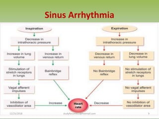
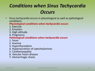
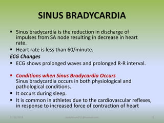
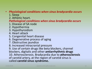
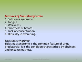
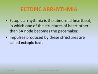
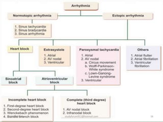
The document discusses different types of arrhythmia, which refers to irregular heartbeat. It describes normotopic arrhythmia where the sinus node is the pacemaker, including sinus arrhythmia, sinus tachycardia, and sinus bradycardia. Ectopic arrhythmia occurs when other parts of the heart act as the pacemaker, such as atrial or ventricular muscle. The document also covers heart block, paroxysmal tachycardia, atrial flutter, and artificial pacemakers used to treat arrhythmias.
Arrhythmia is also known as irregular heart beats. If SA node is not the pacemaker, any other part of the heart such as atrial muscle, AV node and ventricular muscle becomes the pacemaker. the beats may be fast, slow or miss beats.
Arrhythmia refers to irregular heartbeat that can be too fast, too slow, or have extra or missed beats. It occurs due to the heart's natural pacemaker (sinus node) not controlling heartbeat or other parts of the heart taking over as pacemaker. Arrhythmia is classified as normotopic if the sinus node is still pacemaker or ectopic if another area is pacemaker. Common types of normotopic arrhythmia include sinus arrhythmia which varies heartbeat with breathing, and sinus tachycardia or bradycardia which are fast or slow heart rates. Ectopic arrhythmia occurs when other areas generate impulses, like atrial flutter which causes rapid ineffective atrial contractions
This document discusses different types of arrhythmia including normotopic arrhythmia like sinus arrhythmia, sinus tachycardia, and sinus bradycardia. It also discusses ectopic arrhythmia which occurs when structures other than the SA node act as the pacemaker, leading to conditions like heart block, extrasystole, paroxysmal tachycardia, atrial flutter, atrial fibrillation, and ventricular fibrillation. Abnormal pacemakers can also develop from the AV node, atrial musculature, or ventricular musculature. Current of injury affects the ECG pattern through axis deviation when parts of the ventricle are affected.
If you feel irregular heart beats, this is what it could meanNarayanahealthcare
Arrhythmia is a disorder of the heart pulse or heart rhythm such as beating too fast (tachycardia) or beating too slow (bradycardia), or irregular heartbeat. Premature or extra beats are the most common type of arrhythmia. This usually feels like a fluttering in the chest or a feeling of a skipped heartbeat.
An arrhythmia is an irregular heartbeat that occurs when the heart's electrical signals don't work properly, causing the heart to beat too fast, too slow, or irregularly. Some key types discussed include sinus tachycardia, sinus bradycardia, premature atrial contractions, atrial flutter, atrial fibrillation, supraventricular tachycardia, ventricular tachycardia, and heart block. Causes can include direct damage to the conduction system, inflammation, electrolyte imbalances, and drugs. Treatment depends on the specific type but may include medications, catheter ablation, cardioversion, or pacemaker implantation.
This document provides an overview of heart conduction and various types of arrhythmias or disturbances in heart rhythm. It begins with a description of normal heart conduction and then defines arrhythmia. Various mechanisms of arrhythmia are described including increased automaticity, triggered automaticity, and reentry. Arrhythmias are classified based on heart rate as tachyarrhythmias or bradyarrhythmias, and based on site of impulse generation as supraventricular or ventricular arrhythmias. Specific types of arrhythmias are defined including sinus arrhythmias, atrial arrhythmias like atrial fibrillation and flutter, junctional arrhythmias, and ventricular arrhythmias. Treatment options for arrhythmias include medications
Cardiac arrhythmias are abnormalities in the heart's rhythm that can cause symptoms ranging from palpitations to sudden death. The two main types are bradycardias, which are slow heart rates below 60 bpm, and tachycardias, which are fast heart rates over 100 bpm. Arrhythmias can arise from problems in the sinus node, atria, AV junction, or ventricles due to issues with automaticity or re-entry of electrical impulses. Common arrhythmias include sinus tachycardia/bradycardia, premature beats, atrial fibrillation, and heart blocks. Treatment depends on the specific arrhythmia and symptoms but may include lifestyle changes, medications
This document provides information on various cardiac dysrhythmias, including their causes, types, and management. It discusses junctional rhythms arising from the AV junction and their two major types - disturbances in automaticity and disturbances in conduction. Various types of AV blocks are described including first, second (Mobitz I and II), and third degree heart block. Other arrhythmias covered include bundle branch blocks, ventricular arrhythmias, pre-excitation syndromes like Wolf-Parkinson-White syndrome, and sudden cardiac death. The management of dysrhythmias through pharmacotherapy, defibrillation, ablation, and pacemakers is also summarized.
The document discusses various types of cardiac arrhythmias including their definitions, causes, clinical manifestations and management. It describes normal sinus rhythm and defines arrhythmias as any change from the normal heart rhythm. Common arrhythmias discussed include sinus tachycardia, sinus bradycardia, premature atrial complexes, premature ventricular complexes, atrial flutter, atrial fibrillation and ventricular tachycardia. It provides EKG images to demonstrate the different arrhythmias and compares characteristics of supraventricular and ventricular arrhythmias. Causes, clinical significance and treatment approaches for different arrhythmias are also summarized.
This document discusses different types of arrhythmias, or irregular heart rhythms, including tachyarrhythmias (fast rhythms) and bradyarrhythmias (slow rhythms). It describes specific conditions like ventricular fibrillation, atrial fibrillation, various types of heart block, and paroxysmal supraventricular tachycardia. The document also lists factors that can predispose patients to arrhythmias and notes that immediately life-threatening rhythms include ventricular fibrillation, pulseless ventricular tachycardia, and ventricular asystole.
Arrhythmia refers to an irregular heartbeat that can be too fast, too slow, or have an extra or missed beat. Arrhythmias are classified as normotopic or ectopic based on whether the sinoatrial node is the pacemaker. Normotopic arrhythmias include sinus arrhythmia, sinus tachycardia, and sinus bradycardia. Ectopic arrhythmias occur when another part of the heart acts as the pacemaker, such as atrial flutter, atrial fibrillation, or ventricular fibrillation. An artificial pacemaker can be implanted to regulate abnormal heart rhythms through electrodes that deliver electrical pulses to the heart.
ECG analysis on normal sinus rhythm and atrial arrhythmias.pptxcvkrishnapriya575
ECG play a vital role in healthcare industry. Analyzing a ECG is an hectic procedure hence this slide provide simple view about an ECG analysis on normal sinus rhythm and atrial arrhythmiasThe importance of ECG in the healthcare industry cannot be overstated. It is a crucial diagnostic tool that helps doctors and other medical professionals to accurately assess a patient's cardiac health. However, analyzing an ECG can be a complicated and time-consuming process, which is why this slide has been created to provide a simplified overview of ECG analysis for normal sinus rhythm and atrial arrhythmias. With this information, healthcare providers can quickly and easily interpret ECG results and make informed decisions about patient care.The importance of ECG in the healthcare industry cannot be overstated. It is a crucial diagnostic tool that helps doctors and other medical professionals to accurately assess a patient's cardiac health. However, analyzing an ECG can be a complicated and time-consuming process, which is why this slide has been created to provide a simplified overview of ECG analysis for normal sinus rhythm and atrial arrhythmias. With this information, healthcare providers can quickly and easily interpret ECG results and make informed decisions about patient care.
Atrial fibrillation occurs when disorganized electrical signals in the atria cause the atria to quiver rather than contract in a coordinated manner. This leads to an irregular and rapid heartbeat. Complications include an increased risk of stroke from blood clots, heart failure from reduced cardiac output, and myocardial infarction from increased cardiac demand. Treatment involves rate control with medications, rhythm control with cardioversion or antiarrhythmic drugs, and anticoagulation to prevent stroke.
Digoxin is a cardiac glycoside extracted from plants that is used to treat heart conditions like atrial fibrillation and congestive heart failure. It works by increasing calcium levels in heart cells, strengthening contractions and decreasing heart rate. Therapeutic doses are narrow, so monitoring serum digoxin levels is important. Common side effects include nausea, fatigue, and cardiac arrhythmias at toxic levels. It interacts with many other drugs, so dose adjustments may be needed.
An arrhythmia is any abnormal heart rate or rhythm that arises from problems with the heart's electrical system. The document classifies and describes different types of arrhythmias including sinus node dysfunction, atrial arrhythmias like atrial fibrillation, and ventricular arrhythmias like ventricular tachycardia. Treatment depends on the specific arrhythmia but may include medications, catheter ablation, pacemakers, or defibrillators. Exercise is an important part of rehabilitation and can help reduce risk factors when done according to an individualized prescription.
This document provides information about cardiac arrhythmias. It defines arrhythmias as any abnormal heart rate or rhythm and normal sinus rhythm as 60-100 bpm originating from the sinus node. Arrhythmias are classified as sinus, atrial, or ventricular based on origin. Common types include sinus tachycardia/bradycardia, atrial fibrillation, ventricular tachycardia, and heart blocks. Symptoms vary depending on type but can include palpitations, dizziness, and fainting. Diagnosis involves ECG, echocardiogram, and Holter monitoring. Treatment depends on type and may include medications, ablation, pacemakers, or defibrillators.
IDENTIFICATION AND APPROACH TO BRADYARRHYTHMIAS .pptxDr Dravid m c
Explanation of SA Nodal and AV nodal block , ECG changes , identification clinical features and presentation of patients to emergency department, their approach and medical linea of treatment
This document discusses heart arrhythmias, which are irregular or abnormal heartbeats. It defines different types of arrhythmias including tachycardia, bradycardia, atrial fibrillation, supraventricular tachycardia, and more. It discusses what causes arrhythmias, risk factors, potential symptoms, diagnostic tests used to evaluate arrhythmias like EKGs, and treatment options. Common treatments include medications, medical procedures, lifestyle changes, and in some cases devices like pacemakers.
Atrial flutter is a type of abnormal heart rhythm caused by rapid electrical signals in the atria. It causes the atria to contract very quickly, between 200-400 beats per minute, while allowing some signals to pass to the ventricles so they contract between 120-180 times per minute. Risk factors include pre-existing heart conditions like hypertension or cardiomyopathy. Treatment involves rate or rhythm control through medication or cardioversion, as well as anticoagulation due to risks of blood clots and ablation to disrupt the abnormal electrical pathways.
The cardiac cycle describes the sequence of mechanical events regulated by the heart's electrical activity. The sinoatrial node acts as the natural pacemaker, initiating each heartbeat around 60-80 times per minute by rapidly depolarizing cardiac muscle cells. Electrical impulses then travel to the atrioventricular node and throughout the myocardium, causing atrial contraction. The impulse then reaches the ventricles, triggering ventricular systole. Arrhythmias are irregular heartbeats that can range from harmless to life-threatening, such as ventricular fibrillation where the ventricles quiver ineffectively instead of pumping blood.
Definition: Cardiac arrhythmias refer to abnormal heart rhythms, where the heartbeat may be too slow (bradycardia), too fast (tachycardia), or irregular.
These irregularities disrupt the normal electrical signaling in the heart.
This document discusses different types of heart block and cardiac arrhythmias. It defines heart block as a decreased or stopped conduction of impulses through the AV nodal area. There are three classifications of heart block - first, second, and third degree - which are differentiated based on how many atrial impulses are conducted to the ventricles. Cardiac arrhythmias occur when there is a disorder in the formation or conduction of electrical impulses in the heart, altering the heart rate or rhythm. Arrhythmias are classified based on where they originate - the sinus node, atria, junctional area, or ventricles. Common arrhythmias discussed include sinus tachycardia, atrial fib
This document discusses different types of heart block and arrhythmias. It defines first, second, and third degree heart block and their characteristic ECG patterns. It also covers different types of arrhythmias including sinus, atrial, junctional, and ventricular arrhythmias. For each type of arrhythmia, it discusses causes, ECG characteristics, clinical manifestations, and potential treatments. The document provides an overview of heart block and various cardiac arrhythmias for medical professionals.
Heart arrhythmias result from disturbances in the rate, regularity, or origin of the heart's electrical signals. They are divided into two groups - one including life-threatening ventricular fibrillation and tachycardia requiring immediate defibrillation, and another including less dangerous arrhythmias that may still require treatment. Ventricular arrhythmias like ventricular tachycardia and fibrillation involve abnormal electrical signals originating in the ventricles. These can cause symptoms like fainting, chest pain, or shortness of breath and carry risks like inadequate blood flow leading to cardiac arrest if not terminated quickly.
Anatomy, physiology & patophysiology of the cardiovascularCarlos Galiano
This document provides an overview of the anatomy, physiology, and pathophysiology of the cardiovascular system as it relates to anesthesia. It discusses the structure and function of the heart and coronary circulation. It then covers topics such as cardiac cycle, hemodynamics, effects of the autonomic nervous system, and cardiovascular disorders including heart failure, hypertension, ischemic heart disease, and cardiac tamponade. For each topic, it provides details on pathophysiology and considerations for anesthesia management.
Myocardial ischemia occurs when heart muscle does not receive enough oxygen due to narrowing of the coronary arteries. The main causes are atherosclerosis and coronary thrombosis. Symptoms include chest pain known as angina. Diagnosis involves ECG, stress testing, and cardiac catheterization. Treatment aims to increase blood flow through medications like nitrates and calcium channel blockers, or procedures like angioplasty and bypass surgery. Myocardial infarction results from prolonged ischemia and cell death. It can cause complications like heart failure and arrhythmias. Treatment focuses on restoring blood flow, managing pain and arrhythmias, and preventing further damage.
1. Dysrhythmias are disorders of heart rhythm caused by abnormalities in the heart's electrical conduction system.
2. Common dysrhythmias include sinus tachycardia/bradycardia, premature atrial contractions, atrial fibrillation/flutter, supraventricular tachycardia, ventricular tachycardia/fibrillation, heart blocks, and bundle branch blocks.
3. Dysrhythmias can be caused by underlying conditions like myocardial infarction, electrolyte imbalances, drug toxicity, and more. Management involves treating the underlying cause and using medications, cardioversion, or pacemakers depending on the specific dysrhythmia.
This document discusses acute kidney injury (AKI), formerly called acute renal failure. AKI is a sudden deterioration in renal function that results in the kidneys' inability to maintain fluid and electrolyte homeostasis. It occurs in 2-8% of hospitalized children. AKI is classified as prerenal, intrinsic renal, or postrenal based on pathogenesis. Prerenal AKI is due to decreased renal perfusion without intrinsic kidney damage. Intrinsic renal AKI involves direct kidney damage from issues like ischemia, toxins, or glomerulonephritis. Postrenal AKI is caused by urinary tract obstruction. The diagnosis of AKI involves history, exam, labs, imaging, and sometimes kidney biopsy
This document discusses haematemesis and melaena, which refer to vomiting and passing black tarry stool containing blood from the upper GI tract. Haematemesis with bright red or dark coffee ground blood requires emergency care. Melaena results from oxidation of iron in haemoglobin as it passes through the lower GI tract. Upper GI bleeding has an annual incidence over 100 per 100,000 people and increases with age. Risk factors include NSAID use and hospitalization for other conditions. Patients may experience vomiting blood, black tarry stool, hypovolemic shock, lethargy or abdominal pain. Initial treatment involves stabilization, blood transfusion, risk assessment and endoscopy after stabilization.
Shock is defined as acute circulatory failure resulting in inadequate tissue perfusion and cellular hypoxia. There are several potential causes of shock, including failure of the heart to pump effectively, mechanical impediments to blood flow, loss of circulatory volume, or abnormalities in the peripheral circulation. The clinical features of shock vary depending on the specific type but may include signs of low blood pressure, increased heart rate, pale and cold skin, confusion or loss of consciousness, and metabolic acidosis in severe cases. Proper assessment of tissue perfusion through monitoring factors like skin signs, lactate levels, and urine output is important for diagnosis.
The document discusses cardiac arrhythmias, including their definition, causes, types, and prevalence in certain populations. It provides details on the heart's conduction system and normal conduction pathway. Common types of arrhythmias mentioned include atrial fibrillation, atrial flutter, and ventricular tachycardia. The document notes that while arrhythmias are common after coronary artery bypass surgery, some studies have found a reduced incidence in runners and athletes. It also discusses a study that found an association between participation in strength-based sports like football and atrial fibrillation.
Tetanus is a medical condition caused by a toxin produced by Clostridium tetani bacteria. It causes prolonged contraction of skeletal muscle fibers. Common causes of infection include contaminated wounds, burns, and puncture wounds. The toxin acts at the spinal cord to cause muscle contractions from minor stimuli. Symptoms include lockjaw, neck stiffness, and painful muscle spasms. Treatment involves antibiotics, tetanus immunoglobulin, muscle relaxants, and maintaining respiration and nutrition during recovery.
Acute respiratory failure occurs when the respiratory system fails to maintain adequate gas exchange. There are two main types: hypoxemic respiratory failure, characterized by low oxygen levels (PaO2) with normal or low carbon dioxide (PaCO2) levels; and ventilatory (hypercapnic) respiratory failure, characterized by high PaCO2 levels. Hypoxemic failure is most common and can result from conditions that impair gas exchange like pneumonia or pulmonary edema. Ventilatory failure involves impaired ventilation and can be caused by conditions that obstruct airflow like COPD. Diagnosis involves blood gas analysis and imaging. Treatment focuses on supporting oxygenation and ventilation through oxygen supplementation, ventilation support, and treating the underlying cause.
This document discusses haematemesis and melaena, which refer to vomiting and passing black stool containing blood from upper gastrointestinal bleeding. Haematemesis involves vomiting bright red or dark blood, while melaena involves black tarry stool due to blood degradation. Common causes of upper GI bleeding include peptic ulcers and esophageal varices. Initial treatment involves stabilization, risk assessment, and upper endoscopy to determine the source of bleeding. Endoscopic therapies like clips, cautery, and injections can control active or recent bleeding. Ongoing intravenous PPIs after endoscopy may prevent rebleeding. Surgery is considered if bleeding persists despite endoscopic treatment. Long-term prevention involves lifestyle changes and maintenance P
Erysipelas is a bacterial skin infection caused by streptococci that extends into cutaneous lymphatics. It presents with a fiery red plaque with sharply demarcated advancing margins and local signs of inflammation. Risk factors include skin trauma, lymphatic impairment, and immunosuppression. Treatment involves antibiotics like penicillin for 10-20 days. Recurrence is common in those with predisposing skin conditions, and long-term antibiotic prophylaxis may be used. Complications are rare but include abscess, gangrene, and thrombophlebitis. With treatment, prognosis is generally excellent.
Dr. Nikhil Oza presented on AIDS (Acquired Immuno Deficiency Syndrome). AIDS is caused by the HIV virus which breaks down the immune system. It occurs globally and can affect all ages/groups. The virus is found in blood and genital secretions. Common transmission routes are unprotected sex and needle sharing. The virus has a long incubation period before symptoms appear. Symptoms progress from asymptomatic infection to AIDS-related illnesses like pneumonia. Diagnosis involves antibody testing. Treatment involves antiretroviral drugs to suppress the virus. Prevention strategies include safe sex, needle safety, and maternal screening/treatment.
share - Lions, tigers, AI and health misinformation, oh my!.pptxTina Purnat
• Pitfalls and pivots needed to use AI effectively in public health
• Evidence-based strategies to address health misinformation effectively
• Building trust with communities online and offline
• Equipping health professionals to address questions, concerns and health misinformation
• Assessing risk and mitigating harm from adverse health narratives in communities, health workforce and health system
Recomendações da OMS sobre cuidados maternos e neonatais para uma experiência pós-natal positiva.
Em consonância com os ODS – Objetivos do Desenvolvimento Sustentável e a Estratégia Global para a Saúde das Mulheres, Crianças e Adolescentes, e aplicando uma abordagem baseada nos direitos humanos, os esforços de cuidados pós-natais devem expandir-se para além da cobertura e da simples sobrevivência, de modo a incluir cuidados de qualidade.
Estas diretrizes visam melhorar a qualidade dos cuidados pós-natais essenciais e de rotina prestados às mulheres e aos recém-nascidos, com o objetivo final de melhorar a saúde e o bem-estar materno e neonatal.
Uma “experiência pós-natal positiva” é um resultado importante para todas as mulheres que dão à luz e para os seus recém-nascidos, estabelecendo as bases para a melhoria da saúde e do bem-estar a curto e longo prazo. Uma experiência pós-natal positiva é definida como aquela em que as mulheres, pessoas que gestam, os recém-nascidos, os casais, os pais, os cuidadores e as famílias recebem informação consistente, garantia e apoio de profissionais de saúde motivados; e onde um sistema de saúde flexível e com recursos reconheça as necessidades das mulheres e dos bebês e respeite o seu contexto cultural.
Estas diretrizes consolidadas apresentam algumas recomendações novas e já bem fundamentadas sobre cuidados pós-natais de rotina para mulheres e neonatos que recebem cuidados no pós-parto em unidades de saúde ou na comunidade, independentemente dos recursos disponíveis.
É fornecido um conjunto abrangente de recomendações para cuidados durante o período puerperal, com ênfase nos cuidados essenciais que todas as mulheres e recém-nascidos devem receber, e com a devida atenção à qualidade dos cuidados; isto é, a entrega e a experiência do cuidado recebido. Estas diretrizes atualizam e ampliam as recomendações da OMS de 2014 sobre cuidados pós-natais da mãe e do recém-nascido e complementam as atuais diretrizes da OMS sobre a gestão de complicações pós-natais.
O estabelecimento da amamentação e o manejo das principais intercorrências é contemplada.
Recomendamos muito.
Vamos discutir essas recomendações no nosso curso de pós-graduação em Aleitamento no Instituto Ciclos.
Esta publicação só está disponível em inglês até o momento.
Prof. Marcus Renato de Carvalho
www.agostodourado.com
ABDOMINAL TRAUMA in pediatrics part one.drhasanrajab
Abdominal trauma in pediatrics refers to injuries or damage to the abdominal organs in children. It can occur due to various causes such as falls, motor vehicle accidents, sports-related injuries, and physical abuse. Children are more vulnerable to abdominal trauma due to their unique anatomical and physiological characteristics. Signs and symptoms include abdominal pain, tenderness, distension, vomiting, and signs of shock. Diagnosis involves physical examination, imaging studies, and laboratory tests. Management depends on the severity and may involve conservative treatment or surgical intervention. Prevention is crucial in reducing the incidence of abdominal trauma in children.
8 Surprising Reasons To Meditate 40 Minutes A Day That Can Change Your Life.pptxHolistified Wellness
We’re talking about Vedic Meditation, a form of meditation that has been around for at least 5,000 years. Back then, the people who lived in the Indus Valley, now known as India and Pakistan, practised meditation as a fundamental part of daily life. This knowledge that has given us yoga and Ayurveda, was known as Veda, hence the name Vedic. And though there are some written records, the practice has been passed down verbally from generation to generation.
Adhd Medication Shortage Uk - trinexpharmacy.comreignlana06
The UK is currently facing a Adhd Medication Shortage Uk, which has left many patients and their families grappling with uncertainty and frustration. ADHD, or Attention Deficit Hyperactivity Disorder, is a chronic condition that requires consistent medication to manage effectively. This shortage has highlighted the critical role these medications play in the daily lives of those affected by ADHD. Contact : +1 (747) 209 – 3649 E-mail : sales@trinexpharmacy.com
2. Arrhythmia
Arrhythmia refers to irregular heartbeat or
disturbance in the rhythm of heart.
In arrhythmia, heartbeat may be fast or slow
or there may be an extra beat or a missed
beat.
It occurs in physiological and pathological
conditions.
12/25/2018 2
studyforum911@hotmail.com
3. CLASSIFICATION
• In arrhythmia, SA node may or may not be the
pacemaker.
• If SA node is not the pacemaker, any other part of
the heart such as atrial muscle, AV node and
ventricular muscle becomes the pacemaker.
Accordingly, arrhythmia is classified into two types:
A. Normotopic arrhythmia
B. Ectopic arrhythmia
12/25/2018 3
studyforum911@hotmail.com
4. Normotopic Arrhythmia
• Normotopic arrhythmia is the irregular
heartbeat, in which SA node is the pacemaker.
Normotopic arrhythmia is of three types:
1. Sinus arrhythmia
2. Sinus tachycardia
3. Sinus bradycardia.
12/25/2018 4
studyforum911@hotmail.com
5. SINUS ARRHYTHMIA
Sinus arrhythmia is a normal rhythmical increase and decrease in
heart rate, in relation to respiration. It is also called respiratory
sinus arrhythmia (RSA).
Normal sinus rhythm means the normal heartbeat with SA node as
the pacemaker. Normal heart rate is 72 per minute.
However, under physiological conditions, in a normal healthy
person, heart rate varies according to the phases of respiratory
cycle.
Heart rate increases during inspiration and decreases during
expiration.
ECG Changes
Only the duration of R-R interval varies rhythmically according to
phases of respiration.
It is shortened during inspiration and prolonged during expiration
12/25/2018 5
studyforum911@hotmail.com
6. Cause
Sinus arrhythmia is due to fluctuation in the discharge of impulses
from SA node.
During inspiration, the lungs are inflated and the intrathoracic
pressure
decreases.
This increases the venous return.
Inflation of lungs stimulates the stretch receptors of lungs, which
send impulses to vasodilator area (cardioinhibitory center) through
afferent fbers of vagus. It leads to
reflex inhibition of vasodilator area and reduction in vagal tone.
Because of these two factors, heart rate increases.
Simultaneously, increased venous return initiates Bainbridge reflex
that causes increase in heart rate.
12/25/2018 6
studyforum911@hotmail.com
7. Conti…
• During expiration, the lungs are deflated and
intrathoracic pressure increases.
• This decreases the venous return.
• During deflation of lungs, the stretch receptors are not
stimulated and vasodilator area is not inhibited.
• So, vagal tone increases, resulting in decreased heart
rate. Simultaneously, decreased venous return
abolishes Bainbridge reflex. It also decreases the heart
rate.
12/25/2018 7
studyforum911@hotmail.com
8. SINUS TACHYCARDIA
Sinus tachycardia is the increase in discharge of
impulses from SA node, resulting in increase in heart
rate.
Discharge of impulses from SA node is very rapid and
the heart rate increases up to 100/minute and
sometimes up to 150/minute.
ECG Changes
ECG is normal in sinus tachycardia, except for short R-R
intervals because of increased heart rate
12/25/2018 8
studyforum911@hotmail.com
10. Conditions when Sinus Tachycardia
Occurs
• Sinus tachycardia occurs in physiological as well as pathological
conditions.
Physiological conditions when tachycardia occurs
1. Exercise
2. Emotion
3. High altitude
4. Pregnancy.
Pathological conditions when tachycardia occurs
1. Fever
2. Anemia
3. Hyperthyroidism
4. Hypersecretion of catecholamines
5. Cardiomyopathy
6. Valvular heart disease
7. Hemorrhagic shock.
12/25/2018 10
studyforum911@hotmail.com
11. • Features of Sinus Tachycardia
• 1. Palpitations (sensation of feeling the
heartbeat)
2. Dizziness
3. Fainting
4. Shortness of breath
5. Chest discomfort (angina).
12/25/2018 11
studyforum911@hotmail.com
12. SINUS BRADYCARDIA
Sinus bradycardia is the reduction in discharge of
impulses from SA node resulting in decrease in heart
rate.
Heart rate is less than 60/minute.
ECG Changes
ECG shows prolonged waves and prolonged R-R interval.
Conditions when Sinus Bradycardia Occurs
Sinus bradycardia occurs in both physiological and
pathological conditions.
It occurs during sleep.
It is common in athletes due to the cardiovascular reflexes,
in response to increased force of contraction of heart
12/25/2018 12
studyforum911@hotmail.com
13. • Physiological conditions when sinus bradycardia occurs
1. Sleep
2. Athletic heart.
Pathological conditions when sinus bradycardia occurs
1. Disease of SA node
2. Hypothermia
3. Hypothyroidism
4. Heart attack
5. Congenital heart disease
6. Degenerative process of aging
7. Obstructive jaundice
8. Increased intracranial pressure
9. Use of certain drugs like beta blockers, channel
blockers, digitalis and other antiarrhythmic drugs
10. Atherosclerosis. Bradycardia due to atherosclerosis
of carotid artery, at the region of carotid sinus is
called carotid sinus syndrome.
12/25/2018 13
studyforum911@hotmail.com
14. Features of Sinus Bradycardia
1. Sick sinus syndrome
2. Fatigue
3. Weakness
4. Shortness of breath
5. Lack of concentration
6. Difficulty in exercising.
Sick sinus syndrome
Sick sinus syndrome is the common feature of sinus
bradycardia. It is the condition characterized by dizziness
and unconsciousness.
12/25/2018 14
studyforum911@hotmail.com
15. ECTOPIC ARRHYTHMIA
• Ectopic arrhythmia is the abnormal heartbeat,
in which one of the structures of heart other
than SA node becomes the pacemaker.
• Impulses produced by these structures are
called ectopic foci.
12/25/2018 15
studyforum911@hotmail.com
17. HEART BLOCK
• Heart block is the blockage of impulses generated
by SA node in the conductive system.
• Because of the blockage, the impulses cannot
reach the cardiac musculature, resulting in
ectopic arrhythmia. Based on the area affected,
the heart block is classified into two
types.
1. Sinoatrial block
2. Atrioventricular block
12/25/2018 17
studyforum911@hotmail.com
18. Sinoatrial Block – AV Nodal Rhythm
Sinoatrial block is the failure of impulse transmission from SA node
to AV node. It is also called sinus block.
During sinoatrial block, heart stops beating. Immediately, AV node
takes over the pacemaker function and produces the impulses.
This leads to AV nodal (atrioventricular) rhythm.
Sinoatrial block is due to the defect in internodal fbers and it occurs
suddenly.
Initially, the heart stops for a while.
Then after few seconds, the AV node becomes the pacemaker and
the heart starts beating with decreased rate of 40 to 60/minute.
Impulses may be discharged from any part of AV node, viz.
12/25/2018 18
studyforum911@hotmail.com
19. Atrioventricular Block (AV Block)
Atrioventricular block is the heart block in which the
impulses are not transmitted from atria (from AV
node) to ventricles because of defective conductive
system.
Atrioventricular block is of two categories:
1. Incomplete heart block
2. Complete heart block
12/25/2018 19
studyforum911@hotmail.com
20. • Incomplete Heart Block
Incomplete heart block is the condition in which the
transmission of impulses from atria to ventricles is
slowed down and not blocked completely. Impulses
reach ventricles late.
Incomplete heart block is of four types:
i. First degree heart block
ii. Second degree heart block
iii. Wenckebach phenomenon
iv. Bundle branch block
12/25/2018 20
studyforum911@hotmail.com
21. PAROXYSMAL TACHYCARDIA
Paroxysmal tachycardia is the sudden attack of increased heart rate
due to ectopic foci arising from atria, AV node or ventricle. It is also
called BouveretHoffmann syndrome.
Increase in heart rate due to ectopic foci arising from either atria or
AV node is called supraventricular tachycardia (SVT).
Symptoms include palpitations, chest pain, rapid breathing and
dizziness.
Paroxysmal tachycardia is of three types:
1. Atrial paroxysmal tachycardia
2. AV nodal paroxysmal tachycardia
3. Ventricular paroxysmal tachycardia.
12/25/2018 21
studyforum911@hotmail.com
22. Atrial Flutter
Atrial flutter is an arrhythmia characterized by rapid ineffective atrial
contractions, caused by ectopic foci originating from atrial musculature.
It is often associated with atrial paroxysmal tachycardia.
Both the atria beat rapidly like the wings of a bird, hence the name atrial
flutter.
Atrial rate is about 250 to 350/minute.
Maximum number of impulses conducted by AV node is about 230 to 240
/minute.
So, during atrial flutter, the second degree of heart block occurs. The ratio
between atrial beats and ventricular beats is 2 : 1 or sometimes 3 : 1.
Atrial flutter is common in patients suffering from
cardiovascular diseases such as hypertension and coronary artery disease.
12/25/2018 22
studyforum911@hotmail.com
23. Artificial Pacemaker
Artificial pacemaker is a small electronic device that is surgically
implanted to regulate abnormal heartbeat.
It contains a battery powered pulse generator, that produces
electrical impulses capable of stimulating the heart.
This pacemaker is implanted under the skin over the chest of the
patient.
Pulses generated by this device are transmitted to the heart
through electrodes.
Electrodes connected to the device are inserted and passed
through a vein and positioned in the heart chambers.
The device has a lithium battery that may last for 10 to 15 years.
The outer casing of the pacemaker is usually made of titanium,
which is rarely rejected by body’s immune system.
12/25/2018 23
studyforum911@hotmail.com