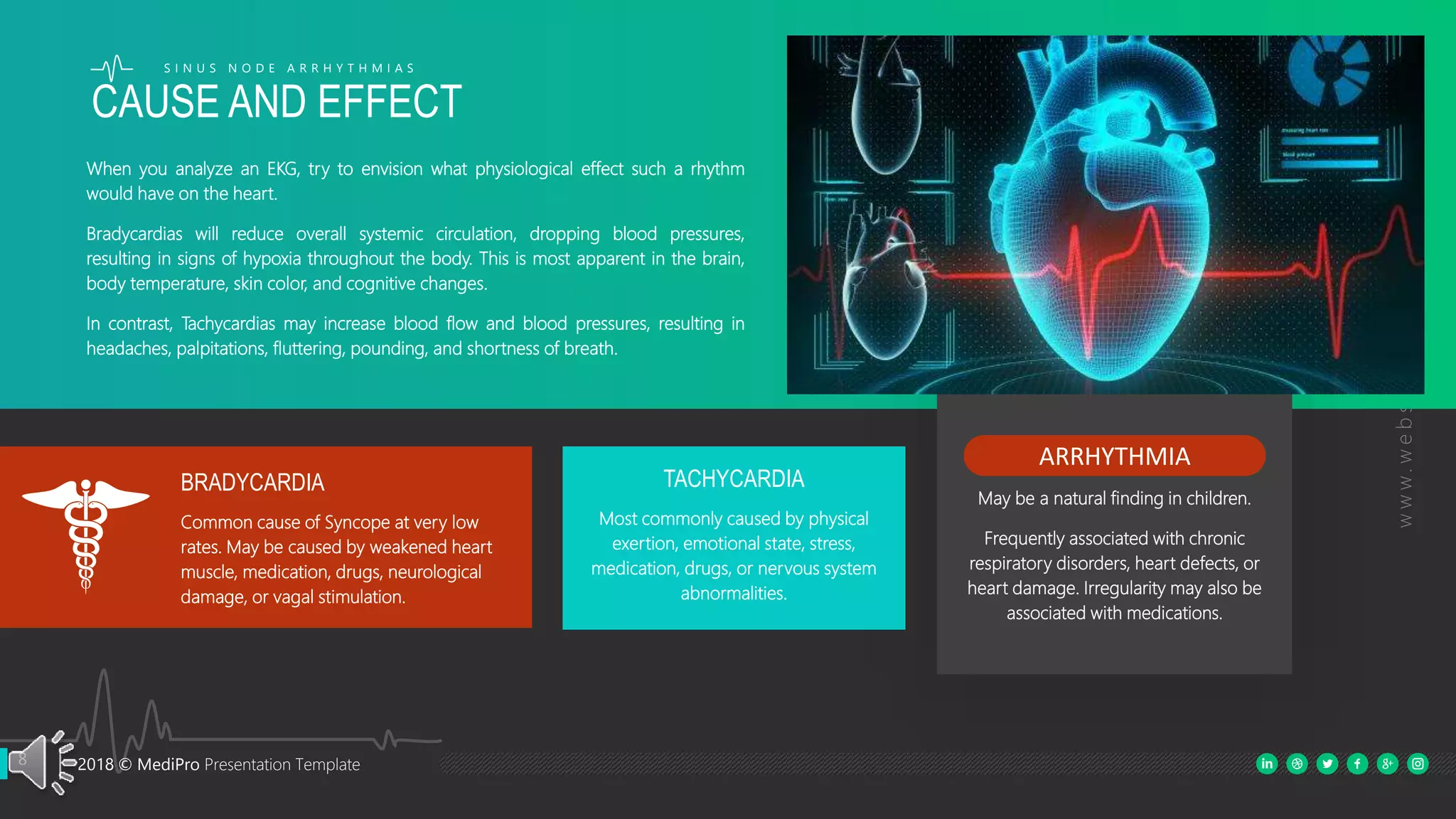
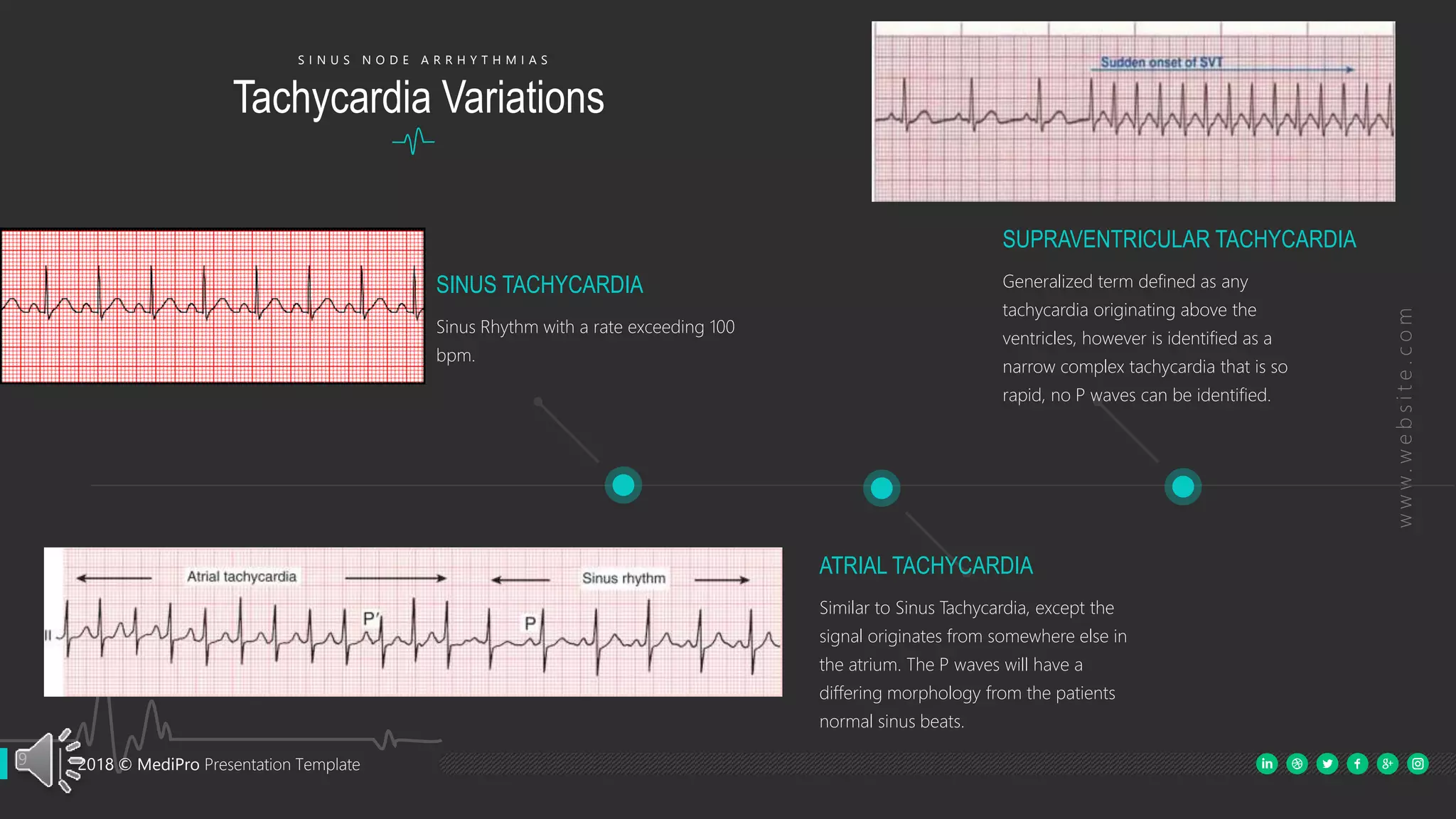
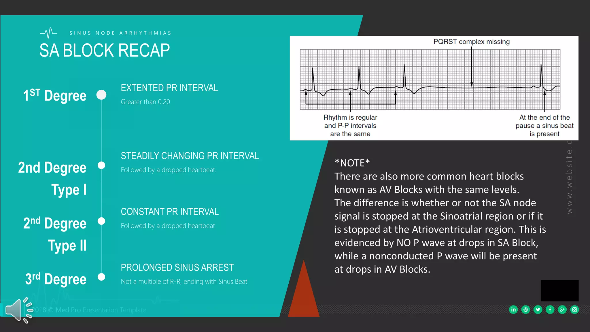
Basic EKG Interpretation
CHAPTER 4: SINUS NODE ARRHYTHMIAS
Accompanying Textbook:
ECG Interpretation Made Incredibly Easy, 5th Ed.
© 2011 by Lippincott Williams & Wilkins. All rights reserved
Instructed by: Douglas J Michel, PMD
2 2018 © MediPro Presentation Template
www.website.com
PRIMARY SINUS RHYTHMS
S I N U S N O D E A R R H Y T H M I A S
Sinus Bradycardia is a sinus rhythm
originating in the SA node, at a rate less
than 60 bpm.
SINUS BRADYCARDIA
Originating in the SA Node, this otherwise
sinus rhythm is the only SA rhythm that is
irregular. Sinus Arrhythmia has a heart
rate within normal limits, and often varies
with a patients respiratory cycle.
SINUS ARRHYTHMIA
Sinus Tachycardia is a sinus rhythm
originating in the SA node, at a rate
greater than 100 bpm.
SINUS TACHYCARDIA
SINUS
ARRHYTHMIA
SINUS
BRADYCARDIA
SINUS
TACHYCARDIA
3 2018 © MediPro Presentation Template
www.website.com
3 2018 © MediPro Presentation Template
A normal P wave should be upright, precede the QRS complex, approximately 2-3
mm high, with a duration of 0.06-0.12 seconds. Their shape should be generally
rounded in appearance.
PR INTERVAL: 0.12-0.20
QRS INTERVAL: 0.06-0.12
QT INTERVAL: 0.36-0.44
T WAVES – Normal in appearance
RHYTHM - Regular
SINUS BRADYCARDIA – Under 60 BPM
SINUS TACHYCARDIA – Over 100 BPM
SINUS NODE RHYTHM
CHARACTERISTICS
SINUS
RHYTHMS SINUS
BRADYCARDIA
LESS THAN 60 BPM
SINUS
TACHYCARDIA
OVER 100 BPM
4 2018 © MediPro Presentation Template
www.website.com
The defining characteristic of Sinus Arrhythmia is the irregular rhythm.
PR INTERVAL: 0.12-0.20
QRS INTERVAL: 0.06-0.12
QT INTERVAL: 0.36-0.44
T WAVES – Normal in appearance
RHYTHM – Irregular
While it may vary by provider, another standard rule of thumb in distinguishing
between Sinus rhythm and Sinus Arrhythmia is a heart rate variation of more than 20
bpm between the minimum heart rate and maximum heart rates within a 2 minute
ECG strip.
SINUS ARRHYTHMIA
S I N U S N O D E A R R H Y T H M I A S
5 2018 © MediPro Presentation Template
www.website.com
ATRIAL STANDSTILL
SINUS PAUSE / ARREST
Sinus Pause; 1-2 beats are not
formed, no P waves are present
during the pause.
Sinus Arrest: 3 or more beats are
not formed with NO P waves.
Usually ends with a junctional
escape beat.
SECOND DEGREE TYPE I
SA BLOCK
Steadily increasing or decreasing
PR interval until a heartbeat is
dropped.
No P waves are present because
the SA node impulse is blocked.
SECOND DEGREE TYPE II
SA BLOCK
Constant PR interval leading up to
a dropped beat.
R-R of first and last beat after the
pause are multiples of the intrinsic
R-R interval
3RD DEGREE SA BLOCK
Rhythm is regular except for
pauses.
Indefinite non R-R multiple pause
Differs from sinus arrest due to
ending in a sinus beat.
SINOATRIAL
BLOCKS
S I N U S N O D E A R R H Y T H M I A S
Sinus Arrest vs Sinus Block
Sinus arrest is due to the
sudden cessation of the
generation of impulses by SA
node for more than 2 s.
Sinus block is due to the
blockage of the transmission of
impulses from SA node to the
atria.
SA node
Impulses are not generated by
the SA node.
Impulses are generated by the
SA node.
Causes
Sinus arrest is due to,
•Hypoxia
•Myocardial ischemia
•Hyperkalemia
•Adverse effects of some drugs
Causes of sinus block are,
•Sick sinus syndrome
•Increased vagal stimulation
•Myocarditis and myocardial
infarction
•Use of digoxin and beta
blockers
Treatment
If there is an increase in the
vagal stimulation, the patient
should be observed for at least
a day. Any underlying pathology
has to be aptly treated.
Symptomatic sinus block has to
be treated by the implantation
of an artificial pacemaker.
6 2018 © MediPro Presentation Template
www.website.com
Sick Sinus Syndrome generally presents as bradycardia, mixed with
intermittent pausing, SA block, and sudden paroxysmal
tachyarrhythmias.
Brady Tachy Syndrome – a form of SSS in which the presenting SSS is
intermixed with paroxysmal atrial flutters or atrial tachycardias.
The high degree of irregularity associated with Sick Sinus Syndrome
is frequently confused with atrial fibrillation. The key difference to
look for lies in the presence of P waves and patterns in the
alternating cycles. Additionally, be vigilant for P wave and PR
changes corresponding to changing rhythms.
Atrial Fibrillation by definition is irregularly irregular, hence no actual
pattern exists within the rhythm.
The Sinus Node Dysfunction
Sick Sinus Syndrome
S I N U S N O D E A R R H Y T H M I A S
7 2018 © MediPro Presentation Template
www.website.com
Understanding how these different rhythms may manifest in
patients often helps in difficult rhythm identification.
Depending on whether your position has you in direct
contact or care of a patient, the patients presentation and
symptoms are key to determining urgency as well as
treatment and monitoring.
Matching Symptoms with
Rhythms
S I N U S N O D E A R R H Y T H M I A S
Signs
And Symptoms
Observe your patient!
In all aspects of patient care, the phrase “treat the patient, not the monitor”
holds true. One patient with a heart rate of 45 bpm may appear in perfect
health, such as professional athletes. Another may be clammy, diaphoretic,
dizzy, or experiencing altered states of consciousness. Similarly, you would
not implement emergency measures on a patient whose leads have fallen
off just because the monitor shows what appears to be Asystole.
8 2018 © MediPro Presentation Template
www.website.com
Most commonly caused by physical
exertion, emotional state, stress,
medication, drugs, or nervous system
abnormalities.
TACHYCARDIA
ARRHYTHMIA
May be a natural finding in children.
Frequently associated with chronic
respiratory disorders, heart defects, or
heart damage. Irregularity may also be
associated with medications.
Common cause of Syncope at very low
rates. May be caused by weakened heart
muscle, medication, drugs, neurological
damage, or vagal stimulation.
BRADYCARDIA
CAUSE AND EFFECT
S I N U S N O D E A R R H Y T H M I A S
When you analyze an EKG, try to envision what physiological effect such a rhythm
would have on the heart.
Bradycardias will reduce overall systemic circulation, dropping blood pressures,
resulting in signs of hypoxia throughout the body. This is most apparent in the brain,
body temperature, skin color, and cognitive changes.
In contrast, Tachycardias may increase blood flow and blood pressures, resulting in
headaches, palpitations, fluttering, pounding, and shortness of breath.
9 2018 © MediPro Presentation Template
www.website.com
Sinus Rhythm with a rate exceeding 100
bpm.
SINUS TACHYCARDIA
Similar to Sinus Tachycardia, except the
signal originates from somewhere else in
the atrium. The P waves will have a
differing morphology from the patients
normal sinus beats.
ATRIAL TACHYCARDIA
Generalized term defined as any
tachycardia originating above the
ventricles, however is identified as a
narrow complex tachycardia that is so
rapid, no P waves can be identified.
SUPRAVENTRICULAR TACHYCARDIA
Tachycardia Variations
S I N U S N O D E A R R H Y T H M I A S
10 2018 © MediPro Presentation Template
www.website.com
Rate 40 – 60 bpm
Inverted / Retrograde P waves, narrow to
slightly wide complex, different QRS
morphology.
JUNCTIONAL RHYTHM
Rate 20 – 40 bpm
No P waves, wide complex
IDIOVENTRICULAR RHYTHM
Rate: Any – 60 bpm
Normal P waves, Narrow QRS complex
SINUS BRADYCARDIA
While Tachycardic rhythms can vary in range, Bradycardic rhythms have much more limiting intrinsic
rhythms. While they can exceed these rates, they are most commonly within the listed ranges. The
following are listed in order by pacemaker site origin and intrinsic rates.
Bradycardia Variations
S I N U S N O D E A R R H Y T H M I A S
11 2018 © MediPro Presentation Template
www.website.com
EXTENTED PR INTERVAL
Greater than 0.201ST Degree
STEADILY CHANGING PR INTERVAL
Followed by a dropped heartbeat.2nd Degree
Type I
CONSTANT PR INTERVAL
Followed by a dropped heartbeat2nd Degree
Type II
PROLONGED SINUS ARREST
Not a multiple of R-R, ending with Sinus Beat3rd Degree
SA BLOCK RECAP
S I N U S N O D E A R R H Y T H M I A S
*NOTE*
There are also more common heart blocks
known as AV Blocks with the same levels.
The difference is whether or not the SA node
signal is stopped at the Sinoatrial region or if it
is stopped at the Atrioventricular region. This is
evidenced by NO P wave at drops in SA Block,
while a nonconducted P wave will be present
at drops in AV Blocks.

Basicekgft chapter4 ppt

  • 1.
    Basic EKG Interpretation CHAPTER4: SINUS NODE ARRHYTHMIAS Accompanying Textbook: ECG Interpretation Made Incredibly Easy, 5th Ed. © 2011 by Lippincott Williams & Wilkins. All rights reserved Instructed by: Douglas J Michel, PMD
  • 2.
    2 2018 ©MediPro Presentation Template www.website.com PRIMARY SINUS RHYTHMS S I N U S N O D E A R R H Y T H M I A S Sinus Bradycardia is a sinus rhythm originating in the SA node, at a rate less than 60 bpm. SINUS BRADYCARDIA Originating in the SA Node, this otherwise sinus rhythm is the only SA rhythm that is irregular. Sinus Arrhythmia has a heart rate within normal limits, and often varies with a patients respiratory cycle. SINUS ARRHYTHMIA Sinus Tachycardia is a sinus rhythm originating in the SA node, at a rate greater than 100 bpm. SINUS TACHYCARDIA SINUS ARRHYTHMIA SINUS BRADYCARDIA SINUS TACHYCARDIA
  • 3.
    3 2018 ©MediPro Presentation Template www.website.com 3 2018 © MediPro Presentation Template A normal P wave should be upright, precede the QRS complex, approximately 2-3 mm high, with a duration of 0.06-0.12 seconds. Their shape should be generally rounded in appearance. PR INTERVAL: 0.12-0.20 QRS INTERVAL: 0.06-0.12 QT INTERVAL: 0.36-0.44 T WAVES – Normal in appearance RHYTHM - Regular SINUS BRADYCARDIA – Under 60 BPM SINUS TACHYCARDIA – Over 100 BPM SINUS NODE RHYTHM CHARACTERISTICS SINUS RHYTHMS SINUS BRADYCARDIA LESS THAN 60 BPM SINUS TACHYCARDIA OVER 100 BPM
  • 4.
    4 2018 ©MediPro Presentation Template www.website.com The defining characteristic of Sinus Arrhythmia is the irregular rhythm. PR INTERVAL: 0.12-0.20 QRS INTERVAL: 0.06-0.12 QT INTERVAL: 0.36-0.44 T WAVES – Normal in appearance RHYTHM – Irregular While it may vary by provider, another standard rule of thumb in distinguishing between Sinus rhythm and Sinus Arrhythmia is a heart rate variation of more than 20 bpm between the minimum heart rate and maximum heart rates within a 2 minute ECG strip. SINUS ARRHYTHMIA S I N U S N O D E A R R H Y T H M I A S
  • 5.
    5 2018 ©MediPro Presentation Template www.website.com ATRIAL STANDSTILL SINUS PAUSE / ARREST Sinus Pause; 1-2 beats are not formed, no P waves are present during the pause. Sinus Arrest: 3 or more beats are not formed with NO P waves. Usually ends with a junctional escape beat. SECOND DEGREE TYPE I SA BLOCK Steadily increasing or decreasing PR interval until a heartbeat is dropped. No P waves are present because the SA node impulse is blocked. SECOND DEGREE TYPE II SA BLOCK Constant PR interval leading up to a dropped beat. R-R of first and last beat after the pause are multiples of the intrinsic R-R interval 3RD DEGREE SA BLOCK Rhythm is regular except for pauses. Indefinite non R-R multiple pause Differs from sinus arrest due to ending in a sinus beat. SINOATRIAL BLOCKS S I N U S N O D E A R R H Y T H M I A S Sinus Arrest vs Sinus Block Sinus arrest is due to the sudden cessation of the generation of impulses by SA node for more than 2 s. Sinus block is due to the blockage of the transmission of impulses from SA node to the atria. SA node Impulses are not generated by the SA node. Impulses are generated by the SA node. Causes Sinus arrest is due to, •Hypoxia •Myocardial ischemia •Hyperkalemia •Adverse effects of some drugs Causes of sinus block are, •Sick sinus syndrome •Increased vagal stimulation •Myocarditis and myocardial infarction •Use of digoxin and beta blockers Treatment If there is an increase in the vagal stimulation, the patient should be observed for at least a day. Any underlying pathology has to be aptly treated. Symptomatic sinus block has to be treated by the implantation of an artificial pacemaker.
  • 6.
    6 2018 ©MediPro Presentation Template www.website.com Sick Sinus Syndrome generally presents as bradycardia, mixed with intermittent pausing, SA block, and sudden paroxysmal tachyarrhythmias. Brady Tachy Syndrome – a form of SSS in which the presenting SSS is intermixed with paroxysmal atrial flutters or atrial tachycardias. The high degree of irregularity associated with Sick Sinus Syndrome is frequently confused with atrial fibrillation. The key difference to look for lies in the presence of P waves and patterns in the alternating cycles. Additionally, be vigilant for P wave and PR changes corresponding to changing rhythms. Atrial Fibrillation by definition is irregularly irregular, hence no actual pattern exists within the rhythm. The Sinus Node Dysfunction Sick Sinus Syndrome S I N U S N O D E A R R H Y T H M I A S
  • 7.
    7 2018 ©MediPro Presentation Template www.website.com Understanding how these different rhythms may manifest in patients often helps in difficult rhythm identification. Depending on whether your position has you in direct contact or care of a patient, the patients presentation and symptoms are key to determining urgency as well as treatment and monitoring. Matching Symptoms with Rhythms S I N U S N O D E A R R H Y T H M I A S Signs And Symptoms Observe your patient! In all aspects of patient care, the phrase “treat the patient, not the monitor” holds true. One patient with a heart rate of 45 bpm may appear in perfect health, such as professional athletes. Another may be clammy, diaphoretic, dizzy, or experiencing altered states of consciousness. Similarly, you would not implement emergency measures on a patient whose leads have fallen off just because the monitor shows what appears to be Asystole.
  • 8.
    8 2018 ©MediPro Presentation Template www.website.com Most commonly caused by physical exertion, emotional state, stress, medication, drugs, or nervous system abnormalities. TACHYCARDIA ARRHYTHMIA May be a natural finding in children. Frequently associated with chronic respiratory disorders, heart defects, or heart damage. Irregularity may also be associated with medications. Common cause of Syncope at very low rates. May be caused by weakened heart muscle, medication, drugs, neurological damage, or vagal stimulation. BRADYCARDIA CAUSE AND EFFECT S I N U S N O D E A R R H Y T H M I A S When you analyze an EKG, try to envision what physiological effect such a rhythm would have on the heart. Bradycardias will reduce overall systemic circulation, dropping blood pressures, resulting in signs of hypoxia throughout the body. This is most apparent in the brain, body temperature, skin color, and cognitive changes. In contrast, Tachycardias may increase blood flow and blood pressures, resulting in headaches, palpitations, fluttering, pounding, and shortness of breath.
  • 9.
    9 2018 ©MediPro Presentation Template www.website.com Sinus Rhythm with a rate exceeding 100 bpm. SINUS TACHYCARDIA Similar to Sinus Tachycardia, except the signal originates from somewhere else in the atrium. The P waves will have a differing morphology from the patients normal sinus beats. ATRIAL TACHYCARDIA Generalized term defined as any tachycardia originating above the ventricles, however is identified as a narrow complex tachycardia that is so rapid, no P waves can be identified. SUPRAVENTRICULAR TACHYCARDIA Tachycardia Variations S I N U S N O D E A R R H Y T H M I A S
  • 10.
    10 2018 ©MediPro Presentation Template www.website.com Rate 40 – 60 bpm Inverted / Retrograde P waves, narrow to slightly wide complex, different QRS morphology. JUNCTIONAL RHYTHM Rate 20 – 40 bpm No P waves, wide complex IDIOVENTRICULAR RHYTHM Rate: Any – 60 bpm Normal P waves, Narrow QRS complex SINUS BRADYCARDIA While Tachycardic rhythms can vary in range, Bradycardic rhythms have much more limiting intrinsic rhythms. While they can exceed these rates, they are most commonly within the listed ranges. The following are listed in order by pacemaker site origin and intrinsic rates. Bradycardia Variations S I N U S N O D E A R R H Y T H M I A S
  • 11.
    11 2018 ©MediPro Presentation Template www.website.com EXTENTED PR INTERVAL Greater than 0.201ST Degree STEADILY CHANGING PR INTERVAL Followed by a dropped heartbeat.2nd Degree Type I CONSTANT PR INTERVAL Followed by a dropped heartbeat2nd Degree Type II PROLONGED SINUS ARREST Not a multiple of R-R, ending with Sinus Beat3rd Degree SA BLOCK RECAP S I N U S N O D E A R R H Y T H M I A S *NOTE* There are also more common heart blocks known as AV Blocks with the same levels. The difference is whether or not the SA node signal is stopped at the Sinoatrial region or if it is stopped at the Atrioventricular region. This is evidenced by NO P wave at drops in SA Block, while a nonconducted P wave will be present at drops in AV Blocks.