Downloaded 353 times

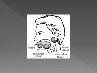
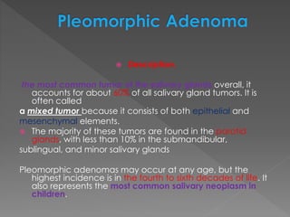

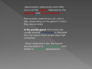
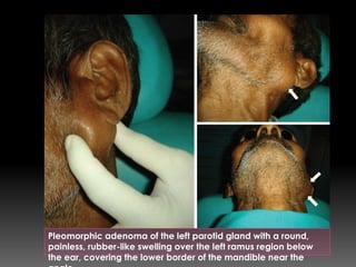
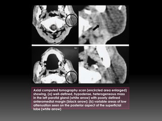


Pleomorphic adenoma is the most common salivary gland tumor, accounting for 60% of cases. It occurs most often in the parotid glands and presents as a painless, firm mass. Histologically, it contains both epithelial and mesenchymal elements arranged in a trabecular pattern within a fibrous stroma. Treatment involves complete surgical removal of the tumor with adequate margins to prevent recurrence due to microscopic projections outside the capsule. Imaging such as CT or MRI is used to identify the location and characteristics of the tumor prior to surgery.


