Sialolithiasis
Introduction
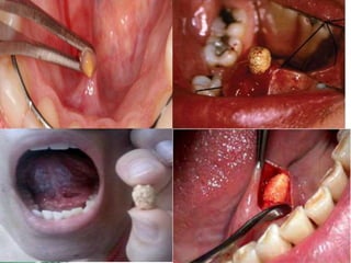
• Sialolithiasis is the presence of calculi in the
salivary glands or ducts.
• Stones will form in the salivary gland or ducts
following the stagnation of saliva; they are
typically composed of calcium phosphate and
hydroxyapatite, as the saliva is rich in calcium
Risk Factors
• Medication, commonly diuretics or anti
cholinergics
• Dehydration
• Gout
• Smoking
• Chronic periodontal disease
• Hyperparathyroidism
Clinical Features
• asymptomatic,
• intermittent facial swelling associated with
eating, which can be painful or painless.
• Symptoms are usually unilateral in nature.
• When the gland is palpated, saliva can be seen
at the duct orifice, along with the presence of
small stones.
• On palpation, a stone may be palpable in the
duct and the gland may feel tender in the
presence of infection.
Investigations
• Most cases of suspected sialolithiasis are
investigated* with either ultrasound or
radiographs.
Management
• Most patients are managed conservatively
with oral hydration, analgesia, and
sialologues, such as lemon juice, which
promote saliva production. Milking /
massaging the gland can help as well.
• If the gland becomes infected and the patient
develops sialedenitis, then antibiotics are
typically indicated.
Definitive Management
• Interventional radiology procedures are most
commonly trialled, which involve fluoroscopic
control such that the stones are visualised in
the duct and then extracted with a basket.
• A surgical approach can be used to remove
some more difficult stones; a transoral
approach can be used if the stones are distal
or a transcervical approach for proximal
stones (or where the transoral approach has
been unsuccessful).
• Gland removal is last resort. Excision of the
parotid or submandibular gland are only
performed for patients with chronically
persisting symptoms.

Sialolithiasis

  • 1.
  • 2.
    Introduction • Sialolithiasis isthe presence of calculi in the salivary glands or ducts.
  • 3.
    • Stones willform in the salivary gland or ducts following the stagnation of saliva; they are typically composed of calcium phosphate and hydroxyapatite, as the saliva is rich in calcium
  • 4.
    Risk Factors • Medication,commonly diuretics or anti cholinergics • Dehydration • Gout • Smoking • Chronic periodontal disease • Hyperparathyroidism
  • 5.
    Clinical Features • asymptomatic, •intermittent facial swelling associated with eating, which can be painful or painless. • Symptoms are usually unilateral in nature. • When the gland is palpated, saliva can be seen at the duct orifice, along with the presence of small stones. • On palpation, a stone may be palpable in the duct and the gland may feel tender in the presence of infection.
  • 7.
    Investigations • Most casesof suspected sialolithiasis are investigated* with either ultrasound or radiographs.
  • 8.
    Management • Most patientsare managed conservatively with oral hydration, analgesia, and sialologues, such as lemon juice, which promote saliva production. Milking / massaging the gland can help as well. • If the gland becomes infected and the patient develops sialedenitis, then antibiotics are typically indicated.
  • 9.
    Definitive Management • Interventionalradiology procedures are most commonly trialled, which involve fluoroscopic control such that the stones are visualised in the duct and then extracted with a basket.
  • 10.
    • A surgicalapproach can be used to remove some more difficult stones; a transoral approach can be used if the stones are distal or a transcervical approach for proximal stones (or where the transoral approach has been unsuccessful).
  • 11.
    • Gland removalis last resort. Excision of the parotid or submandibular gland are only performed for patients with chronically persisting symptoms.