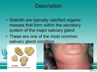
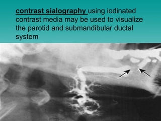
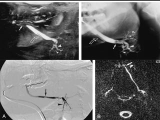
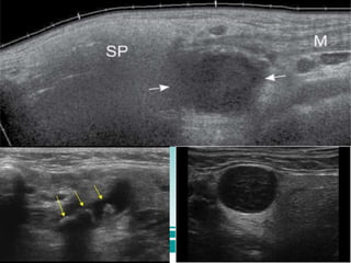
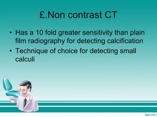
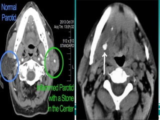
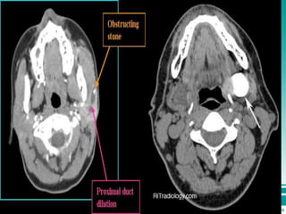
Sialolithiasis
Presented by:- Ayam Chhatkuli
Guided by:- Dr. Manish sir
Description
• Sialolith are typically calcified organic
masses that form within the secretory
system of the major salivary gland
• These are one of the most common
salivary gland condition
Etiology
• Believed to develop from deposition of the
calcium salts around a focus of material
within the duct lumen
• Initiating focus may consist of:-
 Desquamated epithelial cells
 Bacteria
 Foreign bodies or mucus
The etiological factors favouring salivary
stone formation may be classified into two
groups:-
i. Factors favouring saliva retention and
ii. Saliva composition
i. Factors favouring saliva retention
 Irregularities in the duct system
 Local inflammation
 Dehydration
 Medications such as anticholinergics and
diuretics
ii. Saliva composition
 calcium saturation and deficit of
crystallization inhibitors such as phytate
 bacterial infection increases salivary pH
favouring calcium phosphate supersaturation
promoting sialolith formation
 smoking affect the cytotoxic activity of partial
duct obstruction
Clinical features
Age:- wide age range, has been reported in
children also
Sex:- more common in males
Site:- most common in submandibular
gland(80-90%), followed by parotid (5-
15%) and sublingual (2%-5%)
Very rarely occurs in minor salivary gland
Presentation
 Pain
 acute, colicky, periprandial
 intermittent swelling of the affected gland
 Swelling will be evident upon eating
 If only partially obstructed, swelling will
subside when salivary stimulation ceases
 stasis of saliva may lead to infection, fibrosis
and gland atrophy
 gland is often enlarged and tender to
palpation
 fistula, a sinus tract or ulceration may occur
in the tissue covering the stone in chronic
cases
Other complications include acute
sialadenitis, ductal stricture and ductal
dilation
Diagnosis
₴. Plain film radiographs
• inexpensive, readily available, and result
in minimal radiation exposure
₴. AP view of face
• Useful incase of stones in the parotid
gland
• An occusal film placed intraorally adjacent
to the duct may also be useful
£. Conventional sialography
using panoramic occlusal, and panoramic
radiography is useful in cases where there
is a strong clinical suspicion of
inflammation or salivary stone disease
contrast sialography using iodinated
contrast media may be used to visualize
the parotid and submandibular ductal
system
Limitation
o use of ionizing radiation
o dependence on successful ductal
cannulation
o pain during and after the procedure
o potential allergy to the contrast media
Contra indicated in presence of acute
sialadenitis.
₴.Ultrasound
Non invasive, less costly and maybe able to
visualize radiolucent calculi
Limitation
o May not be able for correct assessment of
the number of calculi
o Calculi less than 2mm may not produce an
acoustic shadow
£.Non contrast CT
• Has a 10 fold greater sensitivity than plain
film radiography for detecting calcification
• Technique of choice for detecting small
calculi
£. CBCT
• Advantage of reduced superimposition
and distortion of anatomical structure
• Reduced radiation exposure over medical
CT
£. MRI sialography
It is contraindicated in individuals with
pacemakers or claustrophobia
£. Sialendoscopy
• Access to deeper segment of duct
• Simultaneous visualization and removal of
sialolith
Treatment
• During acute phase of sialolithiasis,
therapy is primarily supportive
• Use of analgesics, hydration, antibiotics
and antipyretics as necessary
• Sialogogues, massage and heat applied to
the affected area may also be useful
Stones at or near the orifice
 Can be removed transorally by milking the
gland
Deeper stones
 Conventional surgery or sialoendoscopy
Interventional sialoendoscopy
 effective even in cases of multiple stones
upto 4-5 mm in diameter
Extracorporeal shock wave
lithotripsy(ESWL)
 allows for fragmantation of large sialolith
of any size or location
Contraindicated in cases of
 complete duct stenosis
 Pregnancy
 Patient with cardiac pacemaker
Sialoadenectomy
Incase of fixed intraparenchymal stones and
failing gland-sparing techniques
Why is there
greater chance of
sialolith formation
in submandibular
gland?
1. The torturous course of whatsons duct
2. The higher calcium and phosphate levels
of secretion contained within
3. The dependent position of the
submandibular glands that leaves them
prone to stasis
4. The increased mucoid nature of secretion
In radiograph
Once the radiopacity is established it must be
distinguished from several entities
A calcified lymph node
An embedded tooth
A foreign body
A phlebolith
Calcification in the facial artery
Myositis osificans
An anatomic stricture( such as hyoid bone)
Sialolithiasis (exam oriented presentation)

Sialolithiasis (exam oriented presentation)

  • 1.
    Sialolithiasis Presented by:- AyamChhatkuli Guided by:- Dr. Manish sir
  • 2.
    Description • Sialolith aretypically calcified organic masses that form within the secretory system of the major salivary gland • These are one of the most common salivary gland condition
  • 3.
    Etiology • Believed todevelop from deposition of the calcium salts around a focus of material within the duct lumen • Initiating focus may consist of:-  Desquamated epithelial cells  Bacteria  Foreign bodies or mucus
  • 4.
    The etiological factorsfavouring salivary stone formation may be classified into two groups:- i. Factors favouring saliva retention and ii. Saliva composition
  • 5.
    i. Factors favouringsaliva retention  Irregularities in the duct system  Local inflammation  Dehydration  Medications such as anticholinergics and diuretics
  • 6.
    ii. Saliva composition calcium saturation and deficit of crystallization inhibitors such as phytate  bacterial infection increases salivary pH favouring calcium phosphate supersaturation promoting sialolith formation  smoking affect the cytotoxic activity of partial duct obstruction
  • 7.
    Clinical features Age:- wideage range, has been reported in children also Sex:- more common in males Site:- most common in submandibular gland(80-90%), followed by parotid (5- 15%) and sublingual (2%-5%) Very rarely occurs in minor salivary gland
  • 8.
    Presentation  Pain  acute,colicky, periprandial  intermittent swelling of the affected gland  Swelling will be evident upon eating  If only partially obstructed, swelling will subside when salivary stimulation ceases
  • 9.
     stasis ofsaliva may lead to infection, fibrosis and gland atrophy  gland is often enlarged and tender to palpation  fistula, a sinus tract or ulceration may occur in the tissue covering the stone in chronic cases Other complications include acute sialadenitis, ductal stricture and ductal dilation
  • 10.
  • 11.
    ₴. Plain filmradiographs • inexpensive, readily available, and result in minimal radiation exposure
  • 12.
    ₴. AP viewof face • Useful incase of stones in the parotid gland • An occusal film placed intraorally adjacent to the duct may also be useful
  • 13.
    £. Conventional sialography usingpanoramic occlusal, and panoramic radiography is useful in cases where there is a strong clinical suspicion of inflammation or salivary stone disease
  • 14.
    contrast sialography usingiodinated contrast media may be used to visualize the parotid and submandibular ductal system
  • 16.
    Limitation o use ofionizing radiation o dependence on successful ductal cannulation o pain during and after the procedure o potential allergy to the contrast media Contra indicated in presence of acute sialadenitis.
  • 17.
    ₴.Ultrasound Non invasive, lesscostly and maybe able to visualize radiolucent calculi Limitation o May not be able for correct assessment of the number of calculi o Calculi less than 2mm may not produce an acoustic shadow
  • 19.
    £.Non contrast CT •Has a 10 fold greater sensitivity than plain film radiography for detecting calcification • Technique of choice for detecting small calculi
  • 22.
    £. CBCT • Advantageof reduced superimposition and distortion of anatomical structure • Reduced radiation exposure over medical CT
  • 23.
    £. MRI sialography Itis contraindicated in individuals with pacemakers or claustrophobia
  • 24.
    £. Sialendoscopy • Accessto deeper segment of duct • Simultaneous visualization and removal of sialolith
  • 26.
    Treatment • During acutephase of sialolithiasis, therapy is primarily supportive • Use of analgesics, hydration, antibiotics and antipyretics as necessary • Sialogogues, massage and heat applied to the affected area may also be useful
  • 27.
    Stones at ornear the orifice  Can be removed transorally by milking the gland Deeper stones  Conventional surgery or sialoendoscopy
  • 28.
    Interventional sialoendoscopy  effectiveeven in cases of multiple stones upto 4-5 mm in diameter
  • 29.
    Extracorporeal shock wave lithotripsy(ESWL) allows for fragmantation of large sialolith of any size or location Contraindicated in cases of  complete duct stenosis  Pregnancy  Patient with cardiac pacemaker
  • 30.
    Sialoadenectomy Incase of fixedintraparenchymal stones and failing gland-sparing techniques
  • 31.
    Why is there greaterchance of sialolith formation in submandibular gland?
  • 32.
    1. The torturouscourse of whatsons duct 2. The higher calcium and phosphate levels of secretion contained within 3. The dependent position of the submandibular glands that leaves them prone to stasis 4. The increased mucoid nature of secretion
  • 33.
    In radiograph Once theradiopacity is established it must be distinguished from several entities A calcified lymph node An embedded tooth A foreign body A phlebolith Calcification in the facial artery Myositis osificans An anatomic stricture( such as hyoid bone)