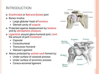
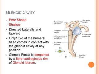
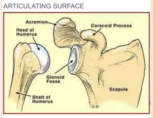
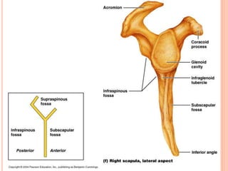
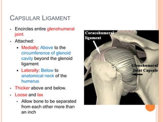
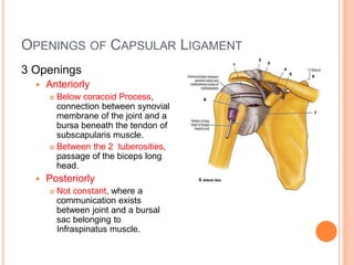
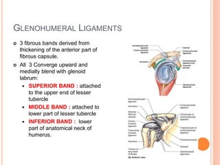
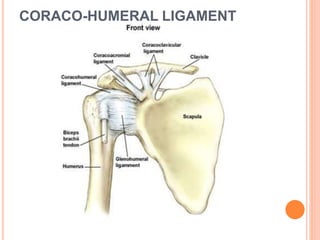
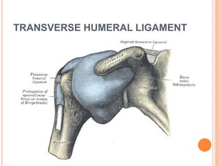
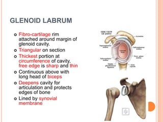
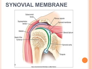
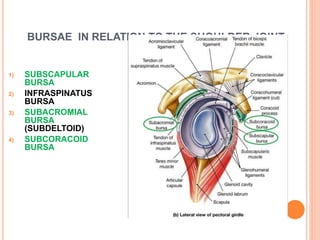
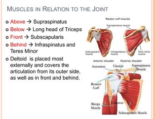
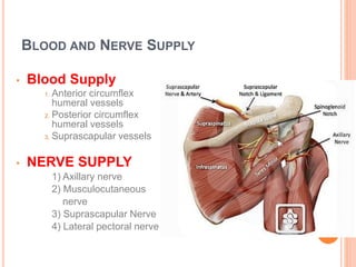
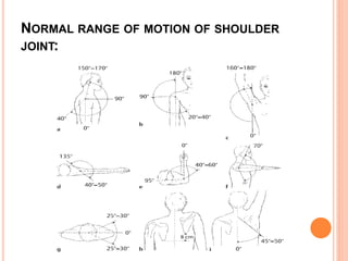
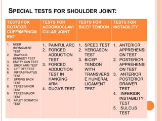
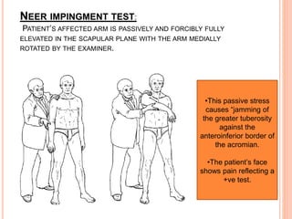
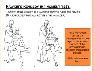
SHOULDER ANATOMY AND TESTS
INTRODUCTION
 Enarthrodial or Ball-and-Socket joint
 Bones Involve
 Large globular head of humerus
 Glenoid cavity of scapula
 Protected against displacement by tendons
and by atmospheric pressure.
 Ligaments around gleno-humeral joint; Limit
the amount of joint movement
 Capsular
 Coracohumeral
 Transverse Humeral
 Glenoid Ligament
 Above protected by arched vault formed by:
 Under surface of coracoid process
 Under surface of acromion process
 Coraco-acromial ligament
GLENOID CAVITY
• Pear Shape
• Shallow
• Directed Laterally and
Upward
• Only1/3rd of the humeral
head comes in contact with
the glenoid cavity at any
position.
• Glenoid Fossa is deepened
by a fibro-cartilaginous rim
of Glenoid labrum.
ARTICULATING SURFACE
CAPSULAR LIGAMENT
• Encircles entire glenohumeral
joint.
• Attached:
 Medially: Above to the
circumference of glenoid
cavity beyond the glenoid
ligament
 Laterally: Below to
anatomical neck of the
humerus
• Thicker above and below.
• Loose and lax
• Allow bone to be separated
from each other more than
an inch
MUSCLES SUPPORTING CAPSULAR LIGAMENT
 Superiorly 
Supraspinatus
 Inferiorly  Long Head of
Triceps
 Posteriorly  Tendons of
Infraspinatus and Teres
Minor
 Anteriorly  Tendon of
Subscapularis
OPENINGS OF CAPSULAR LIGAMENT
3 Openings
 Anteriorly
 Below coracoid Process,
connection between synovial
membrane of the joint and a
bursa beneath the tendon of
subscapularis muscle.
 Between the 2 tuberosities,
passage of the biceps long
head.
 Posteriorly
 Not constant, where a
communication exists
between joint and a bursal
sac belonging to
Infraspinatus muscle.
GLENOHUMERAL LIGAMENTS
 3 fibrous bands derived from
thickening of the anterior part of
fibrous capsule.
 All 3 Converge upward and
medially blend with glenoid
labrum:
 SUPERIOR BAND : attached
to the upper end of lesser
tubercle
 MIDDLE BAND : attached to
lower part of lesser tubercle
 INFERIOR BAND : lower
part of anatomical neck of
humerus.
CORACO-HUMERAL LIGAMENT
TRANSVERSE HUMERAL LIGAMENT
GLENOID LABRUM
 Fibro-cartilage rim
attached around margin of
glenoid cavity.
 Triangular on section
 Thickest portion at
circumference of cavity,
free edge is sharp and thin
 Continuous above with
long head of biceps
 Deepens cavity for
articulation and protects
edges of bone
 Lined by synovial
membrane
SYNOVIAL MEMBRANE
BURSAE IN RELATION TO THE SHOULDER JOINT
1) SUBSCAPULAR
BURSA
2) INFRASPINATUS
BURSA
3) SUBACROMIAL
BURSA
(SUBDELTOID)
4) SUBCORACOID
BURSA
MUSCLES IN RELATION TO THE JOINT
 Above  Supraspinatus
 Below  Long head of Triceps
 Front  Subscapularis
 Behind  Infraspinatus and
Teres Minor
 Deltoid is placed most
externally and covers the
articulation from its outer side,
as well as in front and behind.
BLOOD AND NERVE SUPPLY
• Blood Supply
1. Anterior circumflex
humeral vessels
2. Posterior circumflex
humeral vessels
3. Suprascapular vessels
• NERVE SUPPLY
1) Axillary nerve
2) Musculocutaneous
nerve
3) Suprascapular Nerve
4) Lateral pectoral nerve
SPECIAL TESTS OF SHOULDER JOINT
NORMAL RANGE OF MOTION OF SHOULDER
JOINT:
SPECIAL TESTS FOR SHOULDER JOINT:
TESTS FOR
ROTATOR
CUFF/IMPINGM
ENT
TESTS FOR
ACROMIOCLAVI
CULAR JOINT
TESTS FOR
BICEP TENDON
TESTS FOR
INSTABILITY
1. NEER
IMPINGMENT
TEST
2. HAWKINS
KENNEDY TEST
3. EMPTY CAN TEST
4. DROP ARM TEST
5. LIFT OFF.TEST
6. INFRASPINATUS
TEST
7. SPRING BACK
TEST
8. TERES MINOR
TEST
9. TERES MAJOR
TEST
10. APLEY SCRATCH
TEST
1. PAINFUL ARC
2. FORCED
ADDUCTION
TEST
3. FORCED
ADDUCTION
TEST IN
HANGING
ARM
4. DUGA’S TEST
1. SPEED TEST
2. YERGASON
TEST
3. BICEP
TENDON
WITH
TRANSVERS
E HUMERAL
LIGAMENT
TEST
1. ANTERIOR
APPREHENSI
ON TEST
2. POSTERIOR
APPREHENSI
ON TEST
3. ANTERIOR
POSTERIOR
DRAWER
TEST
4. INFERIOR
INSTABILITY
TEST
5. SULCUS
TEST
TESTS FOR ROTATOR CUFF
AND IMPINGMENT SYNDROME
IMPINGEMENT:
Primary impingment Secondary impingment
Occur because of degenerative
changes to the rotator cuff,the
acromian process,the coracoid
process and anterior tissues from
stress overload.
Occurs due to problem with
muscle dynamics with an upset in
the normal force couple action
leading to muscle imbalance and
abnormal movement patterns at
both the glenohumeral joint and
the scapulothoracic articulation.
Impingement is primary cause of
pain.
It is secondary to altered muscle
dynamics.
Occurs mostly in 40+ age group
people.
Occurs in young patients.(15-
35years old)
It is said to be intrinsic when
rotator cuff degeneration occurs
and extrinsic when the shape of
the acromian and degeneration of
the coracoacromial ligament
occurs.
Commonly seen with joint
instability.
GRADING OF IMPINGEMET:
 Mostly impingement and instability often occurs
together in throwing athletes and accordingly it is
classified as:
GRADE I:
Pure
impingement
with no
instability.(ofte
n seen in older
patients)
GRADE II:
Secondary
impingment
and instability
caused by
chronic
capsular and
labral
microtrauma.
GRADE III:
Secondary
impingement
and instability
caused by
generalized
hypermobility
or laxity.
GRADE IV:
Primary
instability with
no
impingement.
NEER IMPINGMENT TEST:
PATIENT’S AFFECTED ARM IS PASSIVELY AND FORCIBLY FULLY
ELEVATED IN THE SCAPULAR PLANE WITH THE ARM MEDIALLY
ROTATED BY THE EXAMINER.
•This passive stress
causes “jamming of
the greater tuberosity
against the
anteroinferior border of
the acromian.
•The patient’s face
shows pain reflecting a
+ve test.
HAWKIN’S KENNEDY IMPINGMENT TEST:
PATIENT STAND WHILE THE EXAMINER FORWARD FLEXS THE ARM TO
90º AND FORCIBLY MEDIALLY ROTATES THE SHOULDER.
•This movement
pushes the
supraspinatus tendon
against the anterior
surface of the
coracoacromial
ligament and coracoid
process.
•Pain indicates +ve
test.
SUPRASPINATUS TEST/EMPTY CAN TEST:
THIS TEST MAY BE PERFORMED WITH THE PATIENT STANDING OR
SEATED.WITH THE ELBOW EXTENDED, THE PATIENT’S ARM IS HELD AT
90° OF ABDUCTION,30° OF HORIZONTAL FLEXION, AND IN INTERNAL
ROTATION (WITH THUMB FACING DOWN). THE EXAMINER EXERTS
PRESSURE ON THE UPPER ARM DURING THE ABDUCTION AND
HORIZONTAL FLEXION MOTION.
•When this test elicits severe
pain and the patient is
unable to hold his or her arm
abducted 90° against gravity, this
is called a positive empty can
test/supraspinatus tendinitis.
•The superior portions of the
rotator cuff (supraspinatus) are
particularly assessed in internal
rotation (with the thumb down),
and the
•anterior portions in external
rotation.
DROP ARM(CODMAN’S)TEST:
THE PATIENT IS SEATED, AND THE EXAMINER PASSIVELY ABDUCTS THE
PATIENT’S EXTENDED ARM APPROXIMATELY 120°. THE PATIENT IS
ASKED TO HOLD THE ARM IN THIS POSITION WITHOUT SUPPORT AND
THEN SLOWLY ALLOW IT TO DROP.
Weakness in maintaining the position
of the arm, with or
without pain, or sudden dropping of
the arm suggests a rotator cuff
lesion. Most often this is due to a
defect in the supraspinatus. In
pseudoparalysis, the patient will be
unable to lift the affected arm. This
global sign suggests a rotator cuff
disorder.
SUBSCAPULARIS TEST/LIFT OFF TEST:
PATIENT IN STANDING POSITION PLACES THE DORSUM OF THE HAND
ON THE BACK. THE PATIENT THEN LIFTS THE HAND AWAY FROM THE
BACK. IF PATIENT IS ABLE TO DO THEN LOAD PUSHING ON HAND IS
DONE BY THE EXAMINER TO CHECK THE STRENGH.
•A patient with a subscapularis
tear will be unable to do
this.
•Abnormal motion in the scapula
during the test may indicate
scapular instability.
INFRASPINATUS TEST:
COMPARATIVE TESTING OF BOTH SIDES IS BEST. THE PATIENT’S
ARMS SHOULD HANG RELAXED WITH THE ELBOWS FLEXED 90° BUT
NOT QUITE TOUCHING THE TRUNK. THE EXAMINER PLACES HIS OR
HER PALMS ON THE DORSUM OF EACH OF THE PATIENT’S HANDS AND
THEN ASKS THE PATIENT TO EXTERNALLY ROTATE BOTH FOREARMS
AGAINST THE RESISTANCE OF THE EXAMINER’S HANDS.
Pain or weakness in external rotation
indicates a disorder of the infraspinatus
(external rotator).
As infraspinatus tears are usually
painless, weakness in rotation strongly
suggests a tear in the muscle.
This test can also be performed with
the arm abducted 90° and flexed
30° to eliminate involvement of the
deltoid in this motion.
 SPRING BACK TEST:
PATIENT EITHER IN SITTING OR STANDING HOLD THE
ELBOW IN FLEXION AT 90º BY THE SIDE. EXAMINER
PASSIVELY BRING THE SHOULDER TO 90º ABDUCTION
AND LATERALLY ROTATE TO THE END RANGE AND ASK
THE PATIENT TO HOLD THE ARM TO THIS POSITION.
FOR +VE TEST OF INFRASPINATUS WEAKNESS/LESION
PATIENT CANNOT HOLD THE POSITION AND HAND
SPRING BACK ANTERIORLY.
TERES MINOR TEST:
PATIENT LIES PRONE AND PLACES HIS HAND ON THE
OPPOSITE POSTERIOR ILIAC CREST. ASK THE PATIENT
TO EXTEND AND ADDUCT THE MEDIALLY ROTATED ARM
AGAINST RESISTANCE. PAIN OR WEAKNESS INDICATE
+VE TEST.
TERES MAJOR TEST:
THE PATIENT IS STANDING AND RELAXED. THE EXAMINER ASSESSES
THE POSITION OF THE PATIENT’S HANDS FROM BEHIND. THE TERES
MAJOR IS AN INTERNAL ROTATOR. WHERE A CONTRACTURE IS
PRESENT, THE PALM OF THE AFFECTED HAND WILL FACE BACKWARD
COMPARED WITH THE CONTRALATERAL HAND.
APLEY’S SCRTCH TEST:
THE SEATED PATIENT IS ASKED TO TOUCH THE CONTRALATERAL
SUPERIOR MEDIAL CORNER OF THE SCAPULA WITH THE INDEX
FINGER.
Pain elicited in the rotator cuff and failure to
reach the scapula because of restricted
mobility in external rotation and abduction
indicate rotator cuff pathology (most probably
involving the supraspinatus).
ACROMIOCLAVICULAR JOINT
TESTS
TOSSY CLASSIFICATION:
 TOSSY TYPE 1: CONTUSION OF THE
ACROMIOCLAVICULAR JOINT WITHOUT
SIGNIFICANT INJURY TO THE CAPSULE AND
LIGAMENTS.
 TOSSY TYPE 2: SUBLUXATION OF THE
ACROMIOCLAVICULAR JOINT WITH RUPTURE OF
THE ACROMIOCLAVICULAR LIGAMENTS.
 TOSSY TYPE 3: DISLOCATION OF THE
ACROMIOCLAVICULAR JOINTWITH ADDITIONAL
RUPTURE OF THE CORACOCLAVICULAR
LIGAMENTS.
ACROMIOCLAVICULAR JOINT PROBLEM
 MAY BE ELICITED BY ANTERIOR PAIN WITH MOTION AND
TENDERNESS TO PALPATION OVER THE
ACROMIOCLAVICULAR JOINT.

FINDINGS WILL OFTEN INCLUDE PALPABLE BONY THICKENING
OF THE ARTICULAR MARGIN.

TOSSY CLASSIFIES ACROMIOCLAVICULAR JOINT INJURIES
INTO THREE DEGREES OF SEVERITY:
PAINFUL ARC:
THE PATIENT’S ARM IS PASSIVELY AND ACTIVELY ABDUCTED FROM THE
REST POSITION ALONGSIDE THE TRUNK. PAIN IN THE
ACROMIOCLAVICULAR JOINT OCCURS BETWEEN 140°AND 180° OF
ABDUCTION. INCREASING ABDUCTION LEADS TO INCREASING COM-
PRESSION AND CONTORTION IN THE JOINT. (IN AN IMPINGEMENT
SYNDROME OR A ROTATOR CUFF TEAR, BY COMPARISON, PAIN
SYMPTOMS WILL OCCUR BETWEEN 70°
AND 120°.
In the evaluation of the active
and passive ranges of motion,
the patient can often avoid the
painful arc by externally rotating
the arm while abducting it. This
increases the clearance
between the acromion and the
diseased tendinous portion of
the rotator cuff, avoiding
impingement in the range
between 70° and 120°.
FORCED ADDUCTION TEST:
THE 90°-ABDUCTED ARM ON THE AFFECTED SIDE IS FORCIBLY
ADDUCTED ACROSS THE CHEST TOWARD THE NORMAL SIDE.
FORCED ADDUCTION TEST ON HANGING ARM:
THE EXAMINER GRASPS THE UPPER ARM OF THE AFFECTED SIDE
WITH ONE HAND WHILE THE OTHER HAND RESTS ON THE CONTRALATERAL
SHOULDER AND IMMOBILIZES THE SHOULDER GIRDLE. THEN THE EXAMINER
FORCIBLY ADDUCTS THE HANGING AFFECTED ARM BEHIND THE PATIENT’S
BACK AGAINST THE PATIENT’S RESISTANCE.
Pain across the anterior
aspect of the shoulder
suggests
acromioclavicular joint
disease or subacromial
impingement.
DUGA’S TEST:
THE PATIENT IS SEATED OR STANDING AND TOUCHES THE
CONTRALATERAL SHOULDER WITH THE HAND OF THE 90°-
FLEXED ARM OF THE AFFECTED SIDE THEN ATTEMPT TO LOWER
THE ELBOW TO THE CHEST IS MADE.
Acromioclavicular joint pain
suggests joint disease
(osteoarthritis,
instability, disk injury, or
infection).
A differential diagnosis
must exclude anterior
subacromial impingement
BICEP TENDON TEST
THE CLOSE ANATOMIC PROXIMITY OF THE
INTRAARTICULAR PORTION OF THE TENDON
TO THE CORACOACROMIAL ARCH
PREDISPOSES IT TO INVOLVEMENT IN
DEGENERATIVE PROCESSES IN THE
SUBACROMIAL SPACE. A ROTATOR CUFF TEAR
IS OFTEN ACCOMPANIED BY A RUPTURE OR
INJURIES OF THE BICEPS TENDON.
SPEED TEST:
IN SITTING THE EXAMINER RESISTS SHOULDER FORWARD
FLEXION BY THE PATIENT WHILE THE PATIENT’S FOREARM IS IN
SUPINATION. PAIN IN THE REGION OF THE BICIPITAL GROOVE
SUGGESTS A DISORDER OF THE LONG HEAD OF THE BICEPS
TENDON.
YERGASON TEST:
WITH THE PATIENT’S ELBOW FLEXED TO 90º AND STABILIZED AGAINST
THORAX AND WITH FOREARM PRONATED, THE EXAMINER RESISTS
SUPINATION WHILE THE PATIENT ALSO LATERALLY ROTATES THE ARM
AGAINST RESISTANCE. DURING THIS MOVEMENT WHEN THE TENDON
IS FELT IN GROOVE AS “POP OUT” .
•Pain in the bicipital groove is a sign of
a lesion of the biceps tendon, its tendon
sheath, or its ligamentous connection
via the
•transverse ligament.
•The typical provoked pain can be
increased by pressing on the tendon in
the bicipital groove.
BICEP TENDINITIS WITH TRANSVERSE HUMERAL
LIGAMENT TEST:
THE PATIENT IS SEATED WITH THE ARM ABDUCTED 90°, INTERNALLY
ROTATED, AND EXTENDED AT THE ELBOW. FROM THIS POSITION, THE
EXAMINER EXTERNALLY ROTATES THE ARM WHILE PALPATING THE
BICIPITAL GROOVE TO VERIFY WHETHER THE TENDON SNAPS.
•In the presence of
ligamentous insufficiency, this
motion will cause the biceps
tendon to spontaneously
displace out of the bicipital
groove.
•Pain reported without
displacement suggests biceps
•tendinitis.
INSTABILITY TESTS
SHOULDER PAIN MAY BE ATTRIBUTABLE TO AN
UNSTABLE SHOULDER. USUALLY HISTORY OF A PERIOD
OF INTENSIVE SHOULDER USE (SUCH AS COMPETITIVE
SPORTS), AN EPISODE OF REPEATED MINOR TRAUMA
(OVERHEAD USE), OR GENERALIZED LIGAMENT LAXITY.
BOTH YOUNG ATHLETES AND INACTIVE PERSONS ARE
AFFECTED, MEN AND WOMEN ALIKE.
ANTERIOR APPREHENSION TEST:
PATIENT LIE SUPINE OR IN SITTING . ARM IS ABDUCTED TO 90º
AND LATERALLY ROTATED SLOWLY BY THE EXAMINER. WHILE
PERFORMING PATIENT’S EXPRESSIONS ARE NOTED FOR
APPREHENSION/FURTHER RESISTENCE TO ROTATION. THE
TEST IS PERFORMED AT 60°, 90°, AND 120° OF ABDUCTION TO
EVALUATE THE SUPERIOR, MEDIAL, AND INFERIOR
GLENOHUMERAL LIGAMENTS. WITH THE GUIDING HAND, THE
EXAMINER PRESSES THE HUMERAL HEAD IN AN ANTERIOR AND
INFERIOR DIRECTION
Shoulder pain with reflexive
muscle tensing is a sign of an
anterior instability syndrome. This
muscle tension is an attempt by
the patient to prevent imminent
subluxation or dislocation of the
humeral
head.
NOTE:
When the patient complains of sudden stabbing pain
with simultaneous or subsequent paralyzing
weakness in the affected extremity, this is referred
to as the “dead arm sign.” It is attributable to the
transient compression the subluxated humeral head
exerts on the plexus.
 It is important to know that at 45° of abduction, the
test primarily evaluates the medial glenohumeral
ligament and the subscapularis tendon. At or above
90° of abduction, the stabilizing effect of the
subscapularis is neutralized and the test primarily
evaluates the inferior glenohumeral ligament.
POSTERIOR APPREHENSION TEST:
PATIENT LIES SUPINE OR IN SITTING POSITION AND EXAMINER
FORWARD FLEX SHOULDER TO 90º WHILE STABILIZING THE SCAPULA
WITH OTHER HAND. EXAMINER THEN APPLIES A POSTERIOR FORCE
ON THE ELBOW AND MOVES THE ARM IN ADDUCTION AND MEDIALLY
ROTATION.
ANTERIOR AND POSTERIOR DRAWER TEST:
THE PATIENT IS SEATED. THE EXAMINER STANDS BEHIND THE PATIENT.
TO EVALUATE THE RIGHT SHOULDER, THE EXAMINER GRASPS THE
PATIENT’S SHOULDER WITH THE LEFT HAND TO STABILIZE THE
CLAVICLE AND SUPERIOR MARGIN OF THE SCAPULA WHILE USING THE
RIGHT HAND TO MOVE THE HUMERAL HEAD ANTERIORLY AND
POSTERIORLY.
INFERIOR APPREHENSION TEST/FEAGIN TEST:
PATIENT STANDS WITH THE ARM ABDUCTED TO 90º AND ELBOW
EXTENDED AND RESTING ON TOP OF THE EXAMINER’S SHOULDER.
EXAMINER CLASP HIS/HER HANDS AROUND THE PATIENT’S HUMERUS
AND PUSHES THE HUMERUS DOWN AND FORWARD. IN THIS SULCUS
MAY ALSO BE SEEN ABOVE THE CORACOID PROCESS.
SULCUS TEST:
PATIENT STANDS WITH ARM BY THE SIDE AND SHOULDER
MUSCLE RELAXED. THE EXAMINER GRASPS THE PATIENT’S
FOREARM BELOW THE ELBOW AND PULLS THE ARM DISTALLY.
THE PRESENCE OF SULCUS/INDENTATION INFERIOR TO
ACROMIAN IS THE INDICATIVE.
THANK YOU

Shoulder anatomy and examination

  • 1.
  • 2.
    INTRODUCTION  Enarthrodial orBall-and-Socket joint  Bones Involve  Large globular head of humerus  Glenoid cavity of scapula  Protected against displacement by tendons and by atmospheric pressure.  Ligaments around gleno-humeral joint; Limit the amount of joint movement  Capsular  Coracohumeral  Transverse Humeral  Glenoid Ligament  Above protected by arched vault formed by:  Under surface of coracoid process  Under surface of acromion process  Coraco-acromial ligament
  • 3.
    GLENOID CAVITY • PearShape • Shallow • Directed Laterally and Upward • Only1/3rd of the humeral head comes in contact with the glenoid cavity at any position. • Glenoid Fossa is deepened by a fibro-cartilaginous rim of Glenoid labrum.
  • 4.
  • 6.
    CAPSULAR LIGAMENT • Encirclesentire glenohumeral joint. • Attached:  Medially: Above to the circumference of glenoid cavity beyond the glenoid ligament  Laterally: Below to anatomical neck of the humerus • Thicker above and below. • Loose and lax • Allow bone to be separated from each other more than an inch
  • 7.
    MUSCLES SUPPORTING CAPSULARLIGAMENT  Superiorly  Supraspinatus  Inferiorly  Long Head of Triceps  Posteriorly  Tendons of Infraspinatus and Teres Minor  Anteriorly  Tendon of Subscapularis
  • 8.
    OPENINGS OF CAPSULARLIGAMENT 3 Openings  Anteriorly  Below coracoid Process, connection between synovial membrane of the joint and a bursa beneath the tendon of subscapularis muscle.  Between the 2 tuberosities, passage of the biceps long head.  Posteriorly  Not constant, where a communication exists between joint and a bursal sac belonging to Infraspinatus muscle.
  • 9.
    GLENOHUMERAL LIGAMENTS  3fibrous bands derived from thickening of the anterior part of fibrous capsule.  All 3 Converge upward and medially blend with glenoid labrum:  SUPERIOR BAND : attached to the upper end of lesser tubercle  MIDDLE BAND : attached to lower part of lesser tubercle  INFERIOR BAND : lower part of anatomical neck of humerus.
  • 10.
  • 11.
  • 12.
    GLENOID LABRUM  Fibro-cartilagerim attached around margin of glenoid cavity.  Triangular on section  Thickest portion at circumference of cavity, free edge is sharp and thin  Continuous above with long head of biceps  Deepens cavity for articulation and protects edges of bone  Lined by synovial membrane
  • 13.
  • 14.
    BURSAE IN RELATIONTO THE SHOULDER JOINT 1) SUBSCAPULAR BURSA 2) INFRASPINATUS BURSA 3) SUBACROMIAL BURSA (SUBDELTOID) 4) SUBCORACOID BURSA
  • 15.
    MUSCLES IN RELATIONTO THE JOINT  Above  Supraspinatus  Below  Long head of Triceps  Front  Subscapularis  Behind  Infraspinatus and Teres Minor  Deltoid is placed most externally and covers the articulation from its outer side, as well as in front and behind.
  • 16.
    BLOOD AND NERVESUPPLY • Blood Supply 1. Anterior circumflex humeral vessels 2. Posterior circumflex humeral vessels 3. Suprascapular vessels • NERVE SUPPLY 1) Axillary nerve 2) Musculocutaneous nerve 3) Suprascapular Nerve 4) Lateral pectoral nerve
  • 17.
    SPECIAL TESTS OFSHOULDER JOINT
  • 18.
    NORMAL RANGE OFMOTION OF SHOULDER JOINT:
  • 19.
    SPECIAL TESTS FORSHOULDER JOINT: TESTS FOR ROTATOR CUFF/IMPINGM ENT TESTS FOR ACROMIOCLAVI CULAR JOINT TESTS FOR BICEP TENDON TESTS FOR INSTABILITY 1. NEER IMPINGMENT TEST 2. HAWKINS KENNEDY TEST 3. EMPTY CAN TEST 4. DROP ARM TEST 5. LIFT OFF.TEST 6. INFRASPINATUS TEST 7. SPRING BACK TEST 8. TERES MINOR TEST 9. TERES MAJOR TEST 10. APLEY SCRATCH TEST 1. PAINFUL ARC 2. FORCED ADDUCTION TEST 3. FORCED ADDUCTION TEST IN HANGING ARM 4. DUGA’S TEST 1. SPEED TEST 2. YERGASON TEST 3. BICEP TENDON WITH TRANSVERS E HUMERAL LIGAMENT TEST 1. ANTERIOR APPREHENSI ON TEST 2. POSTERIOR APPREHENSI ON TEST 3. ANTERIOR POSTERIOR DRAWER TEST 4. INFERIOR INSTABILITY TEST 5. SULCUS TEST
  • 20.
    TESTS FOR ROTATORCUFF AND IMPINGMENT SYNDROME
  • 21.
    IMPINGEMENT: Primary impingment Secondaryimpingment Occur because of degenerative changes to the rotator cuff,the acromian process,the coracoid process and anterior tissues from stress overload. Occurs due to problem with muscle dynamics with an upset in the normal force couple action leading to muscle imbalance and abnormal movement patterns at both the glenohumeral joint and the scapulothoracic articulation. Impingement is primary cause of pain. It is secondary to altered muscle dynamics. Occurs mostly in 40+ age group people. Occurs in young patients.(15- 35years old) It is said to be intrinsic when rotator cuff degeneration occurs and extrinsic when the shape of the acromian and degeneration of the coracoacromial ligament occurs. Commonly seen with joint instability.
  • 22.
    GRADING OF IMPINGEMET: Mostly impingement and instability often occurs together in throwing athletes and accordingly it is classified as: GRADE I: Pure impingement with no instability.(ofte n seen in older patients) GRADE II: Secondary impingment and instability caused by chronic capsular and labral microtrauma. GRADE III: Secondary impingement and instability caused by generalized hypermobility or laxity. GRADE IV: Primary instability with no impingement.
  • 23.
    NEER IMPINGMENT TEST: PATIENT’SAFFECTED ARM IS PASSIVELY AND FORCIBLY FULLY ELEVATED IN THE SCAPULAR PLANE WITH THE ARM MEDIALLY ROTATED BY THE EXAMINER. •This passive stress causes “jamming of the greater tuberosity against the anteroinferior border of the acromian. •The patient’s face shows pain reflecting a +ve test.
  • 24.
    HAWKIN’S KENNEDY IMPINGMENTTEST: PATIENT STAND WHILE THE EXAMINER FORWARD FLEXS THE ARM TO 90º AND FORCIBLY MEDIALLY ROTATES THE SHOULDER. •This movement pushes the supraspinatus tendon against the anterior surface of the coracoacromial ligament and coracoid process. •Pain indicates +ve test.
  • 25.
    SUPRASPINATUS TEST/EMPTY CANTEST: THIS TEST MAY BE PERFORMED WITH THE PATIENT STANDING OR SEATED.WITH THE ELBOW EXTENDED, THE PATIENT’S ARM IS HELD AT 90° OF ABDUCTION,30° OF HORIZONTAL FLEXION, AND IN INTERNAL ROTATION (WITH THUMB FACING DOWN). THE EXAMINER EXERTS PRESSURE ON THE UPPER ARM DURING THE ABDUCTION AND HORIZONTAL FLEXION MOTION. •When this test elicits severe pain and the patient is unable to hold his or her arm abducted 90° against gravity, this is called a positive empty can test/supraspinatus tendinitis. •The superior portions of the rotator cuff (supraspinatus) are particularly assessed in internal rotation (with the thumb down), and the •anterior portions in external rotation.
  • 26.
    DROP ARM(CODMAN’S)TEST: THE PATIENTIS SEATED, AND THE EXAMINER PASSIVELY ABDUCTS THE PATIENT’S EXTENDED ARM APPROXIMATELY 120°. THE PATIENT IS ASKED TO HOLD THE ARM IN THIS POSITION WITHOUT SUPPORT AND THEN SLOWLY ALLOW IT TO DROP. Weakness in maintaining the position of the arm, with or without pain, or sudden dropping of the arm suggests a rotator cuff lesion. Most often this is due to a defect in the supraspinatus. In pseudoparalysis, the patient will be unable to lift the affected arm. This global sign suggests a rotator cuff disorder.
  • 27.
    SUBSCAPULARIS TEST/LIFT OFFTEST: PATIENT IN STANDING POSITION PLACES THE DORSUM OF THE HAND ON THE BACK. THE PATIENT THEN LIFTS THE HAND AWAY FROM THE BACK. IF PATIENT IS ABLE TO DO THEN LOAD PUSHING ON HAND IS DONE BY THE EXAMINER TO CHECK THE STRENGH. •A patient with a subscapularis tear will be unable to do this. •Abnormal motion in the scapula during the test may indicate scapular instability.
  • 28.
    INFRASPINATUS TEST: COMPARATIVE TESTINGOF BOTH SIDES IS BEST. THE PATIENT’S ARMS SHOULD HANG RELAXED WITH THE ELBOWS FLEXED 90° BUT NOT QUITE TOUCHING THE TRUNK. THE EXAMINER PLACES HIS OR HER PALMS ON THE DORSUM OF EACH OF THE PATIENT’S HANDS AND THEN ASKS THE PATIENT TO EXTERNALLY ROTATE BOTH FOREARMS AGAINST THE RESISTANCE OF THE EXAMINER’S HANDS. Pain or weakness in external rotation indicates a disorder of the infraspinatus (external rotator). As infraspinatus tears are usually painless, weakness in rotation strongly suggests a tear in the muscle. This test can also be performed with the arm abducted 90° and flexed 30° to eliminate involvement of the deltoid in this motion.
  • 29.
     SPRING BACKTEST: PATIENT EITHER IN SITTING OR STANDING HOLD THE ELBOW IN FLEXION AT 90º BY THE SIDE. EXAMINER PASSIVELY BRING THE SHOULDER TO 90º ABDUCTION AND LATERALLY ROTATE TO THE END RANGE AND ASK THE PATIENT TO HOLD THE ARM TO THIS POSITION. FOR +VE TEST OF INFRASPINATUS WEAKNESS/LESION PATIENT CANNOT HOLD THE POSITION AND HAND SPRING BACK ANTERIORLY. TERES MINOR TEST: PATIENT LIES PRONE AND PLACES HIS HAND ON THE OPPOSITE POSTERIOR ILIAC CREST. ASK THE PATIENT TO EXTEND AND ADDUCT THE MEDIALLY ROTATED ARM AGAINST RESISTANCE. PAIN OR WEAKNESS INDICATE +VE TEST.
  • 30.
    TERES MAJOR TEST: THEPATIENT IS STANDING AND RELAXED. THE EXAMINER ASSESSES THE POSITION OF THE PATIENT’S HANDS FROM BEHIND. THE TERES MAJOR IS AN INTERNAL ROTATOR. WHERE A CONTRACTURE IS PRESENT, THE PALM OF THE AFFECTED HAND WILL FACE BACKWARD COMPARED WITH THE CONTRALATERAL HAND.
  • 31.
    APLEY’S SCRTCH TEST: THESEATED PATIENT IS ASKED TO TOUCH THE CONTRALATERAL SUPERIOR MEDIAL CORNER OF THE SCAPULA WITH THE INDEX FINGER. Pain elicited in the rotator cuff and failure to reach the scapula because of restricted mobility in external rotation and abduction indicate rotator cuff pathology (most probably involving the supraspinatus).
  • 32.
  • 33.
    TOSSY CLASSIFICATION:  TOSSYTYPE 1: CONTUSION OF THE ACROMIOCLAVICULAR JOINT WITHOUT SIGNIFICANT INJURY TO THE CAPSULE AND LIGAMENTS.  TOSSY TYPE 2: SUBLUXATION OF THE ACROMIOCLAVICULAR JOINT WITH RUPTURE OF THE ACROMIOCLAVICULAR LIGAMENTS.  TOSSY TYPE 3: DISLOCATION OF THE ACROMIOCLAVICULAR JOINTWITH ADDITIONAL RUPTURE OF THE CORACOCLAVICULAR LIGAMENTS.
  • 34.
    ACROMIOCLAVICULAR JOINT PROBLEM MAY BE ELICITED BY ANTERIOR PAIN WITH MOTION AND TENDERNESS TO PALPATION OVER THE ACROMIOCLAVICULAR JOINT.  FINDINGS WILL OFTEN INCLUDE PALPABLE BONY THICKENING OF THE ARTICULAR MARGIN.  TOSSY CLASSIFIES ACROMIOCLAVICULAR JOINT INJURIES INTO THREE DEGREES OF SEVERITY:
  • 35.
    PAINFUL ARC: THE PATIENT’SARM IS PASSIVELY AND ACTIVELY ABDUCTED FROM THE REST POSITION ALONGSIDE THE TRUNK. PAIN IN THE ACROMIOCLAVICULAR JOINT OCCURS BETWEEN 140°AND 180° OF ABDUCTION. INCREASING ABDUCTION LEADS TO INCREASING COM- PRESSION AND CONTORTION IN THE JOINT. (IN AN IMPINGEMENT SYNDROME OR A ROTATOR CUFF TEAR, BY COMPARISON, PAIN SYMPTOMS WILL OCCUR BETWEEN 70° AND 120°. In the evaluation of the active and passive ranges of motion, the patient can often avoid the painful arc by externally rotating the arm while abducting it. This increases the clearance between the acromion and the diseased tendinous portion of the rotator cuff, avoiding impingement in the range between 70° and 120°.
  • 36.
    FORCED ADDUCTION TEST: THE90°-ABDUCTED ARM ON THE AFFECTED SIDE IS FORCIBLY ADDUCTED ACROSS THE CHEST TOWARD THE NORMAL SIDE. FORCED ADDUCTION TEST ON HANGING ARM: THE EXAMINER GRASPS THE UPPER ARM OF THE AFFECTED SIDE WITH ONE HAND WHILE THE OTHER HAND RESTS ON THE CONTRALATERAL SHOULDER AND IMMOBILIZES THE SHOULDER GIRDLE. THEN THE EXAMINER FORCIBLY ADDUCTS THE HANGING AFFECTED ARM BEHIND THE PATIENT’S BACK AGAINST THE PATIENT’S RESISTANCE. Pain across the anterior aspect of the shoulder suggests acromioclavicular joint disease or subacromial impingement.
  • 37.
    DUGA’S TEST: THE PATIENTIS SEATED OR STANDING AND TOUCHES THE CONTRALATERAL SHOULDER WITH THE HAND OF THE 90°- FLEXED ARM OF THE AFFECTED SIDE THEN ATTEMPT TO LOWER THE ELBOW TO THE CHEST IS MADE. Acromioclavicular joint pain suggests joint disease (osteoarthritis, instability, disk injury, or infection). A differential diagnosis must exclude anterior subacromial impingement
  • 38.
    BICEP TENDON TEST THECLOSE ANATOMIC PROXIMITY OF THE INTRAARTICULAR PORTION OF THE TENDON TO THE CORACOACROMIAL ARCH PREDISPOSES IT TO INVOLVEMENT IN DEGENERATIVE PROCESSES IN THE SUBACROMIAL SPACE. A ROTATOR CUFF TEAR IS OFTEN ACCOMPANIED BY A RUPTURE OR INJURIES OF THE BICEPS TENDON.
  • 39.
    SPEED TEST: IN SITTINGTHE EXAMINER RESISTS SHOULDER FORWARD FLEXION BY THE PATIENT WHILE THE PATIENT’S FOREARM IS IN SUPINATION. PAIN IN THE REGION OF THE BICIPITAL GROOVE SUGGESTS A DISORDER OF THE LONG HEAD OF THE BICEPS TENDON.
  • 40.
    YERGASON TEST: WITH THEPATIENT’S ELBOW FLEXED TO 90º AND STABILIZED AGAINST THORAX AND WITH FOREARM PRONATED, THE EXAMINER RESISTS SUPINATION WHILE THE PATIENT ALSO LATERALLY ROTATES THE ARM AGAINST RESISTANCE. DURING THIS MOVEMENT WHEN THE TENDON IS FELT IN GROOVE AS “POP OUT” . •Pain in the bicipital groove is a sign of a lesion of the biceps tendon, its tendon sheath, or its ligamentous connection via the •transverse ligament. •The typical provoked pain can be increased by pressing on the tendon in the bicipital groove.
  • 41.
    BICEP TENDINITIS WITHTRANSVERSE HUMERAL LIGAMENT TEST: THE PATIENT IS SEATED WITH THE ARM ABDUCTED 90°, INTERNALLY ROTATED, AND EXTENDED AT THE ELBOW. FROM THIS POSITION, THE EXAMINER EXTERNALLY ROTATES THE ARM WHILE PALPATING THE BICIPITAL GROOVE TO VERIFY WHETHER THE TENDON SNAPS. •In the presence of ligamentous insufficiency, this motion will cause the biceps tendon to spontaneously displace out of the bicipital groove. •Pain reported without displacement suggests biceps •tendinitis.
  • 42.
    INSTABILITY TESTS SHOULDER PAINMAY BE ATTRIBUTABLE TO AN UNSTABLE SHOULDER. USUALLY HISTORY OF A PERIOD OF INTENSIVE SHOULDER USE (SUCH AS COMPETITIVE SPORTS), AN EPISODE OF REPEATED MINOR TRAUMA (OVERHEAD USE), OR GENERALIZED LIGAMENT LAXITY. BOTH YOUNG ATHLETES AND INACTIVE PERSONS ARE AFFECTED, MEN AND WOMEN ALIKE.
  • 43.
    ANTERIOR APPREHENSION TEST: PATIENTLIE SUPINE OR IN SITTING . ARM IS ABDUCTED TO 90º AND LATERALLY ROTATED SLOWLY BY THE EXAMINER. WHILE PERFORMING PATIENT’S EXPRESSIONS ARE NOTED FOR APPREHENSION/FURTHER RESISTENCE TO ROTATION. THE TEST IS PERFORMED AT 60°, 90°, AND 120° OF ABDUCTION TO EVALUATE THE SUPERIOR, MEDIAL, AND INFERIOR GLENOHUMERAL LIGAMENTS. WITH THE GUIDING HAND, THE EXAMINER PRESSES THE HUMERAL HEAD IN AN ANTERIOR AND INFERIOR DIRECTION Shoulder pain with reflexive muscle tensing is a sign of an anterior instability syndrome. This muscle tension is an attempt by the patient to prevent imminent subluxation or dislocation of the humeral head.
  • 44.
    NOTE: When the patientcomplains of sudden stabbing pain with simultaneous or subsequent paralyzing weakness in the affected extremity, this is referred to as the “dead arm sign.” It is attributable to the transient compression the subluxated humeral head exerts on the plexus.  It is important to know that at 45° of abduction, the test primarily evaluates the medial glenohumeral ligament and the subscapularis tendon. At or above 90° of abduction, the stabilizing effect of the subscapularis is neutralized and the test primarily evaluates the inferior glenohumeral ligament.
  • 45.
    POSTERIOR APPREHENSION TEST: PATIENTLIES SUPINE OR IN SITTING POSITION AND EXAMINER FORWARD FLEX SHOULDER TO 90º WHILE STABILIZING THE SCAPULA WITH OTHER HAND. EXAMINER THEN APPLIES A POSTERIOR FORCE ON THE ELBOW AND MOVES THE ARM IN ADDUCTION AND MEDIALLY ROTATION.
  • 46.
    ANTERIOR AND POSTERIORDRAWER TEST: THE PATIENT IS SEATED. THE EXAMINER STANDS BEHIND THE PATIENT. TO EVALUATE THE RIGHT SHOULDER, THE EXAMINER GRASPS THE PATIENT’S SHOULDER WITH THE LEFT HAND TO STABILIZE THE CLAVICLE AND SUPERIOR MARGIN OF THE SCAPULA WHILE USING THE RIGHT HAND TO MOVE THE HUMERAL HEAD ANTERIORLY AND POSTERIORLY.
  • 47.
    INFERIOR APPREHENSION TEST/FEAGINTEST: PATIENT STANDS WITH THE ARM ABDUCTED TO 90º AND ELBOW EXTENDED AND RESTING ON TOP OF THE EXAMINER’S SHOULDER. EXAMINER CLASP HIS/HER HANDS AROUND THE PATIENT’S HUMERUS AND PUSHES THE HUMERUS DOWN AND FORWARD. IN THIS SULCUS MAY ALSO BE SEEN ABOVE THE CORACOID PROCESS.
  • 48.
    SULCUS TEST: PATIENT STANDSWITH ARM BY THE SIDE AND SHOULDER MUSCLE RELAXED. THE EXAMINER GRASPS THE PATIENT’S FOREARM BELOW THE ELBOW AND PULLS THE ARM DISTALLY. THE PRESENCE OF SULCUS/INDENTATION INFERIOR TO ACROMIAN IS THE INDICATIVE.
  • 49.