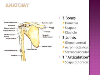
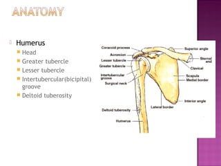
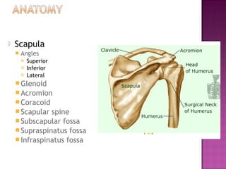
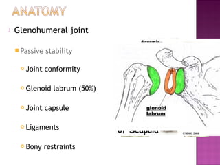
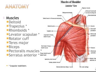
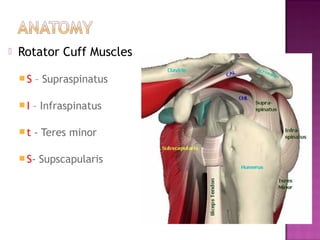
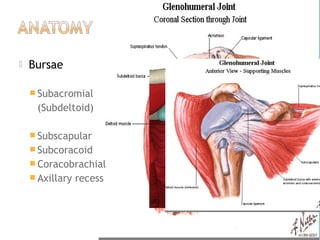
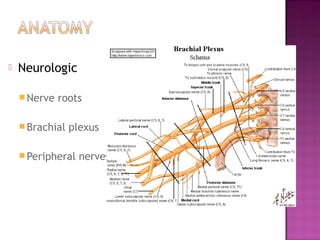
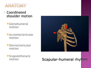
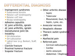
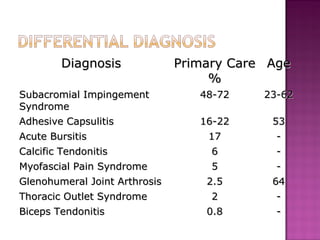
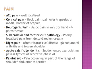
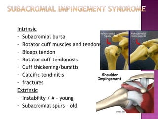
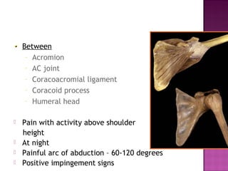
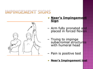
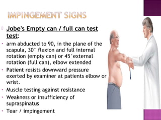
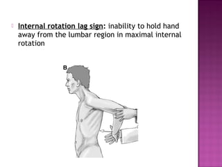
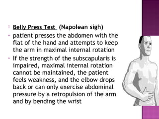
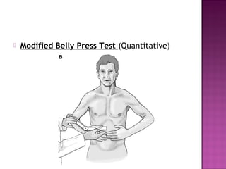
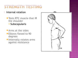
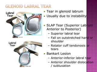
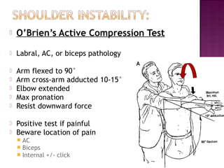
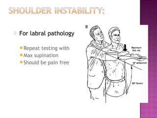
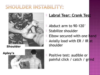
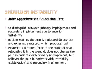
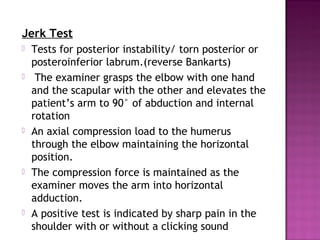
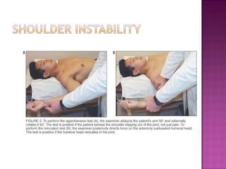
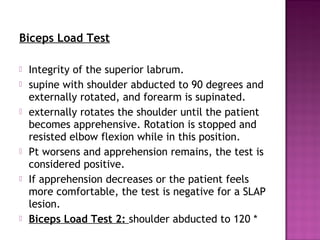
DR. PRUTHVIRAJ NISTANE

Deptt. Of Orthopaedics,Unit -2
  Govt. Medical College and
  Rajindra Hospital, Patiala
   Pertinent anatomy
   Differential diagnosis
   Clinical history
   Physical examination
   Common shoulder injuries
   Shoulder pain: a common
    complaint
    o More common causes in
      adults
    o Peak ages 40-60
    o Impingement syndrome
    o Rotator cuff problems
    o Frozen shoulder
    o 8-13% of all athletic injuries
   3 Bones
     Humerus
     Scapula
     Clavicle
   3 Joints
     Glenohumeral
     Acromioclavicular
     Sternoclavicular
   1 “Articulation”
     Scapulothoracic
   Humerus
     Head
     Greater tubercle
     Lesser tubercle
     Intertubercular(bicipital)
      groove
     Deltoid tuberosity
   Scapula
       Angles
           Superior
           Inferior
           Lateral
     Glenoid
     Acromion
     Coracoid
     Scapular spine
     Subscapular fossa
     Supraspinatus fossa
     Infraspinatus fossa
   Glenohumeral joint

     “Balland socket” vs “Golf
     ball and tee”

     Verymobile at cost of
     stability

     45%    of all dislocations

     Joint
          stability depends on
     multiple factors
   Glenohumeral joint

     Passive   stability

        Joint conformity

        Glenoid labrum (50%)

        Joint capsule

        Ligaments

        Bony restraints
   Muscles
    Deltoid
    Trapezius *
    Rhomboids *
    Levator scapulae *
    Rotator cuff
    Teres major
    Biceps
    Pectoralis muscles *
    Serratus anterior *


       * Scapular stabilizers
   Rotator Cuff Muscles

    S   – Supraspinatus

    I   – Infraspinatus

    t   - Teres minor

     S-   Supscapularis
   Bursae

     Subacromial
     (Subdeltoid)

     Subscapular
     Subcoracoid
     Coracobrachial
     Axillary   recess
   Neurologic

     Nerve   roots

     Brachial   plexus

     Peripheral   nerves
   Coordinated
    shoulder motion

     Glenohumeral
     motion

     Acromioclavicular
     motion

     Sternoclavicular
     motion

     Scapulothoracic
     motion               Scapular-humeral rhythm
   Impingement syndromes                  Other arthritic disease
       Subacromial bursitis               –   Septic
       Rotator cuff tendinopathy
                                           –   Tubercular
       Rotator cuff tear
       Biceps tendinopathy                –   Rheumatoid, Gout, SLE
   Adhesive capsulitis                    –   Septic, Lyme, etc.
   SC joint arthritis, sprain             Avascular necrosis
   AC joint arthritis, sprain             Neoplastic disease
   Glenohumeral joint OA                  Thoracic outlet syndrome
   Instablity
       GH dislocation                     CRPS
       GH subluxation                     Myofascial pain
       Labral tear (e.g. Bankart, SLAP,   Referred pain
        etc.)
   Clavicle fracture                      –   Cervical radiculopathy
   Proximal humerus fracture              –   Cardiac
   Scapular fracture                      –   Aortic aneurysm
                                           –   Abdominal / Diaphragm
Diagnosis             Primary Care Age
                                    %
Subacromial Impingement           48-72    23-62
Syndrome
Adhesive Capsulitis               16-22     53
Acute Bursitis                     17        -
Calcific Tendonitis                 6        -
Myofascial Pain Syndrome            5        -
Glenohumeral Joint Arthrosis       2.5      64
Thoracic Outlet Syndrome            2        -
Biceps Tendonitis                  0.8       -
History
oAge    - 2nd & 3rd decades instability
        4th & 5th decades impingement, frozen shoulder, inflammatory
         joint disease
        6th decade onwards rotator cuff tears , degenerative joint
         disease
oPain
oWeakness
oDeformity
oInstability
oLocking  / Clicking / Clunking
oSport / Occupation
oPrevious treatments
oAlleviating / Exacerbating
oAcute vs. Chronic
oTraumatic vs. Overuse
oHistory of prior injury
oMechanism of injury
•   ACJ pain - well localised
•   Cervical pain - Neck pain, pain over trapezius or
    medial border of scapula
•   Neurogenic Pain - Assoc pain in wrist or hand +/-
    parasthesiae
•   Subacromial and rotator cuff pathology - Poorly
    localised pain from deltoid region usually
•   Night pain - often rotator cuff disease, glenohumeral
    arthritis and frozen shoulder
•   Acute calcific tendonitis - Sudden onset excruciating
    pain is typical of reorptive phase of
•   Painful arc - Pain occurring in part of the range of
    shoulder abduction is termed
   Any history of trauma?
   Was shoulder dislocated?
   How many dislocations since then?
   Was dislocation spontaneous? If atraumatic
    dislocation is there history of joint laxity?
   Painless clicks in the shoulder are common
    and usually have no significance
   Following traumatic event - important to
    exclude brachial plexus injury
   May be due to pain (would examine with
    local anaesthetic joint examination in
    shoulder clinic)
   Restriction of both passive and active
    movements
   Usually associated with frozen shoulder,
    osteoarthritis, rheumatoid arthritis, chronic
    dislocation and cuff tear
 Undressupto waist
     Compare both sides


   Inspection
   Palpation
   Measurements
   Active & passive ROM
   Strength testing
   Special tests
   Distal neuro-vascular
    status, Lymph nodes
   Front & Back
   Above
   Axilla
   Laterally

   Asymmetry
   Attitude
   Obvious deformity
   Swelling
   Skin condition
   Ecchymosis
   Height of shoulder & scapulae
   Muscle atrophy / contractures
     Supraspinatus
     Infraspinatus
     Deltoid
   Contour of shoulder
   Neck
   Epicondyles
   At rest & with movement
   Bony structures
   Joints
   Axilla
   Soft tissues
   Codman’s method
o   Swelling ,crepitus
o   Irregularitis
o   Local temperature
o   Local tenderness
o   Lymph nodes
   Surface Anatomy
               (Anterior)
AC joint
                Manubrium
                Clavicle
 biceps
                SC Joint
                Acromion process
SC joint
                AC Joint
                Deltoid
                Coracoid process
                Pectoralis major
                Trapezius
                Humeral head
                Greater tubercle
                Lesser tubercle
                Biceps (long head)
   Surface Anatomy
                      (Posterior)
Supraspinatus
                       Scapular spine
                       Acromion process
                       Supraspinatus
 Infraspinatus         Infraspinatus
 Inferior angle
 of scapula
                       Deltoid
                       Trapezius
                       Latissumus dorsi
                       Scapula
                          Inferior angle
                          Medial border
30° to coronal plane

   Forward flexion:
    (160 - 180°)
   Anterior deltoid, Pectoralis
    major, coracobrachialis

   Extension:
    (40 - 60°)
   Latissimus dorsi,teres major,
    posterior deltoid

   Adduction:
    (45 °)
   Pectoralis major, latissimus
    dorsi, teres major
   Abduction:
(180°)
   glenohumeral rhythm (2: 1)
    By fixing the scapula with thumb and finger
   try to quantify glenohumeral movement only
   middle deltoid, supraspinatus

 Internal rotation:
    (60 - 90 °)
     subscapularis, pectoralis major, latissimus
     dorsi, teres major; anterior deltoid

   External rotation:                                Apley Scratch Test
    (40-60 °)
     infraspinatus, teres minor; posterior deltoid

   Circumduction
Abduction

•60°-120° painful – impingement
syndrome
•Whole range painful – arthritis of
GH joint,acute tendinitis
•>90° painful – AC joint arthritis
•Upto 30° difficult – rotator cuff
tear
•Restricted movements – Frozen
shoulder, ankylosis
Abduction with          Adduction with
external rotation       internal rotation


           Quick screening test
   scapular motion
•   Scapular dyskinesis (Scapulothoracic
    dysfuntion)

     Compare  scapular motion through ROM on
      both sides
     Wall push-ups


     Symmetrical
     Smooth
   Winging Medial winging results from injury to
    long thoracic nerve,
   Lateral winging results from trapezius
    weakness( accessory spinal nerve ).
   Test & compare both sides
   Be specific to muscle or muscle
    group

   Grade strength on 0 → 5 scale
     0: no contraction
     1: muscle flicker; no movement
     2: motion, but not against gravity
     3: motion against gravity, but not
      resistance
     4: motion against resistance
     5: normal strength
   Apparent lengths – from seventh cervical
    spine to tip of radial styloid
   True length – angle of acromion – tip of
    lateral epicondyle - tip of radial styloid
   Wasting
   Anterior and posterior axillary folds –
    Bryants sigh
   Girth of axilla - Callaways test
   Hamilton ruler test

   Duga’s test
Intrinsic
– Subacromial bursa
– Rotator cuff muscles and tendons tears
– Biceps tendon
– Rotator cuff tendonosis
– Cuff thickening/bursitis
– Calcific tendinitis
– fractures
Extrinsic
– Instability / # – young
– Subacromial spurs – old
Between
     – Acromion
     – AC joint
     – Coracoacromial ligament
     – Coracoid process
     – Humeral head


   Pain with activity above shoulder
    height
   At night
   Painful arc of abduction – 60-120 degrees
   Positive impingement signs
Neer’s Impingement
  Sign

– Arm fully pronated and
  placed in forced flexion

– Trying to impinge
  subacromial structures
  with humeral head

– Pain is positive test

  Neer’s Impingement test
Hawkin’s Sign

– Arm is forward
  flexed to 90
  degrees, then
  forcibly internally
  rotated

– Trying to impinge
  subacromial
  structures with
  humeral head

– Pain is positive test
   Jobe's Empty can / full can test
    test:
•   arm abducted to 90, in the plane of the
    scapula, 30° flexion and full internal
    rotation (empty can) or 45°external
    rotation (full can), elbow extended
•   Patient resists downward pressure
    exerted by examiner at patients elbow or
    wrist.
•   Muscle testing against resistance
•   Weakness or insufficiency of
    supraspinatus
•   Tear / impingement
   Internal Rotation Resistance Stress
       Test

      to differentiate between internal and
       classic impingement
      Shoulder in 90 degrees of abduction
       and 80 degrees of external rotation.
      A manual isometric muscle test is
       performed for external rotation and
       compared with one for internal
       rotation in the same position.
More weakness in internal rotation - internal impingement
more weakness in external rotation - classic outlet impingemen
   Gerber Subcoracoid Impingement
    Test

   impingement between the rotator
    cuff and the coracoid process
   Shoulder flexed 90 degrees and
    adducted 10 to 20 degrees across
    the body to bring the lesser
    tuberosity into contact with the
    coracoid.
   Pain indicates coracoid
    impingement
   Partial thickness tear
   Full (Complete) thickness tear
   Supraspinatus > Infraspinatus > subscapularis
   Reduced function and night pain
   Painful arc, gap may be felt
   May be due to:
       Impingement
       Degeneration
       Overuse
       Trauma

   Partial tears
       Conservative
   Complete tears
       Surgery
Rent Sign/Test
 Tests for a torn rotator cuff or rotator cuff
  impingement
 Patient seated,palpate anterior to the
  anterior edge of the acromion with one hand
  while holding the patient’s flexed elbow with
  the other
 The examiner passively extends the shoulder
  while slowly rotating the shoulder into
  external and internal rotation. The greater
  tuberosity will be prominent and a
  depression of about 1 finger width will be
  felt if a rotator cuff tear is present.
Rent Sign/Test
   Resisted abduction with arms by side

   Jobe's Empty can / full can test test: arm
    abducted to 90°, 30° flexion, in the plane
    of the scapula and full internal rotation
    (empty can) or 45°external rotation (full
    can), elbow extended
   Patient resists downward pressure exerted by
    examiner at patients elbow or wrist.
Drop-Arm Test

Abducted arm slowly
lowered
 – May be able to lower
   arm slowly to 90°
   (deltoid function)
 – Arm will then drop to
   side if rotator cuff tear
Positive test
 – patient unable to
   lower arm further with
   control
   Strength Testing

     Tests Supraspinatus
     Attempt to isolate from
     deltoid

     Sitting
     Arms straight out
     Elbows locked straight
     Thumbs down
     Arm at 30 degrees
      (in scapular plane)
     Attempts to elevate
      arms against resistance
   Resisted ER with the arms by side

   Drop test:
   Shoulder in 90 degrees of
    abduction and at almost full
    external rotation with the elbow
    flexed at 90 degrees. The patient
    is asked to maintain this position

   The sign is positive if a lag or
    “drop” occurs
   External Rotation Stress Test / swing
    door test

   patient's arms by his or her side in neutral
    flexion and abduction, the shoulders are
    externally rotated 45 to 60 degrees.
   The examiner applies force against the dorsum
    of the hands, attempting to rotate the shoulders
    internally back to neutral while the patient is
    asked to resist.
   Pain and weakness
   External Rotation Lag Sign

   The patient is asked to maintain the
    position of maximal external rotation
    actively as the examiner maintaining
    support of the arm at the elbow.
   The sign is positive when a lag or angular
    drop
Hornblower’s Sign (Patte Test)
 strength of the teres minor
 Abduct the patient’s arm to 90 degrees in
  the scapular plane.
 Flexe the elbow to 90 degrees, and the
  patient is asked to laterally rotate the
  shoulder.
 A positive test occurs with weakness and/or
  pain
Hornblower’s Sign (Patte Test)
   External rotation

       Tests RTC muscles that ER
        the shoulder
           Infraspinatus
           Teres minor


       Arms at the sides

       Elbows flexed to 90 degrees

       Externally rotates arms
        against resistance
   Gerber's lift off test:
   dorsum of the hand is placed
    against the lower back.
    If the patient is unable to lift the
    dorsum of the hand off the back
    and push examiner's hand away
    from 'hand behind back position'
    the test is positive
   Internal rotation lag sign: inability to hold hand
    away from the lumbar region in maximal internal
    rotation
   Belly Press Test (Napolean sigh)
•   patient presses the abdomen with the
    flat of the hand and attempts to keep
    the arm in maximal internal rotation
•   If the strength of the subscapularis is
    impaired, maximal internal rotation
    cannot be maintained, the patient
    feels weakness, and the elbow drops
    back or can only exercise abdominal
    pressure by a retropulsion of the arm
    and by bending the wrist
   Modified Belly Press Test (Quantitative)
   Internal rotation

     TestsRTC muscle that IR
     the shoulder
      Subscapularis


     Arms  at the sides
     Elbows flexed to 90
      degrees
     Internally rotates arms
      against resistance
   Injury to long head of
    biceps tendon
   Typically an overuse
    injury
     Repetitive   (overhead)
      lifting
     Impingement
 AERS test:
 (Abduction External Rotation Supination test)
. Pt feels pain on resisted supination in this position.

   Tenderness over antero-superior deltoid bulge on
    shoulder rotations
   Forward flex shoulder to
    about 90°
   Abduct shoulder to about
    10°
   Elbow - extended
   Arm in full supination

   Apply downward force to
    distal arm

   Pain is positive test
   Bicepital tendinitis, SLAP

   Weakness without pain:
    muscle weakness or
    rupture
   Elbow flexed to 90°
   Start in pronated position

   Active supination & flexion
    against resistance
   Palpate biceps tendon

   Pain or painful pop is
    positive test
     Tendonosis
     Subluxation
   Deltoid: resisted abduction over 30º

   Serratus anterior: "Winging" test
Failure to keep humeral
head centered in glenoid
Dislocation
 – Complete disruption of
   joint congruity or
   alignment
Subluxation
 – Partial or incomplete
   dislocation
Laxity
 – Slackness or
   looseness in joint
 – May be normal or
   abnormal
   Sulcus Sign

   Arm relaxed in
    neutral position
   Arm pulled downward
    at wrist

   Positive test is a
    visible sulcus at infra-
    acromial area
     Compare to
     contralateral side
   Feagin maneuver -
   Apprehension Test
   Shoulder abducted to 90°
   Slight stress to humeral head
    directed in anterior direction
   While externally rotating
    shoulder
   Positive test is apprehension
    due to feeling of instability or
    impending dislocation
       Beware if false positives

     Augmentation      test – apply
        anteriorly directed force by
        extending shoulder
   Relocation Test

   After a positive
    apprehension
   Apply posteriorly
    directed force over
    externally rotated
    humeral head

   Positive test is relief of
    apprehension
   Anterior release test
subluxation tests: (Load and Shift Test)

   Anterior subluxation test: abduction and external
    rotation "apprehension test" with thumb posteriorly
    and fingers anteriorly over humeral head

                                     Shoulder
                                     Latchman


   Posterior subluxation test: internal rotation,
    adduction, flexion and push posteriorly
   Apprehension Test
Flex and internally rotate the shoulder

   Circumduction Test
Circumduction in abduction
Tear in glenoid labrum
Usually due to instability

SLAP Tear (Superior Labrum
Anterior to Posterior)
– Superior labral tear
– Fall on outstretched hand or
  shoulder
– Rotator cuff tendonosis or
  tears
Bankart Lesion
– Anterior-inferior labral tear
– Anterior shoulder dislocation
  / subluxation
   O’Brien’s Active Compression Test

   Labral, AC, or biceps pathology

   Arm flexed to 90°
   Arm cross-arm adducted 10-15°
   Elbow extended
   Max pronation
   Resist downward force

   Positive test if painful
   Beware location of pain
       AC
       Biceps
       Internal +/- click
   For labral pathology

     Repeat testing with
     Max supination
     Should be pain free
   Labral Tear: Crank Test

                    Abduct arm to 90-120°
                    Stabilize shoulder
                    Elbow secured with one hand
                    Axially load with ER / IR at
  Shoulder           shoulder
             .


Apley’s
                    Positive test: audible or
                     painful click / catch / grind
   Jobe Apprehension-Relocation Test

   to distinguish between primary impingement and
    secondary impingement due to anterior
    instability
   patient supine, the arm is abducted 90 degrees
    and externally rotated, which produces pain
   Posteriorly directed force to the humeral head,
    relocating it in the glenoid, does not change the
    pain in patients with primary impingement, but
    relieves the pain in patients with instability
    (subluxation) and secondary impingement
Jerk Test
   Tests for posterior instability/ torn posterior or
    posteroinferior labrum.(reverse Bankarts)
    The examiner grasps the elbow with one hand
    and the scapular with the other and elevates the
    patient’s arm to 90° of abduction and internal
    rotation
   An axial compression load to the humerus
    through the elbow maintaining the horizontal
    position.
   The compression force is maintained as the
    examiner moves the arm into horizontal
    adduction. 
   A positive test is indicated by sharp pain in the
    shoulder with or without a clicking sound
Jerk Test
Biceps Load Test

   Integrity of the superior labrum.
   supine with shoulder abducted to 90 degrees and
    externally rotated, and forearm is supinated.
   externally rotates the shoulder until the patient
    becomes apprehensive. Rotation is stopped and
    resisted elbow flexion while in this position.
   Pt worsens and apprehension remains, the test is
    considered positive.
   If apprehension decreases or the patient feels
    more comfortable, the test is negative for a SLAP
    lesion. 
   Biceps Load Test 2: shoulder abducted to 120 *
Biceps Load Test
   Capsule sticks to humeral head
   IR  Flexion  ER
   GLOBAL RESTRICTION ROM
   NIGHT PAIN
   Cause often unknown – but beware diabetes
   Self limiting condition (6mth-2yrs).

   Painful
   Restrictive
   Resolution
– Typically due to fall
        onto tip of shoulder
        (acromion)
      – Arm tucked into side
•   Pain with activity (esp
    overhead, or weight
    training)
•   AC Jt tender
•   Crepitus/clicking
•   Deformity

     – Treatment depends
       on type
   Arm flexed to 90°
   Arm adducted to > 45°
   Hyperadduct shoulder
    (down on elbow)

   Positive test is pain in AC
    joint
   Watch out for false-
    positives
       Where is the pain?
AC Shear Test
 To test for acromioclavicular pathology
 The patient is in sitting position while the
  examiner cups his or her hands over the
  deltoid muscle with one hand on the clavicle
  and the other on the spine of the scapula.
 The examiner then squeezes the heels of the
  hand together.
 Abnormal movement is a positive test.
AC Shear Test
   Where is the pain?          Neurology
     over deltoid – think      C5 deltoid (S, M),
      shoulder                   biceps (R)
     over traps – think        C6 thumb (S), biceps
      neck                       (M), BrRad (R)
     Radicular pain –          C7 digit 3(S), triceps
      think neck                 (M, R)
                                C8 digit 5(S), FDP/S
                                 (M)
   Examine                     T1 medial elbow (S),
     Cervical   spine ROM       finger abduction (M)
                                Radial nerve
                                Ulnar nerve
                                Median nerve
   Assess cervical spine to see if neck
    movements recreate shoulder symptoms
   In full extension of C spine nose parallels the
    floor and in full flexion chin should rest on
    chest
   Lateral rotation approx 80o
   Lateral flexion 40o
   Sensation dermatomes C4 toT2
   Power around elbow, wrist and hand
   Shoulder power tested separately
   Test peripheral nerves, esp. Axillary nerve
   Biceps and triceps reflexes
   Tests for thoracix inlet syndrome
   Radial / Ulnar pulses
   AC joint
   Subacromial space
   Glenohumeral joint
   Biceps tendon (long head)
   Plain XR
     AP (IR and ER)
     Axillary lateral view
     Supraspinatus outlet
      view
     AC joint
   U/S scan
     Cuff tears,
      tendinopathy, bursitis
     Undercalls cuff
      pathology
     Can inject at the
      same time
   Bone scan                 MRI
     Esp
        if referring for        Cuff   tears
     ACJ surgery              MRA
   CT arthrogram           Labral pathology and
     Particularly
                 useful        instability
     for recurrent
     instability
                              Arthrogram
                              Arthroscopy
                              Joint aspiration
 THANK   YOU !!!!

Shoulder examionation

  • 1.
    DR. PRUTHVIRAJ NISTANE Deptt.Of Orthopaedics,Unit -2 Govt. Medical College and Rajindra Hospital, Patiala
  • 2.
    Pertinent anatomy  Differential diagnosis  Clinical history  Physical examination  Common shoulder injuries
  • 3.
    Shoulder pain: a common complaint o More common causes in adults o Peak ages 40-60 o Impingement syndrome o Rotator cuff problems o Frozen shoulder o 8-13% of all athletic injuries
  • 4.
    3 Bones  Humerus  Scapula  Clavicle  3 Joints  Glenohumeral  Acromioclavicular  Sternoclavicular  1 “Articulation”  Scapulothoracic
  • 5.
    Humerus  Head  Greater tubercle  Lesser tubercle  Intertubercular(bicipital) groove  Deltoid tuberosity
  • 6.
    Scapula  Angles  Superior  Inferior  Lateral  Glenoid  Acromion  Coracoid  Scapular spine  Subscapular fossa  Supraspinatus fossa  Infraspinatus fossa
  • 7.
    Glenohumeral joint  “Balland socket” vs “Golf ball and tee”  Verymobile at cost of stability  45% of all dislocations  Joint stability depends on multiple factors
  • 8.
    Glenohumeral joint  Passive stability  Joint conformity  Glenoid labrum (50%)  Joint capsule  Ligaments  Bony restraints
  • 9.
    Muscles Deltoid Trapezius * Rhomboids * Levator scapulae * Rotator cuff Teres major Biceps Pectoralis muscles * Serratus anterior *  * Scapular stabilizers
  • 10.
    Rotator Cuff Muscles S – Supraspinatus I – Infraspinatus t - Teres minor  S- Supscapularis
  • 11.
    Bursae  Subacromial (Subdeltoid)  Subscapular  Subcoracoid  Coracobrachial  Axillary recess
  • 12.
    Neurologic  Nerve roots  Brachial plexus  Peripheral nerves
  • 13.
    Coordinated shoulder motion  Glenohumeral motion  Acromioclavicular motion  Sternoclavicular motion  Scapulothoracic motion Scapular-humeral rhythm
  • 14.
    Impingement syndromes Other arthritic disease  Subacromial bursitis – Septic  Rotator cuff tendinopathy – Tubercular  Rotator cuff tear  Biceps tendinopathy – Rheumatoid, Gout, SLE  Adhesive capsulitis – Septic, Lyme, etc.  SC joint arthritis, sprain Avascular necrosis  AC joint arthritis, sprain Neoplastic disease  Glenohumeral joint OA Thoracic outlet syndrome  Instablity  GH dislocation CRPS  GH subluxation Myofascial pain  Labral tear (e.g. Bankart, SLAP, Referred pain etc.)  Clavicle fracture – Cervical radiculopathy  Proximal humerus fracture – Cardiac  Scapular fracture – Aortic aneurysm – Abdominal / Diaphragm
  • 15.
    Diagnosis Primary Care Age % Subacromial Impingement 48-72 23-62 Syndrome Adhesive Capsulitis 16-22 53 Acute Bursitis 17 - Calcific Tendonitis 6 - Myofascial Pain Syndrome 5 - Glenohumeral Joint Arthrosis 2.5 64 Thoracic Outlet Syndrome 2 - Biceps Tendonitis 0.8 -
  • 16.
  • 17.
    oAge - 2nd & 3rd decades instability 4th & 5th decades impingement, frozen shoulder, inflammatory joint disease 6th decade onwards rotator cuff tears , degenerative joint disease oPain oWeakness oDeformity oInstability oLocking / Clicking / Clunking oSport / Occupation oPrevious treatments oAlleviating / Exacerbating oAcute vs. Chronic oTraumatic vs. Overuse oHistory of prior injury oMechanism of injury
  • 18.
    ACJ pain - well localised • Cervical pain - Neck pain, pain over trapezius or medial border of scapula • Neurogenic Pain - Assoc pain in wrist or hand +/- parasthesiae • Subacromial and rotator cuff pathology - Poorly localised pain from deltoid region usually • Night pain - often rotator cuff disease, glenohumeral arthritis and frozen shoulder • Acute calcific tendonitis - Sudden onset excruciating pain is typical of reorptive phase of • Painful arc - Pain occurring in part of the range of shoulder abduction is termed
  • 19.
    Any history of trauma?  Was shoulder dislocated?  How many dislocations since then?  Was dislocation spontaneous? If atraumatic dislocation is there history of joint laxity?  Painless clicks in the shoulder are common and usually have no significance
  • 20.
    Following traumatic event - important to exclude brachial plexus injury  May be due to pain (would examine with local anaesthetic joint examination in shoulder clinic)
  • 21.
    Restriction of both passive and active movements  Usually associated with frozen shoulder, osteoarthritis, rheumatoid arthritis, chronic dislocation and cuff tear
  • 22.
     Undressupto waist  Compare both sides  Inspection  Palpation  Measurements  Active & passive ROM  Strength testing  Special tests  Distal neuro-vascular status, Lymph nodes
  • 24.
    Front & Back  Above  Axilla  Laterally  Asymmetry  Attitude  Obvious deformity  Swelling  Skin condition  Ecchymosis
  • 25.
    Height of shoulder & scapulae  Muscle atrophy / contractures  Supraspinatus  Infraspinatus  Deltoid  Contour of shoulder  Neck  Epicondyles
  • 27.
    At rest & with movement  Bony structures  Joints  Axilla  Soft tissues  Codman’s method o Swelling ,crepitus o Irregularitis o Local temperature o Local tenderness o Lymph nodes
  • 28.
    Surface Anatomy (Anterior) AC joint  Manubrium  Clavicle biceps  SC Joint  Acromion process SC joint  AC Joint  Deltoid  Coracoid process  Pectoralis major  Trapezius  Humeral head  Greater tubercle  Lesser tubercle  Biceps (long head)
  • 29.
    Surface Anatomy (Posterior) Supraspinatus  Scapular spine  Acromion process  Supraspinatus Infraspinatus  Infraspinatus Inferior angle of scapula  Deltoid  Trapezius  Latissumus dorsi  Scapula  Inferior angle  Medial border
  • 31.
    30° to coronalplane  Forward flexion: (160 - 180°)  Anterior deltoid, Pectoralis major, coracobrachialis  Extension: (40 - 60°)  Latissimus dorsi,teres major, posterior deltoid  Adduction: (45 °)  Pectoralis major, latissimus dorsi, teres major
  • 32.
    Abduction: (180°) glenohumeral rhythm (2: 1)  By fixing the scapula with thumb and finger try to quantify glenohumeral movement only middle deltoid, supraspinatus  Internal rotation: (60 - 90 °) subscapularis, pectoralis major, latissimus dorsi, teres major; anterior deltoid  External rotation: Apley Scratch Test (40-60 °) infraspinatus, teres minor; posterior deltoid  Circumduction
  • 33.
    Abduction •60°-120° painful –impingement syndrome •Whole range painful – arthritis of GH joint,acute tendinitis •>90° painful – AC joint arthritis •Upto 30° difficult – rotator cuff tear •Restricted movements – Frozen shoulder, ankylosis
  • 34.
    Abduction with Adduction with external rotation internal rotation Quick screening test
  • 35.
    scapular motion • Scapular dyskinesis (Scapulothoracic dysfuntion)  Compare scapular motion through ROM on both sides  Wall push-ups  Symmetrical  Smooth  Winging Medial winging results from injury to long thoracic nerve,  Lateral winging results from trapezius weakness( accessory spinal nerve ).
  • 36.
    Test & compare both sides  Be specific to muscle or muscle group  Grade strength on 0 → 5 scale  0: no contraction  1: muscle flicker; no movement  2: motion, but not against gravity  3: motion against gravity, but not resistance  4: motion against resistance  5: normal strength
  • 38.
    Apparent lengths – from seventh cervical spine to tip of radial styloid  True length – angle of acromion – tip of lateral epicondyle - tip of radial styloid  Wasting  Anterior and posterior axillary folds – Bryants sigh  Girth of axilla - Callaways test
  • 41.
    Hamilton ruler test  Duga’s test
  • 43.
    Intrinsic – Subacromial bursa –Rotator cuff muscles and tendons tears – Biceps tendon – Rotator cuff tendonosis – Cuff thickening/bursitis – Calcific tendinitis – fractures Extrinsic – Instability / # – young – Subacromial spurs – old
  • 44.
    Between – Acromion – AC joint – Coracoacromial ligament – Coracoid process – Humeral head  Pain with activity above shoulder height  At night  Painful arc of abduction – 60-120 degrees  Positive impingement signs
  • 45.
    Neer’s Impingement Sign – Arm fully pronated and placed in forced flexion – Trying to impinge subacromial structures with humeral head – Pain is positive test Neer’s Impingement test
  • 46.
    Hawkin’s Sign – Armis forward flexed to 90 degrees, then forcibly internally rotated – Trying to impinge subacromial structures with humeral head – Pain is positive test
  • 47.
    Jobe's Empty can / full can test test: • arm abducted to 90, in the plane of the scapula, 30° flexion and full internal rotation (empty can) or 45°external rotation (full can), elbow extended • Patient resists downward pressure exerted by examiner at patients elbow or wrist. • Muscle testing against resistance • Weakness or insufficiency of supraspinatus • Tear / impingement
  • 48.
    Internal Rotation Resistance Stress Test  to differentiate between internal and classic impingement  Shoulder in 90 degrees of abduction and 80 degrees of external rotation.  A manual isometric muscle test is performed for external rotation and compared with one for internal rotation in the same position. More weakness in internal rotation - internal impingement more weakness in external rotation - classic outlet impingemen
  • 49.
    Gerber Subcoracoid Impingement Test  impingement between the rotator cuff and the coracoid process  Shoulder flexed 90 degrees and adducted 10 to 20 degrees across the body to bring the lesser tuberosity into contact with the coracoid.  Pain indicates coracoid impingement
  • 51.
    Partial thickness tear  Full (Complete) thickness tear  Supraspinatus > Infraspinatus > subscapularis  Reduced function and night pain  Painful arc, gap may be felt  May be due to:  Impingement  Degeneration  Overuse  Trauma  Partial tears  Conservative  Complete tears  Surgery
  • 52.
    Rent Sign/Test  Testsfor a torn rotator cuff or rotator cuff impingement  Patient seated,palpate anterior to the anterior edge of the acromion with one hand while holding the patient’s flexed elbow with the other  The examiner passively extends the shoulder while slowly rotating the shoulder into external and internal rotation. The greater tuberosity will be prominent and a depression of about 1 finger width will be felt if a rotator cuff tear is present.
  • 53.
  • 54.
    Resisted abduction with arms by side  Jobe's Empty can / full can test test: arm abducted to 90°, 30° flexion, in the plane of the scapula and full internal rotation (empty can) or 45°external rotation (full can), elbow extended  Patient resists downward pressure exerted by examiner at patients elbow or wrist.
  • 56.
    Drop-Arm Test Abducted armslowly lowered – May be able to lower arm slowly to 90° (deltoid function) – Arm will then drop to side if rotator cuff tear Positive test – patient unable to lower arm further with control
  • 57.
    Strength Testing  Tests Supraspinatus  Attempt to isolate from deltoid  Sitting  Arms straight out  Elbows locked straight  Thumbs down  Arm at 30 degrees (in scapular plane)  Attempts to elevate arms against resistance
  • 58.
    Resisted ER with the arms by side  Drop test:  Shoulder in 90 degrees of abduction and at almost full external rotation with the elbow flexed at 90 degrees. The patient is asked to maintain this position  The sign is positive if a lag or “drop” occurs
  • 59.
    External Rotation Stress Test / swing door test  patient's arms by his or her side in neutral flexion and abduction, the shoulders are externally rotated 45 to 60 degrees.  The examiner applies force against the dorsum of the hands, attempting to rotate the shoulders internally back to neutral while the patient is asked to resist.  Pain and weakness
  • 61.
    External Rotation Lag Sign  The patient is asked to maintain the position of maximal external rotation actively as the examiner maintaining support of the arm at the elbow.  The sign is positive when a lag or angular drop
  • 62.
    Hornblower’s Sign (PatteTest)  strength of the teres minor  Abduct the patient’s arm to 90 degrees in the scapular plane.  Flexe the elbow to 90 degrees, and the patient is asked to laterally rotate the shoulder.  A positive test occurs with weakness and/or pain
  • 63.
  • 64.
    External rotation  Tests RTC muscles that ER the shoulder  Infraspinatus  Teres minor  Arms at the sides  Elbows flexed to 90 degrees  Externally rotates arms against resistance
  • 65.
    Gerber's lift off test:  dorsum of the hand is placed against the lower back.  If the patient is unable to lift the dorsum of the hand off the back and push examiner's hand away from 'hand behind back position' the test is positive
  • 66.
    Internal rotation lag sign: inability to hold hand away from the lumbar region in maximal internal rotation
  • 67.
    Belly Press Test (Napolean sigh) • patient presses the abdomen with the flat of the hand and attempts to keep the arm in maximal internal rotation • If the strength of the subscapularis is impaired, maximal internal rotation cannot be maintained, the patient feels weakness, and the elbow drops back or can only exercise abdominal pressure by a retropulsion of the arm and by bending the wrist
  • 68.
    Modified Belly Press Test (Quantitative)
  • 69.
    Internal rotation  TestsRTC muscle that IR the shoulder  Subscapularis  Arms at the sides  Elbows flexed to 90 degrees  Internally rotates arms against resistance
  • 70.
    Injury to long head of biceps tendon  Typically an overuse injury  Repetitive (overhead) lifting  Impingement
  • 71.
     AERS test: (Abduction External Rotation Supination test) . Pt feels pain on resisted supination in this position.  Tenderness over antero-superior deltoid bulge on shoulder rotations
  • 72.
    Forward flex shoulder to about 90°  Abduct shoulder to about 10°  Elbow - extended  Arm in full supination  Apply downward force to distal arm  Pain is positive test  Bicepital tendinitis, SLAP  Weakness without pain: muscle weakness or rupture
  • 73.
    Elbow flexed to 90°  Start in pronated position  Active supination & flexion against resistance  Palpate biceps tendon  Pain or painful pop is positive test  Tendonosis  Subluxation
  • 74.
    Deltoid: resisted abduction over 30º  Serratus anterior: "Winging" test
  • 76.
    Failure to keephumeral head centered in glenoid Dislocation – Complete disruption of joint congruity or alignment Subluxation – Partial or incomplete dislocation Laxity – Slackness or looseness in joint – May be normal or abnormal
  • 77.
    Sulcus Sign  Arm relaxed in neutral position  Arm pulled downward at wrist  Positive test is a visible sulcus at infra- acromial area  Compare to contralateral side
  • 78.
    Feagin maneuver -
  • 79.
    Apprehension Test  Shoulder abducted to 90°  Slight stress to humeral head directed in anterior direction  While externally rotating shoulder  Positive test is apprehension due to feeling of instability or impending dislocation  Beware if false positives  Augmentation test – apply anteriorly directed force by extending shoulder
  • 80.
    Relocation Test  After a positive apprehension  Apply posteriorly directed force over externally rotated humeral head  Positive test is relief of apprehension  Anterior release test
  • 81.
    subluxation tests: (Loadand Shift Test)  Anterior subluxation test: abduction and external rotation "apprehension test" with thumb posteriorly and fingers anteriorly over humeral head Shoulder Latchman  Posterior subluxation test: internal rotation, adduction, flexion and push posteriorly
  • 82.
    Apprehension Test Flex and internally rotate the shoulder  Circumduction Test Circumduction in abduction
  • 83.
    Tear in glenoidlabrum Usually due to instability SLAP Tear (Superior Labrum Anterior to Posterior) – Superior labral tear – Fall on outstretched hand or shoulder – Rotator cuff tendonosis or tears Bankart Lesion – Anterior-inferior labral tear – Anterior shoulder dislocation / subluxation
  • 84.
    O’Brien’s Active Compression Test  Labral, AC, or biceps pathology  Arm flexed to 90°  Arm cross-arm adducted 10-15°  Elbow extended  Max pronation  Resist downward force  Positive test if painful  Beware location of pain  AC  Biceps  Internal +/- click
  • 85.
    For labral pathology  Repeat testing with  Max supination  Should be pain free
  • 86.
    Labral Tear: Crank Test  Abduct arm to 90-120°  Stabilize shoulder  Elbow secured with one hand  Axially load with ER / IR at Shoulder shoulder . Apley’s  Positive test: audible or painful click / catch / grind
  • 87.
    Jobe Apprehension-Relocation Test  to distinguish between primary impingement and secondary impingement due to anterior instability  patient supine, the arm is abducted 90 degrees and externally rotated, which produces pain  Posteriorly directed force to the humeral head, relocating it in the glenoid, does not change the pain in patients with primary impingement, but relieves the pain in patients with instability (subluxation) and secondary impingement
  • 88.
    Jerk Test  Tests for posterior instability/ torn posterior or posteroinferior labrum.(reverse Bankarts)   The examiner grasps the elbow with one hand and the scapular with the other and elevates the patient’s arm to 90° of abduction and internal rotation  An axial compression load to the humerus through the elbow maintaining the horizontal position.  The compression force is maintained as the examiner moves the arm into horizontal adduction.   A positive test is indicated by sharp pain in the shoulder with or without a clicking sound
  • 89.
  • 91.
    Biceps Load Test  Integrity of the superior labrum.  supine with shoulder abducted to 90 degrees and externally rotated, and forearm is supinated.  externally rotates the shoulder until the patient becomes apprehensive. Rotation is stopped and resisted elbow flexion while in this position.  Pt worsens and apprehension remains, the test is considered positive.  If apprehension decreases or the patient feels more comfortable, the test is negative for a SLAP lesion.   Biceps Load Test 2: shoulder abducted to 120 *
  • 92.
  • 94.
    Capsule sticks to humeral head  IR  Flexion  ER  GLOBAL RESTRICTION ROM  NIGHT PAIN  Cause often unknown – but beware diabetes  Self limiting condition (6mth-2yrs).  Painful  Restrictive  Resolution
  • 96.
    – Typically dueto fall onto tip of shoulder (acromion) – Arm tucked into side • Pain with activity (esp overhead, or weight training) • AC Jt tender • Crepitus/clicking • Deformity – Treatment depends on type
  • 98.
    Arm flexed to 90°  Arm adducted to > 45°  Hyperadduct shoulder (down on elbow)  Positive test is pain in AC joint  Watch out for false- positives  Where is the pain?
  • 100.
    AC Shear Test To test for acromioclavicular pathology  The patient is in sitting position while the examiner cups his or her hands over the deltoid muscle with one hand on the clavicle and the other on the spine of the scapula.  The examiner then squeezes the heels of the hand together.  Abnormal movement is a positive test.
  • 101.
  • 102.
    Where is the pain?  Neurology  over deltoid – think  C5 deltoid (S, M), shoulder biceps (R)  over traps – think  C6 thumb (S), biceps neck (M), BrRad (R)  Radicular pain –  C7 digit 3(S), triceps think neck (M, R)  C8 digit 5(S), FDP/S (M)  Examine  T1 medial elbow (S),  Cervical spine ROM finger abduction (M)  Radial nerve  Ulnar nerve  Median nerve
  • 103.
    Assess cervical spine to see if neck movements recreate shoulder symptoms  In full extension of C spine nose parallels the floor and in full flexion chin should rest on chest  Lateral rotation approx 80o  Lateral flexion 40o
  • 104.
    Sensation dermatomes C4 toT2  Power around elbow, wrist and hand  Shoulder power tested separately  Test peripheral nerves, esp. Axillary nerve  Biceps and triceps reflexes  Tests for thoracix inlet syndrome  Radial / Ulnar pulses
  • 106.
    AC joint  Subacromial space  Glenohumeral joint  Biceps tendon (long head)
  • 107.
    Plain XR  AP (IR and ER)  Axillary lateral view  Supraspinatus outlet view  AC joint
  • 108.
    U/S scan  Cuff tears, tendinopathy, bursitis  Undercalls cuff pathology  Can inject at the same time
  • 109.
    Bone scan  MRI  Esp if referring for  Cuff tears ACJ surgery  MRA  CT arthrogram  Labral pathology and  Particularly useful instability for recurrent instability  Arthrogram  Arthroscopy  Joint aspiration
  • 110.
     THANK YOU !!!!

Editor's Notes

  • #16 Evaluation of shoulder pain - Applied Evidence Journal of Family Practice ,   July, 2002  by J. Herbert Stevenson ,   Thomas Trojian