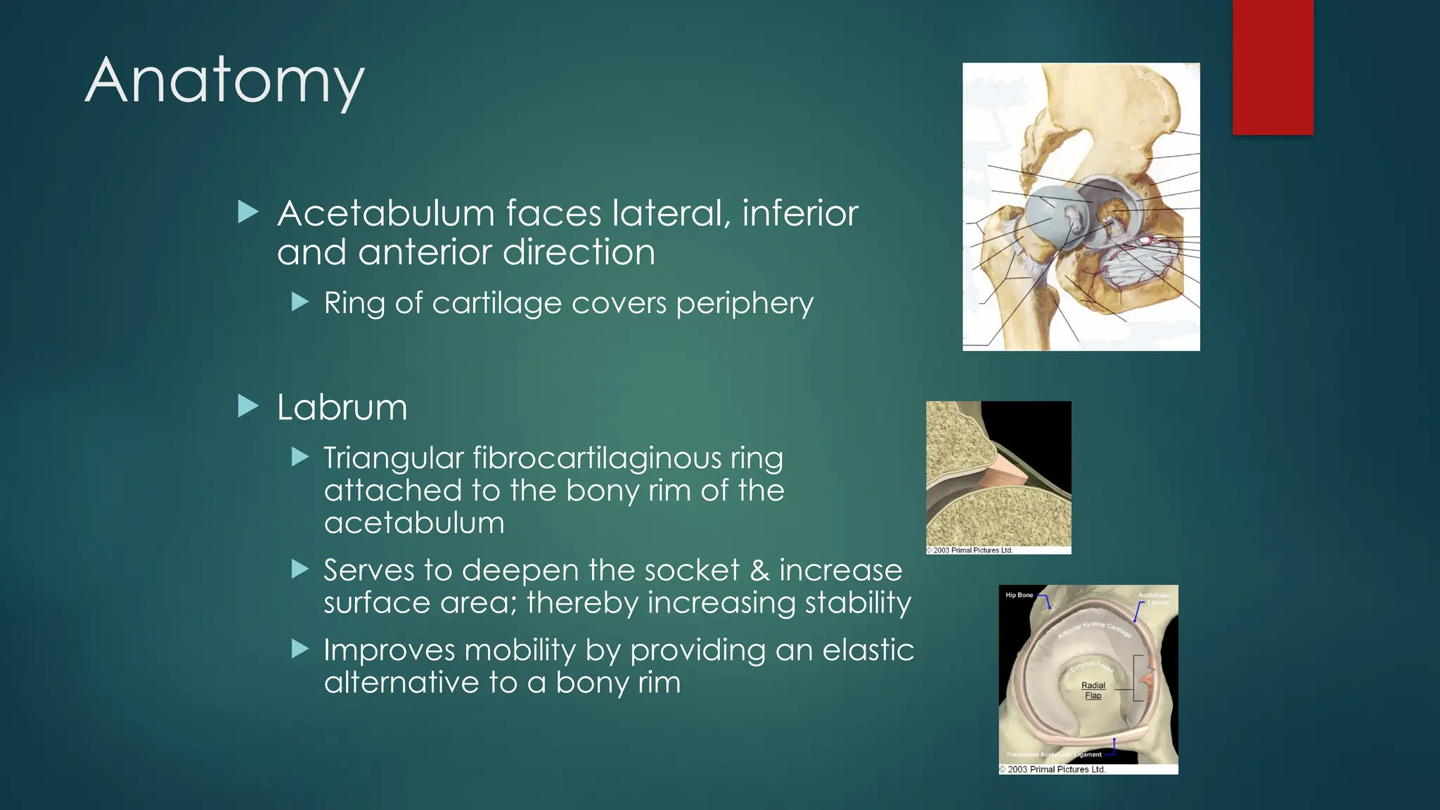
Mobilization for Hip
Joint
BY DR/ KHALED ALSAYANI
 The most common form of hip dysfunction is osteoarthritis, which
affects nearly 80% of individuals by the age of 75 years.
 Osteoporosis can lead to hip fractures and hospitalization for older
individuals.
Anatomy
 Ball & Socket joint
 Convex femoral head: 2/3
covered with cartilage
 Head of femur points in
anterior, medial, superior
direction
Anatomy
 Acetabulum faces lateral, inferior
and anterior direction
 Ring of cartilage covers periphery
 Labrum
 Triangular fibrocartilaginous ring
attached to the bony rim of the
acetabulum
 Serves to deepen the socket & increase
surface area; thereby increasing stability
 Improves mobility by providing an elastic
alternative to a bony rim
Angle of inclination
Angle of anteversion
Anatomy
 fat pad located in the acetabular fossa
 Lubrication
 Shock absorber
 Protects ligamentum teres
Anatomy
 Joint capsule shaped like a
cylindrical sleeve – 4 sets of fibers:
 Longitudinal
 Oblique
 Arcuate
 Circular
 Deep fibers of rectus femoris strengthen
capsule anteriorly
Anatomy
 Ligaments
 Ligamentum Teres
 Iliofemoral ligament
 Pubofemoral ligament
 Ischiofemoral ligament
Arthrokinematics & ROM
 Flexion/Extension:120°/20°
 Spin movement of the head of
the femur
 Abduction: 45°
 Head of the femur glides inferior
 Adduction: 30°
 Head of the femur glides superior
 Internal Rotation: 30°
 Head of the femur glides posterior
 External Rotation: 45°
 Head of the femur glides anterior
 Resting position
 30° flexion, 30° abduction, 20° ER
 Close packed position
 Extension, abduction, internal rotation
Muscles
Flexors:
 Psoas Major
 Psoas Minor
 Iliacus
 Pectineus
 Rectus Femoris
Extensors:
 Gluteus Maximus
 Semitendinosus
 Semimembranosus
 Biceps Femoris (long head)
Adductors:
 Adductor Magnus
 Adductor Longus
 Adductor Brevis
 Gracilis
 Pectineus
Abductors:
 Gluteus Medius
 Tensor Fascia Latae
Internal Rotators:
 Tensor Fascia Latae
 Gluteus Minimus
External Rotators:
 Gluteus Maximus
 Gemellus Superior
 Gemellus Inferior
 Obturator Externus
 Obturator Internus
 Quadratus Femoris
 Piriformis
JOINT MOBILIZATION
Hip distraction
Hip Inferior Glide
Hip Anterior Glide
Hip Posterior Glide
Hip Medial Glide
Hip Lateral Glide

4- Hip mobilization Hip mobilization.pptx

  • 1.
    Mobilization for Hip Joint BYDR/ KHALED ALSAYANI
  • 2.
     The mostcommon form of hip dysfunction is osteoarthritis, which affects nearly 80% of individuals by the age of 75 years.  Osteoporosis can lead to hip fractures and hospitalization for older individuals.
  • 3.
    Anatomy  Ball &Socket joint  Convex femoral head: 2/3 covered with cartilage  Head of femur points in anterior, medial, superior direction
  • 4.
    Anatomy  Acetabulum faceslateral, inferior and anterior direction  Ring of cartilage covers periphery  Labrum  Triangular fibrocartilaginous ring attached to the bony rim of the acetabulum  Serves to deepen the socket & increase surface area; thereby increasing stability  Improves mobility by providing an elastic alternative to a bony rim
  • 5.
  • 6.
  • 7.
    Anatomy  fat padlocated in the acetabular fossa  Lubrication  Shock absorber  Protects ligamentum teres
  • 8.
    Anatomy  Joint capsuleshaped like a cylindrical sleeve – 4 sets of fibers:  Longitudinal  Oblique  Arcuate  Circular  Deep fibers of rectus femoris strengthen capsule anteriorly
  • 9.
    Anatomy  Ligaments  LigamentumTeres  Iliofemoral ligament  Pubofemoral ligament  Ischiofemoral ligament
  • 10.
    Arthrokinematics & ROM Flexion/Extension:120°/20°  Spin movement of the head of the femur  Abduction: 45°  Head of the femur glides inferior  Adduction: 30°  Head of the femur glides superior  Internal Rotation: 30°  Head of the femur glides posterior  External Rotation: 45°  Head of the femur glides anterior
  • 11.
     Resting position 30° flexion, 30° abduction, 20° ER  Close packed position  Extension, abduction, internal rotation
  • 12.
    Muscles Flexors:  Psoas Major Psoas Minor  Iliacus  Pectineus  Rectus Femoris Extensors:  Gluteus Maximus  Semitendinosus  Semimembranosus  Biceps Femoris (long head)
  • 13.
    Adductors:  Adductor Magnus Adductor Longus  Adductor Brevis  Gracilis  Pectineus Abductors:  Gluteus Medius  Tensor Fascia Latae
  • 14.
    Internal Rotators:  TensorFascia Latae  Gluteus Minimus External Rotators:  Gluteus Maximus  Gemellus Superior  Gemellus Inferior  Obturator Externus  Obturator Internus  Quadratus Femoris  Piriformis
  • 18.
  • 19.
  • 20.
  • 21.
  • 22.
  • 23.
  • 24.

Editor's Notes

  • #4 The acetabulum faces lateral, inferior and anterior. A ring of cartilage, which, on the caudal part is interrupted by the acetabular notch, covers the peripheral part. The acetabular notch opens to the capsule. It is covered by the transverse ligament and it forms a passage for the ligamentum teres and the obturator artery branch. The middle, deeper part of the acetabulum (acetabular fossa) is not in contact with the femur head.
  • #7 In the acetabular fossa lays a fat pad, called the pulvinar acetabuli. It is covered with a synovial membrane. Function: Lubrication Shock absorber Protects ligamentum teres The hip is like a hydraulic system. During the swing phase, a decrease in atmospheric pressure will suck fluid from the iliac bursa into the pulvinar acetabuli. During the stance phase, the fluid gets squeezed out. The acetabular fossa is a shallow depression in the floor of the acetabulum, above the acetabular notch. The fossa is lined with a fibroelastic fat pad, which in turn is covered with synovial membrane. The membrane is attached to the medial aspect of the transverse ligament and the margins of the acetabular fossa, enveloping the ligament of the head of the femur, where it extends up to the edge of the pit (fovea) on the femoral head. © 2003 Primal Pictures Ltd.
  • #8 The joint capsule is shaped like a cylindrical sleeve. It runs from the iliac bone to the upper end of the femur. It has 4 sets of fibers: Longitudinal fibers, which keep the joint surfaces together. Oblique fibers, with a similar function as the longitudinal fibers/ Arcuate fibers, which only attach to the acetabular rim Circular fibers, which have no bony attachment, and are thickest in the center of the sleeve. The capsule inserts medially into the acetabular ring, the transverse ligament and the base of the peripheral surface of the labrum. Laterally the capsule inserts into the base of the neck. Deep fibers of the rectus femoris strengthen the capsule anteriorly. Synovial folds (frenulae) are found superior and inferior, to accommodate for ab/adduction.
  • #9 Ligamentum teres From the acetabular notch to the head of the femur. Although very strong, it plays a trivial mechanical role. Contributes to the vascular supply of the femoral head via the artery of the ligamentum teres, a posterior branch of the obturator artery (runs underneath the transverse ligament before entering the ligament) Iliofemoral ligament From the ventral edge of the ilium (caudal of the AIIS) to the intertrochanteric line. The central part is relatively thin, while the superior and inferior part are very strong (8-10mm thick). Pubofemoral ligament The iliofemoral and pubofemoral ligament resemble a Z shape. Between the 2 ligaments, the capsule is thinner. Between the capsule and the iliopsoas tendon is a bursa. Ischiofemoral ligament Located posteriorly. From the dorsal edge of the acetabulum to the medial greater trochanter. The anterior and posterior hip ligaments wind around the collum in a clock wise direction. Extension “winds” or tightens these ligaments, while flexion “unwinds’ or relaxes them. In erect posture, the ligaments are under moderate tension. In external rotation, the anterior fibers are taut, the posterior fibers are slack. In internal rotation, the anterior fibers are slack, the posterior fibers are taut.
  • #11 Position of reference: position denoting a “balanced pelvis” for assessment of hip abduction/adduction ROM – generally in a supine position A line between the 2 ASIS is perpendicular to the line from ASIS through patella