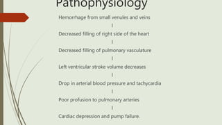
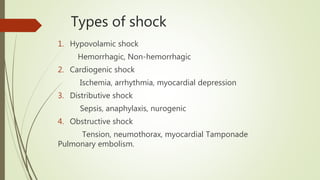
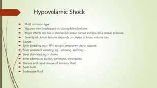
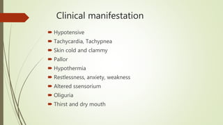
Shock
ď‚´Submitted By :-
Prity Mala
Roll no. :-21
Basic B.Sc nursing
3rd year
Submitted to :-
Mrs. Mamta kujur
Associated professor
College of nursing
RIMS, Ranchi
Quiz
1. Most common type of shock ?
a) Cardiogenic
b) Hypovolamic
c) Anaphylactic
2. Which is not a stage of shock?
a) Progressive
b) Compensatory
c) Initial
d) Reversible
3.Shock results from :
a) Decreased blood flow to the tissue
b) Increase blood flow to the tissue
c) Adequate perfusion
d) Both a and b
4.Which is not the cause of cardiogenic shock?
a) CHF
b) Pulmonary embolism
c) Spinal cord injury
d) Cardiomyopathy
Contents
ď‚´ Introduction
ď‚´ Definition of shock
ď‚´ Stages of shock
ď‚´ Pathophysiology
ď‚´ Types of shock
ď‚´ Diagnostic evaluation
ď‚´ Prognosis of shock
ď‚´ Prevention
ď‚´ Recent research
ď‚´ Summary
ď‚´ Evaluation
ď‚´ References
ď‚´ Bibliography
Introduction
 Shock results from an inadequate profusion of the body’s cells
with oxygenated blood.
ď‚´ Decreased blood flow = Decreased oxygen to the cells = Cell
death = orgain failure = death
ď‚´ Mortality rate = 20%
Definition
ď‚´ Shock is defined as a complex, life threatening condition or
syndrome characterised by inadequate blood flow to the tissue
and cell of the body.
ď‚´ Shock may be defined as inadequate delivery of oxygen and
nutrients to maintain normal tissue and cellular functions.
( Schwartz’s)
Stages of shock
There are 4 stages :-
1. Initial stage :- The cardiac output is insufficient to supply the normal
nutritional needs of tissue but not low enough to cause serious symptoms.
2. Compansetory stage :- The cardiac output is reduced further but due to
compansetory vasoconstriction, the blood pressure tends to remain within the
normal range blood flow to the skin and kidney decrease while blood flow to the
CNS and myocardium is maintained.
Cont..
3. Progressive stage :- The unfavourable change become more
and more apparent falling blood pressure, increased vasoconstriction,
increased heart rate and oliguria. If compansetory mechanism are unable
to cope with the reduce output shock becomes prograssively more
severe and passed onto.
4. Irreversible stage :- In this stage of shock no type of therapy can
save the patients life, Blood pressure decreased, Blood volume can be
normal in this stage, fluid transfusion may restore blood pressure only
temporary, Blood pressure decline until death occurres.
Pathophysiology
Hemorrhage from small venules and veins
l
Decreased filling of right side of the heart
l
Decreased filling of pulmonary vasculature
l
Left ventricular stroke volume decreases
l
Drop in arterial blood pressure and tachycardia
l
Poor profusion to pulmonary arteries
l
Cardiac depression and pump failure.
Types of shock
1. Hypovolamic shock
Hemorrhagic, Non-hemorrhagic
2. Cardiogenic shock
Ischemia, arrhythmia, myocardial depression
3. Distributive shock
Sepsis, anaphylaxis, nurogenic
4. Obstructive shock
Tension, neumothorax, myocardial Tamponade
Pulmonary embolism.
Hypovolamic Shock
ď‚´ Most common type
ď‚´ Occures from inadequate circulating blood volume.
ď‚´ Mejor effects are due to decreased cardiac output and low intra cardiac pressure.
ď‚´ Severity of clinical features depends on degree of blood volume loss.
ď‚´ Causes:-
ď‚´ Sever bleeding, eg :- PPH, ectopic pregnancy, uterus rupture.
ď‚´ Sever persistent vomiting, eg :- prolong vomiting.
ď‚´ Sever diarrhoea, eg :- cholera
ď‚´ Sever edemas or ascities, peritonitis, pancreatitis.
ď‚´ Diuresis and rapid remove of amniotic fluid.
ď‚´ Sever burn.
ď‚´ Inadequate fluid.
Clinical manifestation
ď‚´ Hypotensive
ď‚´ Tachycardia, Tachypnea
ď‚´ Skin cold and clammy
ď‚´ Pallor
ď‚´ Hypothermia
ď‚´ Restlessness, anxiety, weakness
ď‚´ Altered ssensorium
ď‚´ Oliguria
ď‚´ Thirst and dry mouth
Medical Management
> ABCs
> Resuscitation :
ď‚´ Vasopressor are used only as a temporary method to
restore the BP until fluid resuscitation take place.
ď‚´ Immediate control the bleeding
ď‚´Infusion of fluids is the fundamental treatment
ď‚´Crystalloid fluids normal saline.
> Drugs:
ď‚´Sedativs
ď‚´Chronotropic agents
ď‚´Intropic agents
Nursing management
ď‚´ Closely monitored patient at risk for fluid deficit.
ď‚´ Ensure safe administer of prescribed fluid and medications,
and document effects.
ď‚´ Monitor and promptly report sign of complecations and
effects of treatment, monitor patient closely for advers effects.
ď‚´ Monitor for cardiovascular ovreload, signs of difficulty in
breathing and pulmonary edema.
ď‚´ Reduce fear and anxiety about the need for the oxygen mask
by giving explanation and frequent reassurance.
Cardiogenic shock
 Cardiogenic shock occurres when the heart’s ability to pump blood is
impaired.
This is a condition that results from inadequate profusion of body
tissue with oxygenated blood that is insufficient to sustain life, Cardiogenic
output is decreased.
Causes:-
ď‚´ Acute myocardial infarction resulting in massive damage to myocardium.
ď‚´ Pulmonary embolism
ď‚´ Cardiac tamponade Cardiomyopathy
ď‚´ Chronic congestive heart failure.
Clinical manifestation
ď‚´ Same as Hypovolamic shock
ď‚´ Low blood pressure
ď‚´ Rapid pulse
ď‚´ Dyarrhythmia, chest pain
ď‚´ Respiratory distress
ď‚´ Pale skin
ď‚´ Low urine output
ď‚´ Multi-orgain failure
ď‚´ Left and right ventricular failure
Medical Management
ď‚´ Cardiac monitoring, pulse oximetry.
ď‚´ Airway clearens, Suplemental oxygen, vasodilators.
ď‚´ Fluid therapy :- norepinephrine, Dopamine.
ď‚´ Drug Therapy :- Diuretic, Dopamine, Dobutamine,
Heparin, sedative, PCI or thrombolytic, mainly intropic agents.
ď‚´ IABP is utilized if medical therapy is ineffective .
IABP (Intra Aortic Balloon Pump)
Nursing management
ď‚´ Identify patient at risk for Cardiogenic shock .
ď‚´ Promote adequate oxygenatiom of the heart muscle and
decrease cardiac workload.
ď‚´ Monitor hemodynamic and cardiac status.
ď‚´ Provide for safe and accurate administration of IV fluids and
medications.
ď‚´ Monitor for desire effect or side effects.
ď‚´ Perform frequent check of neurovascular status of lower
extremities.
 Take an active role in insuring patient’s safety and comfort
and in reducing anxiety.
Distributive shock
ď‚´ Distributive shock is a condition in which abnormal
distribution of blood flow in the smallest blood vessels results
in inadequate supply of blood to the body’s tissues and
organs.
ď‚´ Types of distributive shock :-
1. Septic shock
2. Anaphylactic shock
3. Neurogenic shock
Septic shock
ď‚´ It is the most common type of shock and caused by widespread infections
due to gram positive and gram negative becteria and viruses.
ď‚´Causes:-
ď‚´ UTI, abortion
ď‚´ Sever burn
ď‚´ CSOM
ď‚´ Due to chronic diseases, eg:- diabetes, AIDS
ď‚´ Indwelling lines and catheter
ď‚´ Improper wound care and management
Clinical manifestation
ď‚´ Hyperthermia
ď‚´ Sever headache
ď‚´ Respiratory distress
ď‚´ Decreased cardiac output
ď‚´ Hypotension
ď‚´ Multi-orgain failure
ď‚´ Anuria
Medical Management
ď‚´ Blood,sputum, urine and wound drainage specimen are
collected to identify and eliminate the cause of infection.
ď‚´ Fluid replacement is instituted.
ď‚´ Broad spectrum antibiotics are started.
ď‚´ Aggressive nutritional supplementation (high protein) is
provided, internal feeding are preferred.
Nursing management
ď‚´ Identify patient at risk for septic shock.
ď‚´ Monitor IV lines, arterial and venous puncture sites, surgical
incision, trauma wounds, urinary catheter and pressure ulcer
for sign of infections.
 Reduced patient’s temperature when orderd for temperature
higher than 40°c.
ď‚´ Administer prescribed iv fluids and medications.
ď‚´ Monitor hemodynamic status, fluid intake and output and
nutritional status.
ď‚´ Monitor daily weight and serum albumin and prealbumin
level to determine daily protein requirement.
Anaphylactic shock
ď‚´ Anaphylactic shock is a life threatening systemic
hypersensitive reaction contact with an allergen.
Causes:-
ď‚´ Drug: penicillin
ď‚´ Blood transfusion
ď‚´ Stings and snake bite
ď‚´ New clothses
ď‚´ Dusting smokes
ď‚´ Suddenly climate changes
Clinical manifestation
ď‚´ Cuogh, whizzing, laryngeal edema, bronchospasm
ď‚´ Hypotension, tachycardia, palpitation
ď‚´ Syncope
ď‚´ Urticaria Angeodema pruritus
ď‚´ Nausea, vomiting
ď‚´ Seizure
ď‚´ Respiratory depression , cardiovascular collapse
ď‚´ Coma
Medical Management
ď‚´ Open the airway by tilting the head, Breathing and circulation
should be stablished carrying BLS if needed.
ď‚´ Administration of epinephrine subcuteniously.
ď‚´ If the respiratory or cardiovascular region fail to improve
within 5 minutes of administration, a 2nd dose should be
given.
ď‚´ Additional drug therapy :
After the administration of epinephrine the other drugs to be
administered are:
Antihistamine, corticosteroids. ( If clinical improvements
occur)
Nursing management
ď‚´ Place the patient in a supine position, with the leg is slightly
elevated.
ď‚´ Monitoring the patients cardiovascular and respiratory status
continuously.
ď‚´ Record blood pressure and heart rate atleast every 5 minutes.
ď‚´ Delivere oxygen at a flowof 5-6 ltr/minut by nesal hood or full
face mask at any time during the episode.
Neurogenic shock
ď‚´ Inability of nervous system to control dilation of blood vessels.
ď‚´ Neurogenic shock results from generalised vasodilation and loss of
vasomotor tone due to:-
1. Massive increase in vascular
capacity.
2. Pulling of blood in periphery
3. Decreased venous return to
the heart.
Causes
ď‚´ Brain traumatic injuries
ď‚´ Brain damage, vasomotor depression
ď‚´ Spinl cord injury
ď‚´ Deep spinal anaesthesia
ď‚´ During LP
ď‚´ Sever pain, hypoglycemia, emotional stress
ď‚´ Drug causing vasomotor center depression
ď‚´ Anti-snake venom
Clinical manifestation
ď‚´ Nervousness
ď‚´ Loss of consciousness
ď‚´ Confusion
ď‚´ Skin warm but dry
ď‚´ Respiratory depression
ď‚´ Hypotension
ď‚´ Decreased BP
ď‚´ Tachycardia
Management
ď‚´ Proper positionig (trendelenburg position) : Displaced
blood from systemic venules into right heart and and
increased cardiac output.
ď‚´ Administration of fluids.
ď‚´ Vasoconstrictor drugs:
Phenylephrine, metaraminol.
Obstructive shock
ď‚´ Flow of blood is obstructed, which impedes circulation and
can result in circulatory arrest.
ď‚´ Several conditions result in this form of shock :-
a) Cardiac tamponade
b) Constrictive pericarditis
c) Tension pneumothorax
d) Massive pulmonary embolism
Treatment
ď‚´ Treatment of choice is pericardial drainage via surgery.
ď‚´ Pulmonary embolism is usually treat with systemic
anticoagulation, but when massive pulmonary embolism
cause right ventricular failure and shock thrombolytic therapy
should be strongly consided.
ď‚´ Thrombolytic therapy :-
Eminase
Retavase
Streptase
Quiz
1. Which is not the sign and symptoms of neurogenic shock ?
a) LOC
b) Nervousness
c) Confusion
d) Dehydration
2. Which is not the cause of septic shock?
a) UTI
b) Abortion
c) Massive infected wund
d) Smokes and dust
Diagnostic evaluation
ď‚´ HgB, WBC, Platelets
ď‚´ Ptt test
ď‚´ ECG
ď‚´ BUN
ď‚´ X-ray
ď‚´ Urinalysis
ď‚´ Wound culture
Prognosis of shock
ď‚´ The prognosis varies with the origin of shock and its duration .
ď‚´ 80% to 90% of young patients survive Hypovolamic shock with
appropriate management.
ď‚´ Cardiogenic shock associated with extensive myocardial infarction
( mortality rate upto75% )
ď‚´ Septic shock : ( mortality rate upto 75%)
ď‚´ Hypovolamic, Anaphylactic and Neurogenic shock are readily treatable
and respond well to medical therapy.
ď‚´ Perfusion of the brain may be the greatest danger during shock.
ď‚´ Therefore urgent treatment are essential for the good prognosis.
Prevention of shock
ď‚´ Primary prevention of shock is an essential focus on nursing intervention :
Hypovolamic shock can be prevented in some instances by closely
monitoring patients who is at risk for fluid deficit and assisting with fluid
replecment before intravascular volume is depleted.
ď‚´ General nursing measure include safe administer of prescribed fluids and
medications and proper documentation, monitoring signs of complecations
and side effects.
Recent research on shock
Utility of Point of care ultrasound in differentiating causes of shock in
resource limited set up.
Done by:- H. Humble Rahulkumar, Parikh Rina Bhavin, K Patel Shreyas, H
Pancholi, krunalkumar, Saxena Atulkumar, Chawada Bansari.
(Department of emergency medicine, Medical college and SSG Hospital
Vadodara, Gujarat, India)
Date of Submission :- 17th july 2018
Date of Acceptance :- 08th oct 2018
Date of publication :- 22nd april 2019
Abstract :-
ď‚´Background :- Delivering early diagnosis of shock in
resource limited setting is challenging, especially with
limited availability of point of care laboratory and
radiological diagnostic facilities. There is growing urgency
to provide point of care diagnosis and treatment for time
sensitive conditions like shock.
ď‚´Aim:- We tried to evaluate the application of point of care
ultrasound Considering different disease cohort and
practice realities in our setup.
ď‚´Setting and Design :- This study was a single center
prospactive diagnostic study to check the diagnostic
accuracy of point of care ultrasound. This study was
approved by ethics committee.
ď‚´ Material and methods :-The study was conducted at the emergency medicine
department of tertiary care government hospital in central Gujarat from November 16th to
October 17th All adults patients with clinical features of shock with systolic blood pressure
<90mmHg and shock index > 1 presenting to emergency department where included as
participants. The results of point of care ultrasound were compared with diagnosis given
by consultant of respective department as per standered departmental practices.
ď‚´ Statistical analysis and results :- A total of 130 patient where enrolled in this study.
Mean time taken to examine by the point of care ultrasound was 12 minutes (range:11-14
minutes). This protocol was able to correctly diagnose 100% of Obstructive shock 96.3% of
Cardiogenic shock, 94.4% of Hypovolamic shock, 80.9% of mixed type of shock and 75% of
distributive type of shock.
ď‚´ Conclusion :- This study highlights the roll of point of care ultrasound for early diagnosis
of shock etiology in emergency medicine department. Diagnosis using point of care
ultrasound significantly agreed with medical diagnosis. It showed good efficacy of point of
care ultrasound to differentiate causes of shock with good accuracy except distributive
shock.
ď‚´ Reference :-
1. Ghane MR, Gharib M, Ebrahimi A, Saeedi M, Akbari-kamrani M, Rezaee M, et al.
Accuracy of early rappid ultra sound in shock examination perform by emergency
physician for diagnosis of shock etiology in critically ill patients. J Emerg trauma
shock 2015, 8; 5-10. Back to cited text no. 1.
2. Perera P, Mailhot T, Riley D, Mandavia D. The RUSH exam: Rapid ultrasound in
shock in the evaluation of the critically ill. Emerg Med Clin North Am 2010;28:29-
56.vii. Back to cited text no. 2
3. PereraP, Mailhot T, Riley D, Mandavia D. The RUSH exam2012 : Rapid ultrasound
in shock in the evaluation of critically ill patient. Ultrasound Clin 2012;7:255-278.
Back to cited text no. 3
Summery
ď‚´ Shock is a circulatory system abnormalities that results in inadequate tissue
profusion.
ď‚´ Hypovolamia is the cause of shock in the majority of trauma patient.
ď‚´ Most type of shock are coused by dysfunction in the heart, blood vessels or
volume of blood.
ď‚´ The most important and first treatment for shock is to recognising the
patient is in shock.
 Treatment focuses on the ABC’s reversible od underlying couses and
prevention of complecations
Evaluation
1. What is shock?
2. How many stages of shock?
3. Explain the nursing management of Hypovolamic shock.
4. What do you mean by septic shock?
5. What is the clinical manifestation of septic shock?
6. What is the cause of Neurogenic shock?
7. What is Obstructive shock?
References
ď‚´ Book
ď‚´ Internet
ď‚´ Discuss with the teacher and friends
Bibliography
ď‚´ Brunner and suddarth, text book of medical surgical Nursing,
twelfth edition, wolter Kluwer, page no. 596-602.
ď‚´ Ansari Javed, A text book of medical surgical nursing, voll-ll
pv publication, page no.
ď‚´ WWW.wikidedia.com
Shock - the life threatening condition

Shock - the life threatening condition

  • 1.
    Shock ď‚´Submitted By :- PrityMala Roll no. :-21 Basic B.Sc nursing 3rd year Submitted to :- Mrs. Mamta kujur Associated professor College of nursing RIMS, Ranchi
  • 2.
    Quiz 1. Most commontype of shock ? a) Cardiogenic b) Hypovolamic c) Anaphylactic 2. Which is not a stage of shock? a) Progressive b) Compensatory c) Initial d) Reversible
  • 3.
    3.Shock results from: a) Decreased blood flow to the tissue b) Increase blood flow to the tissue c) Adequate perfusion d) Both a and b 4.Which is not the cause of cardiogenic shock? a) CHF b) Pulmonary embolism c) Spinal cord injury d) Cardiomyopathy
  • 4.
    Contents ď‚´ Introduction ď‚´ Definitionof shock ď‚´ Stages of shock ď‚´ Pathophysiology ď‚´ Types of shock ď‚´ Diagnostic evaluation ď‚´ Prognosis of shock ď‚´ Prevention ď‚´ Recent research ď‚´ Summary ď‚´ Evaluation ď‚´ References ď‚´ Bibliography
  • 5.
    Introduction  Shock resultsfrom an inadequate profusion of the body’s cells with oxygenated blood.  Decreased blood flow = Decreased oxygen to the cells = Cell death = orgain failure = death  Mortality rate = 20%
  • 6.
    Definition  Shock isdefined as a complex, life threatening condition or syndrome characterised by inadequate blood flow to the tissue and cell of the body.  Shock may be defined as inadequate delivery of oxygen and nutrients to maintain normal tissue and cellular functions. ( Schwartz’s)
  • 8.
    Stages of shock Thereare 4 stages :- 1. Initial stage :- The cardiac output is insufficient to supply the normal nutritional needs of tissue but not low enough to cause serious symptoms. 2. Compansetory stage :- The cardiac output is reduced further but due to compansetory vasoconstriction, the blood pressure tends to remain within the normal range blood flow to the skin and kidney decrease while blood flow to the CNS and myocardium is maintained.
  • 9.
    Cont.. 3. Progressive stage:- The unfavourable change become more and more apparent falling blood pressure, increased vasoconstriction, increased heart rate and oliguria. If compansetory mechanism are unable to cope with the reduce output shock becomes prograssively more severe and passed onto. 4. Irreversible stage :- In this stage of shock no type of therapy can save the patients life, Blood pressure decreased, Blood volume can be normal in this stage, fluid transfusion may restore blood pressure only temporary, Blood pressure decline until death occurres.
  • 10.
    Pathophysiology Hemorrhage from smallvenules and veins l Decreased filling of right side of the heart l Decreased filling of pulmonary vasculature l Left ventricular stroke volume decreases l Drop in arterial blood pressure and tachycardia l Poor profusion to pulmonary arteries l Cardiac depression and pump failure.
  • 11.
    Types of shock 1.Hypovolamic shock Hemorrhagic, Non-hemorrhagic 2. Cardiogenic shock Ischemia, arrhythmia, myocardial depression 3. Distributive shock Sepsis, anaphylaxis, nurogenic 4. Obstructive shock Tension, neumothorax, myocardial Tamponade Pulmonary embolism.
  • 12.
    Hypovolamic Shock ď‚´ Mostcommon type ď‚´ Occures from inadequate circulating blood volume. ď‚´ Mejor effects are due to decreased cardiac output and low intra cardiac pressure. ď‚´ Severity of clinical features depends on degree of blood volume loss. ď‚´ Causes:- ď‚´ Sever bleeding, eg :- PPH, ectopic pregnancy, uterus rupture. ď‚´ Sever persistent vomiting, eg :- prolong vomiting. ď‚´ Sever diarrhoea, eg :- cholera ď‚´ Sever edemas or ascities, peritonitis, pancreatitis. ď‚´ Diuresis and rapid remove of amniotic fluid. ď‚´ Sever burn. ď‚´ Inadequate fluid.
  • 13.
    Clinical manifestation ď‚´ Hypotensive ď‚´Tachycardia, Tachypnea ď‚´ Skin cold and clammy ď‚´ Pallor ď‚´ Hypothermia ď‚´ Restlessness, anxiety, weakness ď‚´ Altered ssensorium ď‚´ Oliguria ď‚´ Thirst and dry mouth
  • 14.
    Medical Management > ABCs >Resuscitation : ď‚´ Vasopressor are used only as a temporary method to restore the BP until fluid resuscitation take place. ď‚´ Immediate control the bleeding ď‚´Infusion of fluids is the fundamental treatment ď‚´Crystalloid fluids normal saline. > Drugs: ď‚´Sedativs ď‚´Chronotropic agents ď‚´Intropic agents
  • 15.
    Nursing management ď‚´ Closelymonitored patient at risk for fluid deficit. ď‚´ Ensure safe administer of prescribed fluid and medications, and document effects. ď‚´ Monitor and promptly report sign of complecations and effects of treatment, monitor patient closely for advers effects. ď‚´ Monitor for cardiovascular ovreload, signs of difficulty in breathing and pulmonary edema. ď‚´ Reduce fear and anxiety about the need for the oxygen mask by giving explanation and frequent reassurance.
  • 16.
    Cardiogenic shock  Cardiogenicshock occurres when the heart’s ability to pump blood is impaired. This is a condition that results from inadequate profusion of body tissue with oxygenated blood that is insufficient to sustain life, Cardiogenic output is decreased. Causes:-  Acute myocardial infarction resulting in massive damage to myocardium.  Pulmonary embolism  Cardiac tamponade Cardiomyopathy  Chronic congestive heart failure.
  • 17.
    Clinical manifestation ď‚´ Sameas Hypovolamic shock ď‚´ Low blood pressure ď‚´ Rapid pulse ď‚´ Dyarrhythmia, chest pain ď‚´ Respiratory distress ď‚´ Pale skin ď‚´ Low urine output ď‚´ Multi-orgain failure ď‚´ Left and right ventricular failure
  • 18.
    Medical Management ď‚´ Cardiacmonitoring, pulse oximetry. ď‚´ Airway clearens, Suplemental oxygen, vasodilators. ď‚´ Fluid therapy :- norepinephrine, Dopamine. ď‚´ Drug Therapy :- Diuretic, Dopamine, Dobutamine, Heparin, sedative, PCI or thrombolytic, mainly intropic agents. ď‚´ IABP is utilized if medical therapy is ineffective .
  • 19.
    IABP (Intra AorticBalloon Pump)
  • 20.
    Nursing management  Identifypatient at risk for Cardiogenic shock .  Promote adequate oxygenatiom of the heart muscle and decrease cardiac workload.  Monitor hemodynamic and cardiac status.  Provide for safe and accurate administration of IV fluids and medications.  Monitor for desire effect or side effects.  Perform frequent check of neurovascular status of lower extremities.  Take an active role in insuring patient’s safety and comfort and in reducing anxiety.
  • 21.
    Distributive shock  Distributiveshock is a condition in which abnormal distribution of blood flow in the smallest blood vessels results in inadequate supply of blood to the body’s tissues and organs.  Types of distributive shock :- 1. Septic shock 2. Anaphylactic shock 3. Neurogenic shock
  • 22.
    Septic shock ď‚´ Itis the most common type of shock and caused by widespread infections due to gram positive and gram negative becteria and viruses. ď‚´Causes:- ď‚´ UTI, abortion ď‚´ Sever burn ď‚´ CSOM ď‚´ Due to chronic diseases, eg:- diabetes, AIDS ď‚´ Indwelling lines and catheter ď‚´ Improper wound care and management
  • 24.
    Clinical manifestation ď‚´ Hyperthermia ď‚´Sever headache ď‚´ Respiratory distress ď‚´ Decreased cardiac output ď‚´ Hypotension ď‚´ Multi-orgain failure ď‚´ Anuria
  • 25.
    Medical Management ď‚´ Blood,sputum,urine and wound drainage specimen are collected to identify and eliminate the cause of infection. ď‚´ Fluid replacement is instituted. ď‚´ Broad spectrum antibiotics are started. ď‚´ Aggressive nutritional supplementation (high protein) is provided, internal feeding are preferred.
  • 26.
    Nursing management  Identifypatient at risk for septic shock.  Monitor IV lines, arterial and venous puncture sites, surgical incision, trauma wounds, urinary catheter and pressure ulcer for sign of infections.  Reduced patient’s temperature when orderd for temperature higher than 40°c.  Administer prescribed iv fluids and medications.  Monitor hemodynamic status, fluid intake and output and nutritional status.  Monitor daily weight and serum albumin and prealbumin level to determine daily protein requirement.
  • 27.
    Anaphylactic shock ď‚´ Anaphylacticshock is a life threatening systemic hypersensitive reaction contact with an allergen. Causes:- ď‚´ Drug: penicillin ď‚´ Blood transfusion ď‚´ Stings and snake bite ď‚´ New clothses ď‚´ Dusting smokes ď‚´ Suddenly climate changes
  • 28.
    Clinical manifestation ď‚´ Cuogh,whizzing, laryngeal edema, bronchospasm ď‚´ Hypotension, tachycardia, palpitation ď‚´ Syncope ď‚´ Urticaria Angeodema pruritus ď‚´ Nausea, vomiting ď‚´ Seizure ď‚´ Respiratory depression , cardiovascular collapse ď‚´ Coma
  • 29.
    Medical Management ď‚´ Openthe airway by tilting the head, Breathing and circulation should be stablished carrying BLS if needed. ď‚´ Administration of epinephrine subcuteniously. ď‚´ If the respiratory or cardiovascular region fail to improve within 5 minutes of administration, a 2nd dose should be given. ď‚´ Additional drug therapy : After the administration of epinephrine the other drugs to be administered are: Antihistamine, corticosteroids. ( If clinical improvements occur)
  • 30.
    Nursing management ď‚´ Placethe patient in a supine position, with the leg is slightly elevated. ď‚´ Monitoring the patients cardiovascular and respiratory status continuously. ď‚´ Record blood pressure and heart rate atleast every 5 minutes. ď‚´ Delivere oxygen at a flowof 5-6 ltr/minut by nesal hood or full face mask at any time during the episode.
  • 31.
    Neurogenic shock ď‚´ Inabilityof nervous system to control dilation of blood vessels. ď‚´ Neurogenic shock results from generalised vasodilation and loss of vasomotor tone due to:- 1. Massive increase in vascular capacity. 2. Pulling of blood in periphery 3. Decreased venous return to the heart.
  • 32.
    Causes ď‚´ Brain traumaticinjuries ď‚´ Brain damage, vasomotor depression ď‚´ Spinl cord injury ď‚´ Deep spinal anaesthesia ď‚´ During LP ď‚´ Sever pain, hypoglycemia, emotional stress ď‚´ Drug causing vasomotor center depression ď‚´ Anti-snake venom
  • 33.
    Clinical manifestation ď‚´ Nervousness ď‚´Loss of consciousness ď‚´ Confusion ď‚´ Skin warm but dry ď‚´ Respiratory depression ď‚´ Hypotension ď‚´ Decreased BP ď‚´ Tachycardia
  • 34.
    Management ď‚´ Proper positionig(trendelenburg position) : Displaced blood from systemic venules into right heart and and increased cardiac output. ď‚´ Administration of fluids. ď‚´ Vasoconstrictor drugs: Phenylephrine, metaraminol.
  • 35.
    Obstructive shock ď‚´ Flowof blood is obstructed, which impedes circulation and can result in circulatory arrest. ď‚´ Several conditions result in this form of shock :- a) Cardiac tamponade b) Constrictive pericarditis c) Tension pneumothorax d) Massive pulmonary embolism
  • 36.
    Treatment ď‚´ Treatment ofchoice is pericardial drainage via surgery. ď‚´ Pulmonary embolism is usually treat with systemic anticoagulation, but when massive pulmonary embolism cause right ventricular failure and shock thrombolytic therapy should be strongly consided. ď‚´ Thrombolytic therapy :- Eminase Retavase Streptase
  • 37.
    Quiz 1. Which isnot the sign and symptoms of neurogenic shock ? a) LOC b) Nervousness c) Confusion d) Dehydration 2. Which is not the cause of septic shock? a) UTI b) Abortion c) Massive infected wund d) Smokes and dust
  • 38.
    Diagnostic evaluation ď‚´ HgB,WBC, Platelets ď‚´ Ptt test ď‚´ ECG ď‚´ BUN ď‚´ X-ray ď‚´ Urinalysis ď‚´ Wound culture
  • 39.
    Prognosis of shock ď‚´The prognosis varies with the origin of shock and its duration . ď‚´ 80% to 90% of young patients survive Hypovolamic shock with appropriate management. ď‚´ Cardiogenic shock associated with extensive myocardial infarction ( mortality rate upto75% ) ď‚´ Septic shock : ( mortality rate upto 75%) ď‚´ Hypovolamic, Anaphylactic and Neurogenic shock are readily treatable and respond well to medical therapy. ď‚´ Perfusion of the brain may be the greatest danger during shock. ď‚´ Therefore urgent treatment are essential for the good prognosis.
  • 40.
    Prevention of shock ď‚´Primary prevention of shock is an essential focus on nursing intervention : Hypovolamic shock can be prevented in some instances by closely monitoring patients who is at risk for fluid deficit and assisting with fluid replecment before intravascular volume is depleted. ď‚´ General nursing measure include safe administer of prescribed fluids and medications and proper documentation, monitoring signs of complecations and side effects.
  • 41.
    Recent research onshock Utility of Point of care ultrasound in differentiating causes of shock in resource limited set up. Done by:- H. Humble Rahulkumar, Parikh Rina Bhavin, K Patel Shreyas, H Pancholi, krunalkumar, Saxena Atulkumar, Chawada Bansari. (Department of emergency medicine, Medical college and SSG Hospital Vadodara, Gujarat, India) Date of Submission :- 17th july 2018 Date of Acceptance :- 08th oct 2018 Date of publication :- 22nd april 2019
  • 42.
    Abstract :- ď‚´Background :-Delivering early diagnosis of shock in resource limited setting is challenging, especially with limited availability of point of care laboratory and radiological diagnostic facilities. There is growing urgency to provide point of care diagnosis and treatment for time sensitive conditions like shock. ď‚´Aim:- We tried to evaluate the application of point of care ultrasound Considering different disease cohort and practice realities in our setup. ď‚´Setting and Design :- This study was a single center prospactive diagnostic study to check the diagnostic accuracy of point of care ultrasound. This study was approved by ethics committee.
  • 43.
    ď‚´ Material andmethods :-The study was conducted at the emergency medicine department of tertiary care government hospital in central Gujarat from November 16th to October 17th All adults patients with clinical features of shock with systolic blood pressure <90mmHg and shock index > 1 presenting to emergency department where included as participants. The results of point of care ultrasound were compared with diagnosis given by consultant of respective department as per standered departmental practices. ď‚´ Statistical analysis and results :- A total of 130 patient where enrolled in this study. Mean time taken to examine by the point of care ultrasound was 12 minutes (range:11-14 minutes). This protocol was able to correctly diagnose 100% of Obstructive shock 96.3% of Cardiogenic shock, 94.4% of Hypovolamic shock, 80.9% of mixed type of shock and 75% of distributive type of shock. ď‚´ Conclusion :- This study highlights the roll of point of care ultrasound for early diagnosis of shock etiology in emergency medicine department. Diagnosis using point of care ultrasound significantly agreed with medical diagnosis. It showed good efficacy of point of care ultrasound to differentiate causes of shock with good accuracy except distributive shock.
  • 44.
    ď‚´ Reference :- 1.Ghane MR, Gharib M, Ebrahimi A, Saeedi M, Akbari-kamrani M, Rezaee M, et al. Accuracy of early rappid ultra sound in shock examination perform by emergency physician for diagnosis of shock etiology in critically ill patients. J Emerg trauma shock 2015, 8; 5-10. Back to cited text no. 1. 2. Perera P, Mailhot T, Riley D, Mandavia D. The RUSH exam: Rapid ultrasound in shock in the evaluation of the critically ill. Emerg Med Clin North Am 2010;28:29- 56.vii. Back to cited text no. 2 3. PereraP, Mailhot T, Riley D, Mandavia D. The RUSH exam2012 : Rapid ultrasound in shock in the evaluation of critically ill patient. Ultrasound Clin 2012;7:255-278. Back to cited text no. 3
  • 45.
    Summery  Shock isa circulatory system abnormalities that results in inadequate tissue profusion.  Hypovolamia is the cause of shock in the majority of trauma patient.  Most type of shock are coused by dysfunction in the heart, blood vessels or volume of blood.  The most important and first treatment for shock is to recognising the patient is in shock.  Treatment focuses on the ABC’s reversible od underlying couses and prevention of complecations
  • 46.
    Evaluation 1. What isshock? 2. How many stages of shock? 3. Explain the nursing management of Hypovolamic shock. 4. What do you mean by septic shock? 5. What is the clinical manifestation of septic shock? 6. What is the cause of Neurogenic shock? 7. What is Obstructive shock?
  • 47.
    References ď‚´ Book ď‚´ Internet ď‚´Discuss with the teacher and friends
  • 48.
    Bibliography ď‚´ Brunner andsuddarth, text book of medical surgical Nursing, twelfth edition, wolter Kluwer, page no. 596-602. ď‚´ Ansari Javed, A text book of medical surgical nursing, voll-ll pv publication, page no. ď‚´ WWW.wikidedia.com