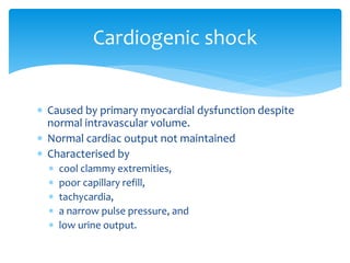
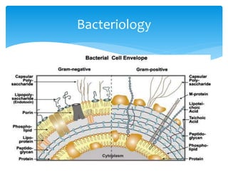
Define and classify shock. Discuss
the current concepts in the
pathophysiology and management
of endotoxic shock
Dr Eretare C. Odjugo
 Introduction
 Definition of terms
 Classification of shock
 Bacteriology
 Aetiology of septic shock
 Pathophysiology of septic shock
 Clinical features of septic shock
 Evaluation
Outline
 Treatment of septic shock
 Prognosis
 Prevention
 Conclusion
 References
Outline
 Infections are a continuous challenge in medical
practice especially in resource limited areas
 The extent of the infection is affected by the persons
co-morbidities, virulence of the infective organism,
hosts immune response and other factors.
Introduction
 Infection: An infection is the invasion of an organism's
body tissues by disease-causing agents, their
multiplication, and the reaction of host tissues to the
infectious agents and the toxins they produce.
 Shock: A state of cellular and tissue hypoxia due to
either reduced oxygen delivery, increased
consumption, inadequate oxygen utilisation or a
combination of these processes.
Terminology
 Sepsis is defined as life-threatening organ dysfunction
due to dysregulated host response to infection.
 Organ dysfunction is defined as an acute change in
total Sequential Organ Failure Assessment (SOFA)
score of 2 points or greater secondary to the infection
cause.
Terminology
 Systemic Inflammatory Response Syndrome (SIRS):
 Adult: Manifestations of SIRS include, but are not limited to:
 Body temperature <36 °C or >38 °C
 Heart rate >90 beats per minute
 Tachypnea (high respiratory rate), with >20 breaths per
minute; or, an arterial PaCO2 less than 4.3 kPa (32 mmHg)
 White blood cell count <4 x 10⁹ cells/L or >12 x 109 cells/L; or
the presence of >10% immature neutrophils (band forms).
Band forms >3% is called bandemia or a "left-shift.”
Terminology
 In children
 Heart rate >2 SD above normal for age in the absence of
stimuli such as pain and drug administration, or unexplained
persistent elevation for >30 minutes to 4 hours.
 Body temperature obtained orally, rectally, <36 °C or >38.5 °C.
 Respiratory rate >2 SD above normal for age or the
requirement for mechanical ventilation not related to
neuromuscular disease or the administration of anesthesia.
 White blood cell count elevated or depressed for age not
related to chemotherapy, or >10% bands plus other immature
forms.
Terminology
 Septic shock: Septic shock is defined by persisting
hypotension requiring vasopressors to maintain a
mean arterial pressure of 65 mm Hg or higher and a
serum lactate level greater than 2 mmol/L (18 mg/dL)
despite adequate volume resuscitation
Terminology
Aetiology
 Hypovolaemic
 Distributive
 Obstructive
 Cardiogenic
Classification of shock
 From loss of blood volume or plasma
 Seen in major bleeding, trauma and burns
 Characterised by
 tachycardia,
 cool clammy extremities,
 hypotension,
 dry skin and mucous membranes, and
 poor turgor.
Hypovolaemic shock
 Caused by an extrinsic or intrinsic impedance to
circulation
 Pulmonary embolism
 Cardiac tamponade
Obstructive shock
 Caused by primary myocardial dysfunction despite
normal intravascular volume.
 Normal cardiac output not maintained
 Characterised by
 cool clammy extremities,
 poor capillary refill,
 tachycardia,
 a narrow pulse pressure, and
 low urine output.
Cardiogenic shock
 Excessive vasodilation
 Impaired blood flow
 Reduced vasomotor resistance or increased
capacitance
 It’s characterised by
 high cardiac output,
 hypotension,
 a large pulse pressure,
 a low diastolic pressure, and
 warm extremities with good capillary refill
Distributive shock
 Septic shock
 Anaphylactic shock
 Neurogenic shock
Distributive shock
Bacteriology
 The normal physiologic response to localized
infection includes activation of host defence
mechanisms that result in the
 influx of activated neutrophils and monocytes,
 release of inflammatory mediators,
 local vasodilation,
 increased endothelial permeability, and
 activation of coagulation pathways.
Pathophysiology
 These responses are brought about by the interaction
between the host responses and the infective agent
 In sepsis, there is an exaggeration of these host
responses.
 leading to
 diffuse endothelial disruption,
 vascular permeability,
 vasodilation, and
 thrombosis of end-organ capillaries
Pathophysiology
 In Gram negative bacteria, the Lipid A moiety of the
lipopolysaccharide molecule leads to cytokine
induction via the lipoteichoic acid.
 For Gram positive bacteria they may also bring about
production of cytokines by secretion of super-
antigens.
Pathophysiology
Sepsis induced ARDS
Septic encephalopathy
Sepsis induced coagulopathy
 This is the system responsible for the progression too
septic shock
 The main components are the vascular response
 Arteriolar vasodilatation
 Increased capacitance
 And Myocardial activity
 Chronotrophic
 Ionotrophic
Circulatory dysfunction in sepsis
 The hypotension is initially compensated by an
increased cardiac output but over time this also fails.
 Myocardial suppression is brought about by
 Beta adrenergic receptor downregulation
 Nitric oxide
 Pulmonary hypertension
Circulatory dysfunction in sepsis
 There is also a challenge with oxygen utilisation at
tissues
 Mitochondrial dysfunction which leads to anaerobic
respiration with resultant lactate production
 Diminished offloading at the tissues
 There is loss of the normal auto-regulatory function of
the autonomic nervous system and thus vital organs
(brain and heart) which normally receive preference do
not
Circulatory dysfunction in sepsis
 Major site of bacteria
 Hypoperfusion leads to failure of gut mucosal barrier
 Translocation of bacteria and endotoxin
 Propagation of sepsis
 Sepsis also causes ileus
GI dysfunction
 Hypoxic hypoxia: Decrease oxygen supply
 Histotoxic hypoxia
 Apoptosis
 Coagulopathy
 Immunosuppression
Organ dysfunction in sepsis
Clinical features
 Temperature abnormalities (fever or hypothermia)
 Elevated pulse rate with warm or cold extremities
 Altered mental status
 Dyspnoea
 Malaise and lethargy
 Localizing symptoms referable to organ systems may
provide useful clues to the etiology of sepsis.
 Full blood count (Leukocytosis with left shift)
 Blood culture (not always positive)
 Serum electrolytes, urea and creatinine
 Liver function test including total protein and albumin
 Clotting profile
 Serum lactate (assessment of perfusion)
 Blood glucose monitoring (predictor of mortality)
 Urinalysis and urine culture
Evaluation
 Specialised investigations to confirm source of
infection
 CT scans/MRIs of various regions
 Plain radiographs
 Ultrasound scans
Evaluation
SOFA scoring
 Based on the current literature, include the following:
 Early recognition
 Source control
 Early and adequate antibiotic therapy
 Early hemodynamic resuscitation and continued
support
 Proper ventilator management with low tidal volume
in patients with acute respiratory distress syndrome
(ARDS)
Management principles
 Resuscitation
 Venous access
 Fluids (Iso-osmotic crystalloids) 20 -40ml/kg over the first hour.
 Respiratory and ventilator support
 Urethral catheterisation
 Correction of anaemia
 Circulatory support
 Antimicrobial therapy
 Source control
 Temperature control
 Nutritional support
Treatment
Haemodynamic support
Aim for MAP >65mmHg
 Fluid
 Crystalloids
 Colloids (4% Albumin)
 Must be monitored for
overload
 Vasopressors
 Norepinephrine
 Epinephrine
 Dopamine
 Phenylephrine
 Ionotropes
 Dopexamine
 Dopamine
 Dobutamine
 Crystalloids ( 30ml/kg over 1st hour)
 Normal saline
 Ringers lactate (not preferred due to interaction with
lactate assays)
 Colloids
 4% Albumin: as effective as normal saline at less volume
more expensive
 Not readily available
Fluids
Vasopressor
First line
 Dopamine 5 – 10mcg/kg/min
 Tachyarhythmias
 May be increased to
20mcg/kg/min
 Norepinephrine 5 – 20mcg/min
 Not weight dependent
 Predictable response
 Shorter hospital stay with less
mortality
 Alpha agonist thus potent
vasoconstrictors
 0.2 – 1.5mcg/kg/min
 Second line
 Synthetic Human Angiotensin 2 (25ml/kg) improves response
when used with 1st line
 Epinephrine potent inotrope
 Causes myocardial and splanchnic ischemia
 Phenylephrine
 Selective potent vasopressor
 Causes reduce myocardial contractility and rate
 ADH
 Reserved for salvage therapy thus should not be used alone
 More potent when acting with nor epinephrine
Vasopressors
 Dobutamine:
 Recommended only when there is hypoperfusion
despite fluid targets and MAP targets have been met.
 Given at 20mcg/kg/min
 Dopamine
Inotropes
 Initial therapy should be combination broad spectrum for
suspected causative agents.
 Daily evaluation for effectiveness
 De-escalation to monotherapy later.
 An aminoglycoside (e.g gentamicin) should be used for
“antibiotic-experienced” patients.
 Cephamycins (Cefotetan) and Carbapenems are preferred for
ESBL producing bacteria (Kliebsiella and E. coli)
 Immunocompromised patients should receive carbapenems
or 4th gen Cephalosporins)
Anti-microbial therapy
 Intra-abdominal infection
 Antibacterials
 Metronidazole – Meropenem
 Imipenem – Cilastin
 Levofloxacin or Ciprofloxacin
 Piperacilin or Tazobactam
 Ceftazidim or Cefepime
 Antifungals
 Caspofungin
 Micafungin
 Fluconazole in C. albicans
Antimicrobial therapy
 The American College of Critical Care Medicine
recommends
 Avoidance of ACTH suppression test
 Use of Hydrocortisone as first line
 Use in patients with suspected adrenal insufficiency
 Do not use dexamethasone therapy for septic shock or
ARDS
 Treatment of septic shock
 200 mg/day in 4 divided doses or
 100mg bolus then 10mg/hour.
 If given for >14 days, it should be tapered off
Corticosteroid use
 Target is between 80 - 110 mg/dl (4.4 – 6.1mmol/l)
 Hyperglycaemia confers a 10% rise in mortality.
Glycaemic control
 In absence of bleeding
 Unfractionated heparin
 LMW heparin can be used.
 Dalteparin used in severe renal dysfunction CrCl
<30ml/min
 Compression stockings and intermittent compression
devices may be used.
DVT prophylaxis
 Treat underlying cause
 Transfuse with plasma or platelet concentrate
 Don’t treat prophylactically
DIC
Treatment of ARDS
 Intubation and
mechanical ventilation
 5 – 8 l/min
 Use of PEEP to prevent
lung injury
 Extra-corporeal
Membrane Oxygenation
 Ubi pus, ibi evacua.
 Antibiotic therapy alone may not suffice for some
causes of septic shock
 The source of infection should be controlled if not all
therapies will eventually fail.
 Abscesses should be drained either percutaneously or
an open drainage.
 Necrotic tissue should be debrided.
Surgery
 Renal dysfunction (AKI) may require dialysis
 For hepatic dysfunction, therapy is mostly supportive
Other modalities
 In the past decades, recovery and survival from septic
shock is on the rise.
 Some countries have as high as 50% (Australia) while
others 30% most developed countries.
 In our setting, septic shock is almost invariably fatal
due to challenges with finance, resources and
personnel.
Prognosis
 Basic measures to prevent nosocomial infections
include the following
 Shortening the hospital stay
 Removing indwelling catheters as early as possible
 Avoiding unnecessary invasive procedures
 Using aseptic techniques
 Prophylactic antibiotics for GI surgeries are also of
benefit
Prevention
 Sepsis and septic shock are major contributors to
surgical morbidity and mortality
 Early recognition and intervention are key to
improving outcomes.
 The best form of treatment still remains prevention
by eliminating risk factors as much as possible
Conclusion
 https://jamanetwork.com/journals/jama/article-
abstract/2598892 accessed last 3/8/20.
 https://emedicine.medscape.com/article/168402-
overview#a1 accessed on 4/8/20
 Surviving Sepsis: Guidelines for management of sepsis and
septic shock 2012
 https://www.uptodate.com/contents/definition-
classification-etiology-and-pathophysiology-of-shock-in-
adults accessed 03/08/20
 Images obtained from https://www.bing.com and are the
properties of the respective copyright holders
References
Thank you

Septic shock.pptx

  • 1.
    Define and classifyshock. Discuss the current concepts in the pathophysiology and management of endotoxic shock Dr Eretare C. Odjugo
  • 2.
     Introduction  Definitionof terms  Classification of shock  Bacteriology  Aetiology of septic shock  Pathophysiology of septic shock  Clinical features of septic shock  Evaluation Outline
  • 3.
     Treatment ofseptic shock  Prognosis  Prevention  Conclusion  References Outline
  • 4.
     Infections area continuous challenge in medical practice especially in resource limited areas  The extent of the infection is affected by the persons co-morbidities, virulence of the infective organism, hosts immune response and other factors. Introduction
  • 6.
     Infection: Aninfection is the invasion of an organism's body tissues by disease-causing agents, their multiplication, and the reaction of host tissues to the infectious agents and the toxins they produce.  Shock: A state of cellular and tissue hypoxia due to either reduced oxygen delivery, increased consumption, inadequate oxygen utilisation or a combination of these processes. Terminology
  • 7.
     Sepsis isdefined as life-threatening organ dysfunction due to dysregulated host response to infection.  Organ dysfunction is defined as an acute change in total Sequential Organ Failure Assessment (SOFA) score of 2 points or greater secondary to the infection cause. Terminology
  • 8.
     Systemic InflammatoryResponse Syndrome (SIRS):  Adult: Manifestations of SIRS include, but are not limited to:  Body temperature <36 °C or >38 °C  Heart rate >90 beats per minute  Tachypnea (high respiratory rate), with >20 breaths per minute; or, an arterial PaCO2 less than 4.3 kPa (32 mmHg)  White blood cell count <4 x 10⁹ cells/L or >12 x 109 cells/L; or the presence of >10% immature neutrophils (band forms). Band forms >3% is called bandemia or a "left-shift.” Terminology
  • 9.
     In children Heart rate >2 SD above normal for age in the absence of stimuli such as pain and drug administration, or unexplained persistent elevation for >30 minutes to 4 hours.  Body temperature obtained orally, rectally, <36 °C or >38.5 °C.  Respiratory rate >2 SD above normal for age or the requirement for mechanical ventilation not related to neuromuscular disease or the administration of anesthesia.  White blood cell count elevated or depressed for age not related to chemotherapy, or >10% bands plus other immature forms. Terminology
  • 10.
     Septic shock:Septic shock is defined by persisting hypotension requiring vasopressors to maintain a mean arterial pressure of 65 mm Hg or higher and a serum lactate level greater than 2 mmol/L (18 mg/dL) despite adequate volume resuscitation Terminology
  • 13.
  • 14.
     Hypovolaemic  Distributive Obstructive  Cardiogenic Classification of shock
  • 15.
     From lossof blood volume or plasma  Seen in major bleeding, trauma and burns  Characterised by  tachycardia,  cool clammy extremities,  hypotension,  dry skin and mucous membranes, and  poor turgor. Hypovolaemic shock
  • 16.
     Caused byan extrinsic or intrinsic impedance to circulation  Pulmonary embolism  Cardiac tamponade Obstructive shock
  • 17.
     Caused byprimary myocardial dysfunction despite normal intravascular volume.  Normal cardiac output not maintained  Characterised by  cool clammy extremities,  poor capillary refill,  tachycardia,  a narrow pulse pressure, and  low urine output. Cardiogenic shock
  • 18.
     Excessive vasodilation Impaired blood flow  Reduced vasomotor resistance or increased capacitance  It’s characterised by  high cardiac output,  hypotension,  a large pulse pressure,  a low diastolic pressure, and  warm extremities with good capillary refill Distributive shock
  • 19.
     Septic shock Anaphylactic shock  Neurogenic shock Distributive shock
  • 20.
  • 21.
     The normalphysiologic response to localized infection includes activation of host defence mechanisms that result in the  influx of activated neutrophils and monocytes,  release of inflammatory mediators,  local vasodilation,  increased endothelial permeability, and  activation of coagulation pathways. Pathophysiology
  • 22.
     These responsesare brought about by the interaction between the host responses and the infective agent  In sepsis, there is an exaggeration of these host responses.  leading to  diffuse endothelial disruption,  vascular permeability,  vasodilation, and  thrombosis of end-organ capillaries Pathophysiology
  • 23.
     In Gramnegative bacteria, the Lipid A moiety of the lipopolysaccharide molecule leads to cytokine induction via the lipoteichoic acid.  For Gram positive bacteria they may also bring about production of cytokines by secretion of super- antigens. Pathophysiology
  • 26.
  • 27.
  • 28.
  • 29.
     This isthe system responsible for the progression too septic shock  The main components are the vascular response  Arteriolar vasodilatation  Increased capacitance  And Myocardial activity  Chronotrophic  Ionotrophic Circulatory dysfunction in sepsis
  • 30.
  • 31.
     The hypotensionis initially compensated by an increased cardiac output but over time this also fails.  Myocardial suppression is brought about by  Beta adrenergic receptor downregulation  Nitric oxide  Pulmonary hypertension Circulatory dysfunction in sepsis
  • 32.
     There isalso a challenge with oxygen utilisation at tissues  Mitochondrial dysfunction which leads to anaerobic respiration with resultant lactate production  Diminished offloading at the tissues  There is loss of the normal auto-regulatory function of the autonomic nervous system and thus vital organs (brain and heart) which normally receive preference do not Circulatory dysfunction in sepsis
  • 33.
     Major siteof bacteria  Hypoperfusion leads to failure of gut mucosal barrier  Translocation of bacteria and endotoxin  Propagation of sepsis  Sepsis also causes ileus GI dysfunction
  • 34.
     Hypoxic hypoxia:Decrease oxygen supply  Histotoxic hypoxia  Apoptosis  Coagulopathy  Immunosuppression Organ dysfunction in sepsis
  • 36.
    Clinical features  Temperatureabnormalities (fever or hypothermia)  Elevated pulse rate with warm or cold extremities  Altered mental status  Dyspnoea  Malaise and lethargy  Localizing symptoms referable to organ systems may provide useful clues to the etiology of sepsis.
  • 37.
     Full bloodcount (Leukocytosis with left shift)  Blood culture (not always positive)  Serum electrolytes, urea and creatinine  Liver function test including total protein and albumin  Clotting profile  Serum lactate (assessment of perfusion)  Blood glucose monitoring (predictor of mortality)  Urinalysis and urine culture Evaluation
  • 38.
     Specialised investigationsto confirm source of infection  CT scans/MRIs of various regions  Plain radiographs  Ultrasound scans Evaluation
  • 39.
  • 40.
     Based onthe current literature, include the following:  Early recognition  Source control  Early and adequate antibiotic therapy  Early hemodynamic resuscitation and continued support  Proper ventilator management with low tidal volume in patients with acute respiratory distress syndrome (ARDS) Management principles
  • 41.
     Resuscitation  Venousaccess  Fluids (Iso-osmotic crystalloids) 20 -40ml/kg over the first hour.  Respiratory and ventilator support  Urethral catheterisation  Correction of anaemia  Circulatory support  Antimicrobial therapy  Source control  Temperature control  Nutritional support Treatment
  • 42.
    Haemodynamic support Aim forMAP >65mmHg  Fluid  Crystalloids  Colloids (4% Albumin)  Must be monitored for overload  Vasopressors  Norepinephrine  Epinephrine  Dopamine  Phenylephrine  Ionotropes  Dopexamine  Dopamine  Dobutamine
  • 43.
     Crystalloids (30ml/kg over 1st hour)  Normal saline  Ringers lactate (not preferred due to interaction with lactate assays)  Colloids  4% Albumin: as effective as normal saline at less volume more expensive  Not readily available Fluids
  • 44.
    Vasopressor First line  Dopamine5 – 10mcg/kg/min  Tachyarhythmias  May be increased to 20mcg/kg/min  Norepinephrine 5 – 20mcg/min  Not weight dependent  Predictable response  Shorter hospital stay with less mortality  Alpha agonist thus potent vasoconstrictors  0.2 – 1.5mcg/kg/min
  • 45.
     Second line Synthetic Human Angiotensin 2 (25ml/kg) improves response when used with 1st line  Epinephrine potent inotrope  Causes myocardial and splanchnic ischemia  Phenylephrine  Selective potent vasopressor  Causes reduce myocardial contractility and rate  ADH  Reserved for salvage therapy thus should not be used alone  More potent when acting with nor epinephrine Vasopressors
  • 46.
     Dobutamine:  Recommendedonly when there is hypoperfusion despite fluid targets and MAP targets have been met.  Given at 20mcg/kg/min  Dopamine Inotropes
  • 47.
     Initial therapyshould be combination broad spectrum for suspected causative agents.  Daily evaluation for effectiveness  De-escalation to monotherapy later.  An aminoglycoside (e.g gentamicin) should be used for “antibiotic-experienced” patients.  Cephamycins (Cefotetan) and Carbapenems are preferred for ESBL producing bacteria (Kliebsiella and E. coli)  Immunocompromised patients should receive carbapenems or 4th gen Cephalosporins) Anti-microbial therapy
  • 48.
     Intra-abdominal infection Antibacterials  Metronidazole – Meropenem  Imipenem – Cilastin  Levofloxacin or Ciprofloxacin  Piperacilin or Tazobactam  Ceftazidim or Cefepime  Antifungals  Caspofungin  Micafungin  Fluconazole in C. albicans Antimicrobial therapy
  • 49.
     The AmericanCollege of Critical Care Medicine recommends  Avoidance of ACTH suppression test  Use of Hydrocortisone as first line  Use in patients with suspected adrenal insufficiency  Do not use dexamethasone therapy for septic shock or ARDS  Treatment of septic shock  200 mg/day in 4 divided doses or  100mg bolus then 10mg/hour.  If given for >14 days, it should be tapered off Corticosteroid use
  • 50.
     Target isbetween 80 - 110 mg/dl (4.4 – 6.1mmol/l)  Hyperglycaemia confers a 10% rise in mortality. Glycaemic control
  • 51.
     In absenceof bleeding  Unfractionated heparin  LMW heparin can be used.  Dalteparin used in severe renal dysfunction CrCl <30ml/min  Compression stockings and intermittent compression devices may be used. DVT prophylaxis
  • 52.
     Treat underlyingcause  Transfuse with plasma or platelet concentrate  Don’t treat prophylactically DIC
  • 53.
    Treatment of ARDS Intubation and mechanical ventilation  5 – 8 l/min  Use of PEEP to prevent lung injury  Extra-corporeal Membrane Oxygenation
  • 54.
     Ubi pus,ibi evacua.  Antibiotic therapy alone may not suffice for some causes of septic shock  The source of infection should be controlled if not all therapies will eventually fail.  Abscesses should be drained either percutaneously or an open drainage.  Necrotic tissue should be debrided. Surgery
  • 55.
     Renal dysfunction(AKI) may require dialysis  For hepatic dysfunction, therapy is mostly supportive Other modalities
  • 57.
     In thepast decades, recovery and survival from septic shock is on the rise.  Some countries have as high as 50% (Australia) while others 30% most developed countries.  In our setting, septic shock is almost invariably fatal due to challenges with finance, resources and personnel. Prognosis
  • 58.
     Basic measuresto prevent nosocomial infections include the following  Shortening the hospital stay  Removing indwelling catheters as early as possible  Avoiding unnecessary invasive procedures  Using aseptic techniques  Prophylactic antibiotics for GI surgeries are also of benefit Prevention
  • 59.
     Sepsis andseptic shock are major contributors to surgical morbidity and mortality  Early recognition and intervention are key to improving outcomes.  The best form of treatment still remains prevention by eliminating risk factors as much as possible Conclusion
  • 60.
     https://jamanetwork.com/journals/jama/article- abstract/2598892 accessedlast 3/8/20.  https://emedicine.medscape.com/article/168402- overview#a1 accessed on 4/8/20  Surviving Sepsis: Guidelines for management of sepsis and septic shock 2012  https://www.uptodate.com/contents/definition- classification-etiology-and-pathophysiology-of-shock-in- adults accessed 03/08/20  Images obtained from https://www.bing.com and are the properties of the respective copyright holders References
  • 61.

Editor's Notes

  • #4 W-
  • #9 When two or more of these criteria are met with or without evidence of infection, patients may be diagnosed with "SIRS." Patients with SIRS and acute organ dysfunction may be termed "severe SIRS."[3][4][9] Note: Fever and an increased white blood cell count are features of the acute-phase reaction, while an increased heart rate is often the initial sign of hemodynamic compromise. An increased rate of breathing may be related to the increased metabolic stress due to infection and inflammation, but may also be an ominous sign of inadequate perfusion resulting in the onset of anaerobic cellular metabolism.
  • #10 Temperature or white blood cell count must be abnormal to qualify as SIRS in pediatric patients.[12] In infants, also includes heart rate less than 10th percentile for age in the absence of vagal stimuli, beta-blockers, or congenital heart disease or unexplained persistent depression for greater than 30 minutes.
  • #18 Myocardiac infarction Cardiomyopathy Valvular heart diseases
  • #21 Gram-positive and gram-negative bacteria induce a variety of proinflammatory mediators, including the cytokines mentioned above, which play a pivotal role in initiating sepsis and shock. Various bacterial cell-wall components are known to release the cytokines, including lipopolysaccharide (LPS; gram-negative bacteria), peptidoglycan (gram-positive and gram-negative bacteria), and lipoteichoic acid (gram-positive bacteria).
  • #30 Hypotension is caused by the redistribution of intravascular fluid volume that results from reduced arterial vascular tone, diminished venous return from venous dilation, and release of myocardial depressant substances.
  • #31 The predominant hemodynamic feature of septic shock is arterial vasodilation. The mechanisms implicated i (1) activation of adenosine triphosphate (ATP)-sensitive potassium channels in vascular smooth muscle cells and (2) activation of NO synthase. The potassium-ATP channels are directly activated by lactic acidosis. NO also activates potassium channels. Potassium efflux from cells results in hyperpolarization, inhibition of calcium influx, and vascular smooth muscle relaxation. [26] The resulting vasodilation can be refractory to endogenous vasoactive hormones (eg, norepinephrine and epinephrine) that are released during shock.
  • #33 The basic pathophysiologic problem seems to be a disparity between oxygen uptake and oxygen demand in the tissues, which may be more pronounced in some areas than in others. This disparity is termed maldistribution of blood flow, either between or within organs, with a resultant defect in the capacity for local extraction of oxygen. 
  • #35 HH: Endothelial damage, ROS, NO and other vasoactive substances Cyto H: Disruption of O2 utilisation by Endotoxin, NO and TNF-A Apoptosis: Accelerated rate of programmed cell death Coagulopathy: Microthrombi and haemorrhages limit supply
  • #38 Lactate levels higher than 2.5 mmol/L are associated with an increase in mortality. The higher the serum lactate, the worse the degree of shock and the higher the mortality.
  • #40 >2 is required for the diagnosis of sepsis
  • #41 The treatment of patients with septic shock has the following major goals: Start adequate antibiotic therapy (proper dosage and spectrum) as early as possible Identify the source of infection, and treat with antimicrobial therapy, surgery, or both (source control) Resuscitate the patient, using supportive measures to correct hypoxia, hypotension, and impaired tissue oxygenation (hypoperfusion) Maintain adequate organ system function, guided by cardiovascular monitoring, and interrupt the progression to multiple organ dysfunction syndrome (MODS)
  • #43 (Recent studies showed the validity of the 70-75 mm Hg lower mean arterial pressure target or 80-85 mm Hg in those patients with preexisting hypertension.)
  • #44 Up to 4 litres of crystalloids can be given
  • #45 Dosages range from 2 to 20 µg/kg/min. A dosage lower than 5 µg/kg/min results in vasodilation of renal, mesenteric, and coronary beds. [11] At a dosage of 5-10 µg/kg/min, beta1 -adrenergic effects induce an increase in cardiac contractility and heart rate. At dosages of about 10 µg/kg/min, alpha-adrenergic effects lead to arterial vasoconstriction and elevation in blood pressure. 
  • #46 Ang II – 20mcg/kg/min and increased in doses of up to 5mcg/kg every 15 minutes. Causes thrombo-embolic phenomena.
  • #50 Do not administer corticosteroids to treat sepsis when shock is not present
  • #54 The goals of mechanical ventilation include the following: Improving gas exchange Reducing work of breathing Avoiding oxygen toxicity Minimizing high airway pressures Avoiding further lung damage Allowing the injured lung to heal