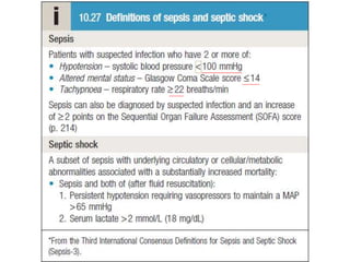
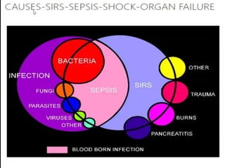
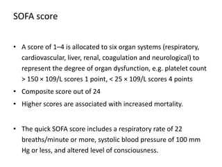
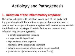
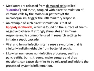
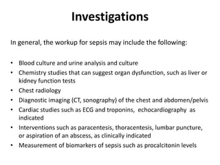
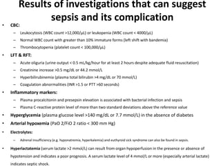
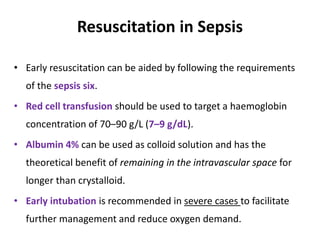
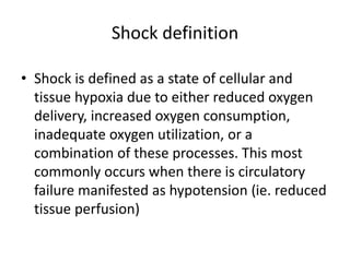
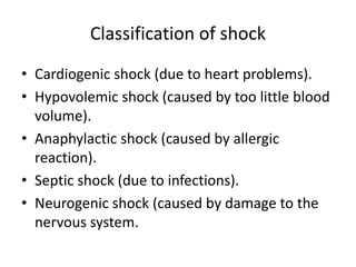
Sepsis
Dr. Jwan Ali Ahmed
Shock definition
• Shock is defined as a state of cellular and
tissue hypoxia due to either reduced oxygen
delivery, increased oxygen consumption,
inadequate oxygen utilization, or a
combination of these processes. This most
commonly occurs when there is circulatory
failure manifested as hypotension (ie. reduced
tissue perfusion)
Classification of shock
• Cardiogenic shock (due to heart problems).
• Hypovolemic shock (caused by too little blood
volume).
• Anaphylactic shock (caused by allergic
reaction).
• Septic shock (due to infections).
• Neurogenic shock (caused by damage to the
nervous system.
Systemic inflammatory reaction
syndrome (SIRS)
Clinically, the Systemic Inflammatory Response Syndrome (SIRS) is the
occurrence of at least two of the following criteria:
1. Fever of more than 38°C (100.4°F) or less than 36°C (96.8°F)
2. Heart rate of more than 90 beats per minute
3. Respiratory rate of more than 20 breaths per minute or arterial
carbon dioxide tension (PaCO2) of less than 32 mm Hg
4. Abnormal white blood cell count (>12,000/µL or < 4,000/µL or
>10% immature [band] forms)
She has sirs but not sepsis
sepsis
Test : Serum lactate
SOFA score
• A score of 1–4 is allocated to six organ systems (respiratory,
cardiovascular, liver, renal, coagulation and neurological) to
represent the degree of organ dysfunction, e.g. platelet count
> 150 × 109/L scores 1 point, < 25 × 109/L scores 4 points
• Composite score out of 24
• Higher scores are associated with increased mortality.
• The quick SOFA score includes a respiratory rate of 22
breaths/minute or more, systolic blood pressure of 100 mm
Hg or less, and altered level of consciousness.
Aetiology and Pathogenesis
1. Initiation of the inflammatory response
The process begins with infection in one part of the body that
triggers a localized inflammatory response. Appropriate source
control and a competent immune system will, in most cases, contain
the infection at this stage. If certain factors are present, the
infection may become systemic.
– a genetic predisposition to sepsis
– a large microbiological load
– high virulence of the organism
– resistance of the organism to treatment
– delay in source control (either surgical or antimicrobial)
– patient factors (immune status, nutrition, frailty)
• Mediators are released from damaged cells (called
‘alarmins’) and these, coupled with direct stimulation of
immune cells by the molecular patterns of the
microorganism, trigger the inflammatory response.
• An example of such direct stimulation is that of
lipopolysaccharide, which is found on the surface of Gram-
negative bacteria. It strongly stimulates an immune
response and is commonly used in research settings to
initiate a septic cascade.
• Viral and fungal infections can cause a syndrome that is
clinically indistinguishable from bacterial sepsis.
• Likewise, numerous non-infective processes, such as
pancreatitis, burns, trauma, major surgery and drug
reactions, can cause alarmins to be released and initiate the
process of systemic inflammation.
2- Propagation of the Inflammatory Response
• Once activated, immune cells such as macrophages release the
inflammatory cytokines interleukin-2 (IL-2), IL-6 and TNF-a, which, in
turn, activate neutrophils.
• Activated neutrophils express adhesion factors and release various
other inflammatory and toxic substances; the net effects are
vasodilatation (via activation of inducible nitric oxide synthase
enzymes) and damage to the endothelium.
• Neutrophils migrate into the interstitial space; fluid and plasma
proteins will also leak through the damaged endothelium, leading to
oedema and intravascular fluid depletion.
3- Activation of the Coagulation system
• Damaged endothelium triggers the coagulation cascade (via
tissue factor, factor VII, and reduced activity of proteins C and
S) and thrombus forms within the microvasculature.
• A vicious circle of endothelial injury, intravascular coagulation
and microvascular occlusion develops, causing more tissue
damage and further release of inflammatory mediators.
• In severe sepsis, intravascular coagulation can become
widespread. This is referred to as disseminated intravascular
coagulation (DIC) and usually heralds the onset of multi-organ
failure.
4- Pathophysiology of Organ Damage
Macrovascular:
– Severe hypovolaemia, vasodilatation or septic cardiomyopathy can
reduce oxygen delivery, causing tissue hypoxia. Paradoxically, most
patients with sepsis have an increased cardiac output and oxygen
delivery.
Microvascular:
– Tissue injury can occur from hypoxia secondary to microvascular
injury and thrombosis. Damaged epithelium permits neutrophils,
proteins and fluid to leak out.
 Shunting:
• Organs fail in sepsis despite supranormal blood flow.
• It is likely that arteriovenous shunt pathways exist within vascular
beds; these shunts open up in septic shock.
 Cellular: Cells are damaged by a number of mechanisms in sepsis:
1) direct injury by microorganisms;
2) injury from toxins produced by immune cells, e.g. O free radicals;
3) mitochondrial injury causing cytopathic hypoxia – cells are
unable to metabolize oxygen;
4) apoptosis – if the cell injury is sufficient, capsase enzymes are
activated within the nucleus and programmed cell death occurs;
5) hypoxia from micro-and macrovascular pathology.
5- Role of Lactate
• Lactate is an excellent biomarker for the severity of sepsis.
• Hyperlactataemia (serum lactate > 2.4 mmol/L or 22 mg/dL) is used
as a marker of severity.
• It is caused by all types of shock and therefore is not specific to
sepsis.
• A lactate level of > 8 mmol/L (>73 mg/dL) is associated with an
extremely high mortality and should trigger immediate escalation.
• Measures to optimize oxygen delivery should be sought, and the
adequacy of resuscitation measured by lactate clearance.
Physiology of hyperlactataemia
6- The anti-inflammatory cascade
• As the inflammatory state develops, a compensatory anti-
inflammatory system is activated involving the release of anti-
inflammatory cytokines such as IL-4 and IL-10 from immune cells.
• While such mechanisms are necessary to keep the inflammatory
response in check, they may lead to a period of immunosuppression
after the initial septic episode.
• Patients recovering from severe sepsis are prone to developing
secondary infections due to a combination of this immunosuppression
and the presence of indwelling devices.
Sepsis mimics
• Pancreatitis
• Drug reactions
• Widespread vasculitis – catastrophic antiphospholipid
syndrome, Goodpasture’s syndrome
• Autoimmune diseases – inflammatory bowel disease,
rheumatoid, arthritis, systemic lupus erythematosus
• Malignancy – carcinoid syndrome
• Haematological conditions – haemophagocytic syndrome,
diffuse lymphoma, thrombotic thrombocytopenic purpura.
• Sepsis is the most common cause of acute systemic inflammation, up
to 20% of patients initially treated for sepsis will have a non-
infectious cause: that is, a sepsis mimic.
• These conditions should be considered where the clinical picture is
not typical, no source of sepsis can be found, or the inflammatory
response seems excessive in the context of local infection.
• Early reconsideration of the diagnosis of sepsis is crucial, as many of
the ‘sepsis mimics’ offer a finite time window for specific
intervention, after which irreversible organ damage will have
occurred.
Non-infective processes, such as pancreatitis,
burns, trauma, major surgery and drug
reactions, can cause alarmins to be released and
initiate the process of systemic inflammation.
Investigations
In general, the workup for sepsis may include the following:
• Blood culture and urine analysis and culture
• Chemistry studies that can suggest organ dysfunction, such as liver or
kidney function tests
• Chest radiology
• Diagnostic imaging (CT, sonography) of the chest and abdomen/pelvis
• Cardiac studies such as ECG and troponins, echocardiography as
indicated
• Interventions such as paracentesis, thoracentesis, lumbar puncture,
or aspiration of an abscess, as clinically indicated
• Measurement of biomarkers of sepsis such as procalcitonin levels
Results of investigations that can suggest
sepsis and its complication
• CBC:
– Leukocytosis (WBC count >12,000/µL) or leukopenia (WBC count < 4000/µL)
– Normal WBC count with greater than 10% immature forms (left shift with bandemia)
– Thrombocytopenia (platelet count < 100,000/µL)
• LFT & RFT:
– Acute oliguria (urine output < 0.5 mL/kg/hour for at least 2 hours despite adequate fluid resuscitation)
– Creatinine increase >0.5 mg/dL or 44.2 mmol/L
– Hyperbilirubinemia (plasma total bilirubin >4 mg/dL or 70 mmol/L)
– Coagulation abnormalities (INR >1.5 or PTT >60 seconds)
• Inflammatory markers:
– Plasma procalcitonin and presepsin elevation is associated with bacterial infection and sepsis
– Plasma C-reactive protein level of more than two standard deviations above the reference value
• Hyperglycemia (plasma glucose level >140 mg/dL or 7.7 mmol/L) in the absence of diabetes
• Arterial hypoxemia (PaO 2/FiO 2 ratio < 300 mm Hg)
• Electrolytes:
– Adrenal insufficiency (e.g. hyponatremia, hyperkalemia) and euthyroid sick syndrome can also be found in sepsis.
• Hyperlactatemia (serum lactate >2 mmol/L) can result from organ hypoperfusion in the presence or absence of
hypotension and indicates a poor prognosis. A serum lactate level of 4 mmol/L or more (especially arterial lactate)
indicates septic shock.
Resuscitation in Sepsis
• Early resuscitation can be aided by following the requirements
of the sepsis six.
• Red cell transfusion should be used to target a haemoglobin
concentration of 70–90 g/L (7–9 g/dL).
• Albumin 4% can be used as colloid solution and has the
theoretical benefit of remaining in the intravascular space for
longer than crystalloid.
• Early intubation is recommended in severe cases to facilitate
further management and reduce oxygen demand.
Antibiotics
• Appropriate antibiotics should be administered as early as
possible
• The antibiotic choice will depend on local patterns of resistance,
patient risk factors and the likely source of infection.
• Microbiological samples (such as blood cultures, urine or CSF)
should be taken, but this should not delay antibiotic
administration, if obtaining samples is difficult.
Drugs used for empiric therapy are
• Imipenem
• Meropenem
• Tigecycline
• Piperacillin-tazobactam
• Ampicillin-sulbactam
• Moxifloxacin
Early source control
• Source control requires an accurate diagnosis; urgent
investigations should be performed as soon as physiological
stability has been established.
• A CT scan of the chest and abdomen with contrast is a high-yield
test in this context.
• Specific points in the history should be reviewed, such as risk
factors for human immunodeficiency virus (HIV), contacts with
tuberculosis and underlying immune status.
Immunocompromised patients will be susceptible to a far
broader spectrum of infectious microorganisms.
Noradrenaline (norepinephrine) for
refractory hypotension
• Central venous access should be established early in the
resuscitation process and a noradrenaline infusion commenced.
• If there is severe hypotension, it is not necessary to wait until 30
mL/kg of fluid has been administered before commencing
noradrenaline; early vasopressor use may improve the outcome
from acute kidney injury.
• Measurement of central and mixed venous oxygen saturations may
provide additional prognostic information.
• Refractory hypotension is due to either inadequate cardiac output
or inadequate systemic vascular resistance (vasoplegia).
• When vasoplegia is suspected, it may be necessary to add
vasopressin (antidiuretic hormone, ADH). This is a potent
vasoconstrictor that may be used to augment noradrenaline
(norepinephrine) in achieving an acceptable MAP.
• Intravenous glucocorticoids are also commonly used in refractory
hypotension. There is little evidence that they improve the overall
outcome, but they do lead to a more rapid reversal of the shocked
state. There is a small increased risk of secondary infection
following glucocorticoid use
Septic cardiomyopathy
• The myocardium can be affected by the septic process, presenting
as either acute left or right ventricular dysfunction.
• A bedside echocardiogram is particularly useful to confirm the
diagnosis, as ECG changes are usually non-specific.
• Dobutamine or adrenaline (epinephrine) can be used to augment
cardiac output, and intravenous calcium should be replaced if
ionized calcium is low.
• Other interventions such as intravenous bicarbonate in profound
metabolic acidosis, high-volume haemofiltration/haemodialysis
and extracorporeal support are sometimes used, but currently
lack evidence of benefit.
Sepsis
Sepsis
Sepsis
Sepsis

Sepsis

  • 1.
  • 2.
    Shock definition • Shockis defined as a state of cellular and tissue hypoxia due to either reduced oxygen delivery, increased oxygen consumption, inadequate oxygen utilization, or a combination of these processes. This most commonly occurs when there is circulatory failure manifested as hypotension (ie. reduced tissue perfusion)
  • 3.
    Classification of shock •Cardiogenic shock (due to heart problems). • Hypovolemic shock (caused by too little blood volume). • Anaphylactic shock (caused by allergic reaction). • Septic shock (due to infections). • Neurogenic shock (caused by damage to the nervous system.
  • 4.
    Systemic inflammatory reaction syndrome(SIRS) Clinically, the Systemic Inflammatory Response Syndrome (SIRS) is the occurrence of at least two of the following criteria: 1. Fever of more than 38°C (100.4°F) or less than 36°C (96.8°F) 2. Heart rate of more than 90 beats per minute 3. Respiratory rate of more than 20 breaths per minute or arterial carbon dioxide tension (PaCO2) of less than 32 mm Hg 4. Abnormal white blood cell count (>12,000/µL or < 4,000/µL or >10% immature [band] forms)
  • 6.
    She has sirsbut not sepsis
  • 7.
  • 8.
    Test : Serumlactate
  • 10.
    SOFA score • Ascore of 1–4 is allocated to six organ systems (respiratory, cardiovascular, liver, renal, coagulation and neurological) to represent the degree of organ dysfunction, e.g. platelet count > 150 × 109/L scores 1 point, < 25 × 109/L scores 4 points • Composite score out of 24 • Higher scores are associated with increased mortality. • The quick SOFA score includes a respiratory rate of 22 breaths/minute or more, systolic blood pressure of 100 mm Hg or less, and altered level of consciousness.
  • 11.
    Aetiology and Pathogenesis 1.Initiation of the inflammatory response The process begins with infection in one part of the body that triggers a localized inflammatory response. Appropriate source control and a competent immune system will, in most cases, contain the infection at this stage. If certain factors are present, the infection may become systemic. – a genetic predisposition to sepsis – a large microbiological load – high virulence of the organism – resistance of the organism to treatment – delay in source control (either surgical or antimicrobial) – patient factors (immune status, nutrition, frailty)
  • 12.
    • Mediators arereleased from damaged cells (called ‘alarmins’) and these, coupled with direct stimulation of immune cells by the molecular patterns of the microorganism, trigger the inflammatory response. • An example of such direct stimulation is that of lipopolysaccharide, which is found on the surface of Gram- negative bacteria. It strongly stimulates an immune response and is commonly used in research settings to initiate a septic cascade. • Viral and fungal infections can cause a syndrome that is clinically indistinguishable from bacterial sepsis. • Likewise, numerous non-infective processes, such as pancreatitis, burns, trauma, major surgery and drug reactions, can cause alarmins to be released and initiate the process of systemic inflammation.
  • 13.
    2- Propagation ofthe Inflammatory Response • Once activated, immune cells such as macrophages release the inflammatory cytokines interleukin-2 (IL-2), IL-6 and TNF-a, which, in turn, activate neutrophils. • Activated neutrophils express adhesion factors and release various other inflammatory and toxic substances; the net effects are vasodilatation (via activation of inducible nitric oxide synthase enzymes) and damage to the endothelium. • Neutrophils migrate into the interstitial space; fluid and plasma proteins will also leak through the damaged endothelium, leading to oedema and intravascular fluid depletion.
  • 15.
    3- Activation ofthe Coagulation system • Damaged endothelium triggers the coagulation cascade (via tissue factor, factor VII, and reduced activity of proteins C and S) and thrombus forms within the microvasculature. • A vicious circle of endothelial injury, intravascular coagulation and microvascular occlusion develops, causing more tissue damage and further release of inflammatory mediators. • In severe sepsis, intravascular coagulation can become widespread. This is referred to as disseminated intravascular coagulation (DIC) and usually heralds the onset of multi-organ failure.
  • 16.
    4- Pathophysiology ofOrgan Damage Macrovascular: – Severe hypovolaemia, vasodilatation or septic cardiomyopathy can reduce oxygen delivery, causing tissue hypoxia. Paradoxically, most patients with sepsis have an increased cardiac output and oxygen delivery. Microvascular: – Tissue injury can occur from hypoxia secondary to microvascular injury and thrombosis. Damaged epithelium permits neutrophils, proteins and fluid to leak out.
  • 17.
     Shunting: • Organsfail in sepsis despite supranormal blood flow. • It is likely that arteriovenous shunt pathways exist within vascular beds; these shunts open up in septic shock.  Cellular: Cells are damaged by a number of mechanisms in sepsis: 1) direct injury by microorganisms; 2) injury from toxins produced by immune cells, e.g. O free radicals; 3) mitochondrial injury causing cytopathic hypoxia – cells are unable to metabolize oxygen; 4) apoptosis – if the cell injury is sufficient, capsase enzymes are activated within the nucleus and programmed cell death occurs; 5) hypoxia from micro-and macrovascular pathology.
  • 20.
    5- Role ofLactate • Lactate is an excellent biomarker for the severity of sepsis. • Hyperlactataemia (serum lactate > 2.4 mmol/L or 22 mg/dL) is used as a marker of severity. • It is caused by all types of shock and therefore is not specific to sepsis. • A lactate level of > 8 mmol/L (>73 mg/dL) is associated with an extremely high mortality and should trigger immediate escalation. • Measures to optimize oxygen delivery should be sought, and the adequacy of resuscitation measured by lactate clearance.
  • 21.
  • 22.
    6- The anti-inflammatorycascade • As the inflammatory state develops, a compensatory anti- inflammatory system is activated involving the release of anti- inflammatory cytokines such as IL-4 and IL-10 from immune cells. • While such mechanisms are necessary to keep the inflammatory response in check, they may lead to a period of immunosuppression after the initial septic episode. • Patients recovering from severe sepsis are prone to developing secondary infections due to a combination of this immunosuppression and the presence of indwelling devices.
  • 23.
    Sepsis mimics • Pancreatitis •Drug reactions • Widespread vasculitis – catastrophic antiphospholipid syndrome, Goodpasture’s syndrome • Autoimmune diseases – inflammatory bowel disease, rheumatoid, arthritis, systemic lupus erythematosus • Malignancy – carcinoid syndrome • Haematological conditions – haemophagocytic syndrome, diffuse lymphoma, thrombotic thrombocytopenic purpura.
  • 24.
    • Sepsis isthe most common cause of acute systemic inflammation, up to 20% of patients initially treated for sepsis will have a non- infectious cause: that is, a sepsis mimic. • These conditions should be considered where the clinical picture is not typical, no source of sepsis can be found, or the inflammatory response seems excessive in the context of local infection. • Early reconsideration of the diagnosis of sepsis is crucial, as many of the ‘sepsis mimics’ offer a finite time window for specific intervention, after which irreversible organ damage will have occurred.
  • 25.
    Non-infective processes, suchas pancreatitis, burns, trauma, major surgery and drug reactions, can cause alarmins to be released and initiate the process of systemic inflammation.
  • 26.
    Investigations In general, theworkup for sepsis may include the following: • Blood culture and urine analysis and culture • Chemistry studies that can suggest organ dysfunction, such as liver or kidney function tests • Chest radiology • Diagnostic imaging (CT, sonography) of the chest and abdomen/pelvis • Cardiac studies such as ECG and troponins, echocardiography as indicated • Interventions such as paracentesis, thoracentesis, lumbar puncture, or aspiration of an abscess, as clinically indicated • Measurement of biomarkers of sepsis such as procalcitonin levels
  • 27.
    Results of investigationsthat can suggest sepsis and its complication • CBC: – Leukocytosis (WBC count >12,000/µL) or leukopenia (WBC count < 4000/µL) – Normal WBC count with greater than 10% immature forms (left shift with bandemia) – Thrombocytopenia (platelet count < 100,000/µL) • LFT & RFT: – Acute oliguria (urine output < 0.5 mL/kg/hour for at least 2 hours despite adequate fluid resuscitation) – Creatinine increase >0.5 mg/dL or 44.2 mmol/L – Hyperbilirubinemia (plasma total bilirubin >4 mg/dL or 70 mmol/L) – Coagulation abnormalities (INR >1.5 or PTT >60 seconds) • Inflammatory markers: – Plasma procalcitonin and presepsin elevation is associated with bacterial infection and sepsis – Plasma C-reactive protein level of more than two standard deviations above the reference value • Hyperglycemia (plasma glucose level >140 mg/dL or 7.7 mmol/L) in the absence of diabetes • Arterial hypoxemia (PaO 2/FiO 2 ratio < 300 mm Hg) • Electrolytes: – Adrenal insufficiency (e.g. hyponatremia, hyperkalemia) and euthyroid sick syndrome can also be found in sepsis. • Hyperlactatemia (serum lactate >2 mmol/L) can result from organ hypoperfusion in the presence or absence of hypotension and indicates a poor prognosis. A serum lactate level of 4 mmol/L or more (especially arterial lactate) indicates septic shock.
  • 28.
    Resuscitation in Sepsis •Early resuscitation can be aided by following the requirements of the sepsis six. • Red cell transfusion should be used to target a haemoglobin concentration of 70–90 g/L (7–9 g/dL). • Albumin 4% can be used as colloid solution and has the theoretical benefit of remaining in the intravascular space for longer than crystalloid. • Early intubation is recommended in severe cases to facilitate further management and reduce oxygen demand.
  • 29.
    Antibiotics • Appropriate antibioticsshould be administered as early as possible • The antibiotic choice will depend on local patterns of resistance, patient risk factors and the likely source of infection. • Microbiological samples (such as blood cultures, urine or CSF) should be taken, but this should not delay antibiotic administration, if obtaining samples is difficult.
  • 30.
    Drugs used forempiric therapy are • Imipenem • Meropenem • Tigecycline • Piperacillin-tazobactam • Ampicillin-sulbactam • Moxifloxacin
  • 32.
    Early source control •Source control requires an accurate diagnosis; urgent investigations should be performed as soon as physiological stability has been established. • A CT scan of the chest and abdomen with contrast is a high-yield test in this context. • Specific points in the history should be reviewed, such as risk factors for human immunodeficiency virus (HIV), contacts with tuberculosis and underlying immune status. Immunocompromised patients will be susceptible to a far broader spectrum of infectious microorganisms.
  • 33.
    Noradrenaline (norepinephrine) for refractoryhypotension • Central venous access should be established early in the resuscitation process and a noradrenaline infusion commenced. • If there is severe hypotension, it is not necessary to wait until 30 mL/kg of fluid has been administered before commencing noradrenaline; early vasopressor use may improve the outcome from acute kidney injury. • Measurement of central and mixed venous oxygen saturations may provide additional prognostic information.
  • 34.
    • Refractory hypotensionis due to either inadequate cardiac output or inadequate systemic vascular resistance (vasoplegia). • When vasoplegia is suspected, it may be necessary to add vasopressin (antidiuretic hormone, ADH). This is a potent vasoconstrictor that may be used to augment noradrenaline (norepinephrine) in achieving an acceptable MAP. • Intravenous glucocorticoids are also commonly used in refractory hypotension. There is little evidence that they improve the overall outcome, but they do lead to a more rapid reversal of the shocked state. There is a small increased risk of secondary infection following glucocorticoid use
  • 35.
    Septic cardiomyopathy • Themyocardium can be affected by the septic process, presenting as either acute left or right ventricular dysfunction. • A bedside echocardiogram is particularly useful to confirm the diagnosis, as ECG changes are usually non-specific. • Dobutamine or adrenaline (epinephrine) can be used to augment cardiac output, and intravenous calcium should be replaced if ionized calcium is low. • Other interventions such as intravenous bicarbonate in profound metabolic acidosis, high-volume haemofiltration/haemodialysis and extracorporeal support are sometimes used, but currently lack evidence of benefit.