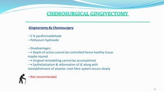
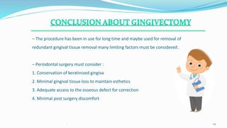
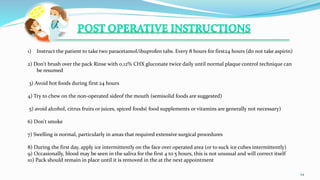
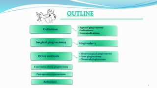
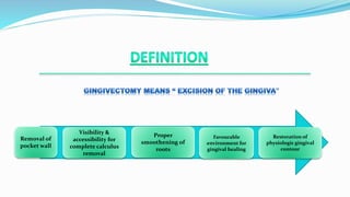
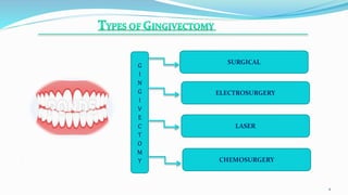
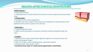
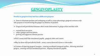
. 1
BDS,MDS(PERIODONTOLOGIST)
.
2
.
Definition
• Types of gingivectomy
• Indications
• Contraindications
Surgical gingivectomy Gingivoplasty
• Electrosurgical gingivectomy
• Laser gingivectomy
• Chemical gingivectomy
Other methods
Conclusion about gingivectomy
References
Post operative instructions
Removal of
pocket wall
Visibility &
accessibility for
complete calculus
removal
Proper
smoothening of
roots
Favourable
environment for
gingival healing
Restoration of
physiologic gingival
contour
.
4
ELECTROSURGERY
SURGICAL
LASER
CHEMOSURGERY
G
I
N
G
I
V
E
C
T
O
M
Y
1. Elimination of suprabony and pseudo pockets , regardless of depth but when wall is
fibrous and firm .
2. Elimination of fibrotic gingival enlargements
3. Elimination of suprabony periodontal or gingival abscess.
4. When base of pockets are at the same level but the margins are at a different level.
5. Create more esthetic forms in which exposure of anatomic crown has not fully
occurred example : defective passive eruption
.
5
1. Infrabony pockets
2. Inadequate zone of attached gingiva
3. Osseous surgery is indicated , examination of shape and morphology of bone
4. When base of pocket is apical to mucogingival junction
5. Aesthetics considerations (ant maxilla )
6. Patients with systemic problems leading to contraindication of surgical procedures example
uncontrolled diabetes , blood dyscrasias
7. Patient suffering from gout taking colchicine .
.
6
.
.
7
The only recommended technique :
Carranza’s clinical periodontology 2nd SAE
• Pocket marking forceps
• Gingivectomy knifes : 1. Kirkland knives (incisions on
facial and lingual surfaces on those distal to the terminal
tooth in arch )
2. Orban periodontal knives (interdental incisions )
3. Bard parker blades (12 and 15)
• Scissors (auxillary )
.
8
.
9
•Explore the pockets
•Pocket marking using a pocket marker
•Curette the granulation tissue
•Remove the calculus remnants , necrotic cementum
•Interrupted or continuous incisions ,beveled at 45 degrees to tooth surface are made starting apically to
marked points and directed coronally to a point between the base of the pocket and crest of the bone.
•Incision should be close to the bone but not exposing it .
•Removal of soft tissue coronal to the bone
•Remove excised pocket wall , clean the area , examine root surface.
•Apically : a band like zone where tissues were attached is seen
•Coronally :calculus remnants , root caries , resorption pattern is seen
•Granulation tissues seen on excised soft tissue.
•Cover with surgical Periodontal - pack
.
10
Figure:- Marking of the depth of the
suprabony pocket with *PMGF.
Figure:- Bleeding point after marking. Figure:- External bevel incision given.
(*PMGF= Goldman fox pocket marker)
.
11
Figure:- The surgical site covered
with periodontal dressing.
Figure:- A complete Gingivectomy
under local anaesthesia.
Figure:- Excised tissue.
-Initial response =
1. blood clot formation,
2. underlying tissue is acutely inflamed and necrotic and soon replaced by the granulation
tissue
– 24 hours later=
1. increased ct cells ( mainly angioblasts)
2. epithelial cells at the margins of the wound start migrating over the granulation tissue.
Epithelial activity reaches a peak in 24 to 36 hours.
– 3 days later =
1. Young fibroblasts are seen
2. highly vascular granulation tissue grows coronally creating free gingival margin and
sulcus
– 2 weeks =
1. Capillaries from vessels of periodontal ligament migrate into connective tissue and
connect with gingival margins
2. After 5 to 14 days, surface epithelialization is generally complete.
3. Complete repair takes about 1 month.
– Connective tissue repair in 7 weeks and the pigmentation is diminished .
.
12
.
.
13
.
14
Similar to gingivectomy but has a different purpose
 Done to eliminate pockets and reshaping as well to create physiologic gingival contours with
sole purpose of recontouring the gingiva in absence of pockets
 Gingival and periodontal diseases often create deformities in gingiva that interfere with
• normal food excursion
• collect plaque and food debris
• prolong and aggravate disease process
NUG causes shelf like interdental papilla , gingival clefts and craters
May be done with periodontal knife , rotary coarse diamond stones or electrodes
Consists of tapering gingival margins , creating escalloped marginal outline , thinning attached
gingiva , creating vertical interdental grooves , shaping interdental papilla .
.
.
15
ADVANTAGES
• Adequate contouring of tissue and controls haemorrhage
– DISADVANTAGES
Poor or noncompatible pacemaker
Unpleasant odour
Bone touching causes irreplaceable damage
Heat causes periodontal damage and tissue loss
On touching cementum burns are produced
– INDICATIONS
– Superficial procedures like
Gingival enlargements
Gingivoplasty
Relocation of muscle and frenum
Incision of periodontal claps and pericoronal abscess
.
16
.
1
17
Removal of gingival enlargements and gingivoplasty is performed with the needle
electrode.
Fully rectified current is used .
Small, ovoid loop or the diamond shaped electrodes are used for festooning.
In all reshaping procedures, electrode is activated and moved in a concise “shaving” motion.
– For hemostasis, the ball electrode is used. Firstly controlled by direct pressure then
coagulating current is used
– For Acute Periodontal Abscess Drainage with needle electrode without exerting painful
pressure is done . Followed by regular procedure
– Frenum and muscles can be relocated using Loop electrode
– Acute Pericornitis – Bent needle electrode is used for incision
1. Delayed healing
2. Greater reduction in gingival height
3. Bone injury
4. Necrosis
5. Sequestration and Loss of bone height
6. furcation exposure
7. tooth mobility
NOT AS FAVOURABLE AS THAT IN SURGICAL GINGIVECTOMY
.
18
.
. 19
The lasers most often used in dentistry are the
– carbon dioxide (CO2 )
– neodymium:yttrium:aluminum-garnet (Nd:YAG)
– with the wavelength of 10,600nm and 1064nm respectively.
The healing is delayed compared with healing after conventional scalpel
gingivectomy.
Requires precautions to avoid reflecting the beam on instrument surfaces,
which could result in injury to neighboring tissues and eyes of the operator
. 20
.
21
Gingivectomy By Chemosurgery
– 5 % paraformaldehyde
– Pottasium hydroxide
– Disadvantages:
– • Depth of action cannot be controlled hence healthy tissue
maybe injured
– • Gingival remodelling cannot be accomplished
– • Epithelialization & reformation of JE along with
reestablishment of alveolar crest fibre system occurs slowly
• Not recommended
.
22
– The procedure has been in use for long time and maybe used for removal of
redundant gingival tissue removal many limiting factors must be considered .
– Periodontal surgery must consider :
1. Conservation of keratinised gingiva
2. Minimal gingival tissue loss to maintain esthetics
3. Adequate access to the osseous defect for correction
4. Minimal post surgery discomfort
. 23
.
24
1) Instruct the patient to take two paracetamol/ibuprofen tabs. Every 8 hours for first24 hours (do not take aspirin)
2) Don't brush over the pack Rinse with 0.12% CHX gluconate twice daily until normal plaque control technique can
be resumed
3) Avoid hot foods during first 24 hours
4) Try to chew on the non-operated sideof the mouth (semisolid foods are suggested)
5) avoid alcohol, citrus fruits or juices, spiced foods( food supplements or vitamins are generally not necessary)
6) Don't smoke
7) Swelling is normal, particularly in areas that required extensive surgical procedures
8) During the first day, apply ice intermittently on the face over operated area (or to suck ice cubes intermittently)
9) Occasionally, blood may be seen in the saliva for the first 4 to 5 hours, this is not unusual and will correct itself
10) Pack should remain in place until it is removed in the at the next appointment
– Carranza’s clinical periodontology Edition 10 and second south Asian edition
.
25
.
26
THEPERIODONTALWORLD

Gingivectomy ppt

  • 1.
  • 2.
    . 2 . Definition • Types ofgingivectomy • Indications • Contraindications Surgical gingivectomy Gingivoplasty • Electrosurgical gingivectomy • Laser gingivectomy • Chemical gingivectomy Other methods Conclusion about gingivectomy References Post operative instructions
  • 3.
    Removal of pocket wall Visibility& accessibility for complete calculus removal Proper smoothening of roots Favourable environment for gingival healing Restoration of physiologic gingival contour
  • 4.
  • 5.
    1. Elimination ofsuprabony and pseudo pockets , regardless of depth but when wall is fibrous and firm . 2. Elimination of fibrotic gingival enlargements 3. Elimination of suprabony periodontal or gingival abscess. 4. When base of pockets are at the same level but the margins are at a different level. 5. Create more esthetic forms in which exposure of anatomic crown has not fully occurred example : defective passive eruption . 5
  • 6.
    1. Infrabony pockets 2.Inadequate zone of attached gingiva 3. Osseous surgery is indicated , examination of shape and morphology of bone 4. When base of pocket is apical to mucogingival junction 5. Aesthetics considerations (ant maxilla ) 6. Patients with systemic problems leading to contraindication of surgical procedures example uncontrolled diabetes , blood dyscrasias 7. Patient suffering from gout taking colchicine . . 6
  • 7.
    . . 7 The only recommendedtechnique : Carranza’s clinical periodontology 2nd SAE
  • 8.
    • Pocket markingforceps • Gingivectomy knifes : 1. Kirkland knives (incisions on facial and lingual surfaces on those distal to the terminal tooth in arch ) 2. Orban periodontal knives (interdental incisions ) 3. Bard parker blades (12 and 15) • Scissors (auxillary ) . 8
  • 9.
    . 9 •Explore the pockets •Pocketmarking using a pocket marker •Curette the granulation tissue •Remove the calculus remnants , necrotic cementum •Interrupted or continuous incisions ,beveled at 45 degrees to tooth surface are made starting apically to marked points and directed coronally to a point between the base of the pocket and crest of the bone. •Incision should be close to the bone but not exposing it . •Removal of soft tissue coronal to the bone •Remove excised pocket wall , clean the area , examine root surface. •Apically : a band like zone where tissues were attached is seen •Coronally :calculus remnants , root caries , resorption pattern is seen •Granulation tissues seen on excised soft tissue. •Cover with surgical Periodontal - pack
  • 10.
    . 10 Figure:- Marking ofthe depth of the suprabony pocket with *PMGF. Figure:- Bleeding point after marking. Figure:- External bevel incision given. (*PMGF= Goldman fox pocket marker)
  • 11.
    . 11 Figure:- The surgicalsite covered with periodontal dressing. Figure:- A complete Gingivectomy under local anaesthesia. Figure:- Excised tissue.
  • 12.
    -Initial response = 1.blood clot formation, 2. underlying tissue is acutely inflamed and necrotic and soon replaced by the granulation tissue – 24 hours later= 1. increased ct cells ( mainly angioblasts) 2. epithelial cells at the margins of the wound start migrating over the granulation tissue. Epithelial activity reaches a peak in 24 to 36 hours. – 3 days later = 1. Young fibroblasts are seen 2. highly vascular granulation tissue grows coronally creating free gingival margin and sulcus – 2 weeks = 1. Capillaries from vessels of periodontal ligament migrate into connective tissue and connect with gingival margins 2. After 5 to 14 days, surface epithelialization is generally complete. 3. Complete repair takes about 1 month. – Connective tissue repair in 7 weeks and the pigmentation is diminished . . 12
  • 13.
  • 14.
    . 14 Similar to gingivectomybut has a different purpose  Done to eliminate pockets and reshaping as well to create physiologic gingival contours with sole purpose of recontouring the gingiva in absence of pockets  Gingival and periodontal diseases often create deformities in gingiva that interfere with • normal food excursion • collect plaque and food debris • prolong and aggravate disease process NUG causes shelf like interdental papilla , gingival clefts and craters May be done with periodontal knife , rotary coarse diamond stones or electrodes Consists of tapering gingival margins , creating escalloped marginal outline , thinning attached gingiva , creating vertical interdental grooves , shaping interdental papilla .
  • 15.
  • 16.
    ADVANTAGES • Adequate contouringof tissue and controls haemorrhage – DISADVANTAGES Poor or noncompatible pacemaker Unpleasant odour Bone touching causes irreplaceable damage Heat causes periodontal damage and tissue loss On touching cementum burns are produced – INDICATIONS – Superficial procedures like Gingival enlargements Gingivoplasty Relocation of muscle and frenum Incision of periodontal claps and pericoronal abscess . 16
  • 17.
    . 1 17 Removal of gingivalenlargements and gingivoplasty is performed with the needle electrode. Fully rectified current is used . Small, ovoid loop or the diamond shaped electrodes are used for festooning. In all reshaping procedures, electrode is activated and moved in a concise “shaving” motion. – For hemostasis, the ball electrode is used. Firstly controlled by direct pressure then coagulating current is used – For Acute Periodontal Abscess Drainage with needle electrode without exerting painful pressure is done . Followed by regular procedure – Frenum and muscles can be relocated using Loop electrode – Acute Pericornitis – Bent needle electrode is used for incision
  • 18.
    1. Delayed healing 2.Greater reduction in gingival height 3. Bone injury 4. Necrosis 5. Sequestration and Loss of bone height 6. furcation exposure 7. tooth mobility NOT AS FAVOURABLE AS THAT IN SURGICAL GINGIVECTOMY . 18
  • 19.
  • 20.
    The lasers mostoften used in dentistry are the – carbon dioxide (CO2 ) – neodymium:yttrium:aluminum-garnet (Nd:YAG) – with the wavelength of 10,600nm and 1064nm respectively. The healing is delayed compared with healing after conventional scalpel gingivectomy. Requires precautions to avoid reflecting the beam on instrument surfaces, which could result in injury to neighboring tissues and eyes of the operator . 20
  • 21.
  • 22.
    Gingivectomy By Chemosurgery –5 % paraformaldehyde – Pottasium hydroxide – Disadvantages: – • Depth of action cannot be controlled hence healthy tissue maybe injured – • Gingival remodelling cannot be accomplished – • Epithelialization & reformation of JE along with reestablishment of alveolar crest fibre system occurs slowly • Not recommended . 22
  • 23.
    – The procedurehas been in use for long time and maybe used for removal of redundant gingival tissue removal many limiting factors must be considered . – Periodontal surgery must consider : 1. Conservation of keratinised gingiva 2. Minimal gingival tissue loss to maintain esthetics 3. Adequate access to the osseous defect for correction 4. Minimal post surgery discomfort . 23
  • 24.
    . 24 1) Instruct thepatient to take two paracetamol/ibuprofen tabs. Every 8 hours for first24 hours (do not take aspirin) 2) Don't brush over the pack Rinse with 0.12% CHX gluconate twice daily until normal plaque control technique can be resumed 3) Avoid hot foods during first 24 hours 4) Try to chew on the non-operated sideof the mouth (semisolid foods are suggested) 5) avoid alcohol, citrus fruits or juices, spiced foods( food supplements or vitamins are generally not necessary) 6) Don't smoke 7) Swelling is normal, particularly in areas that required extensive surgical procedures 8) During the first day, apply ice intermittently on the face over operated area (or to suck ice cubes intermittently) 9) Occasionally, blood may be seen in the saliva for the first 4 to 5 hours, this is not unusual and will correct itself 10) Pack should remain in place until it is removed in the at the next appointment
  • 25.
    – Carranza’s clinicalperiodontology Edition 10 and second south Asian edition . 25
  • 26.