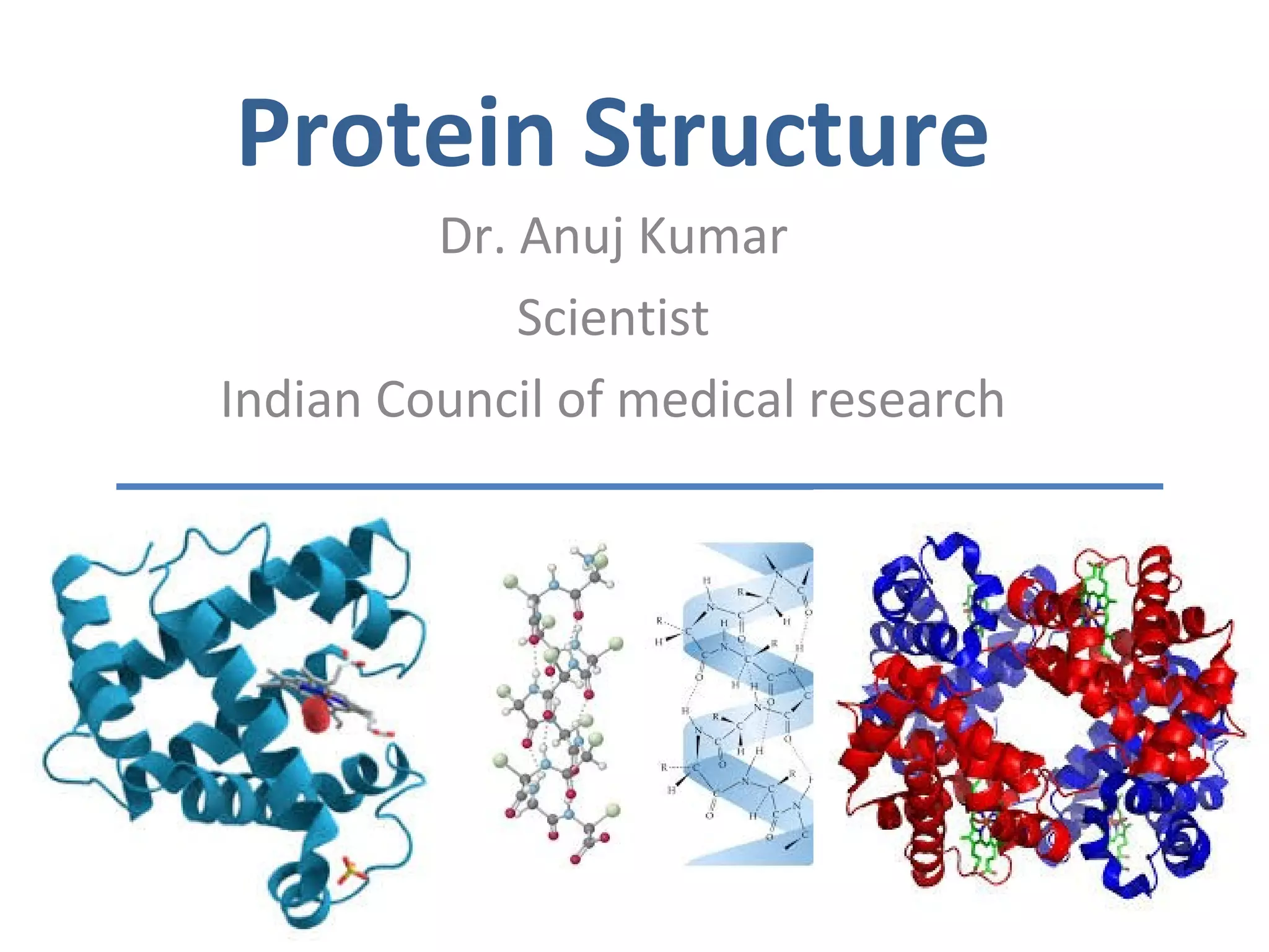
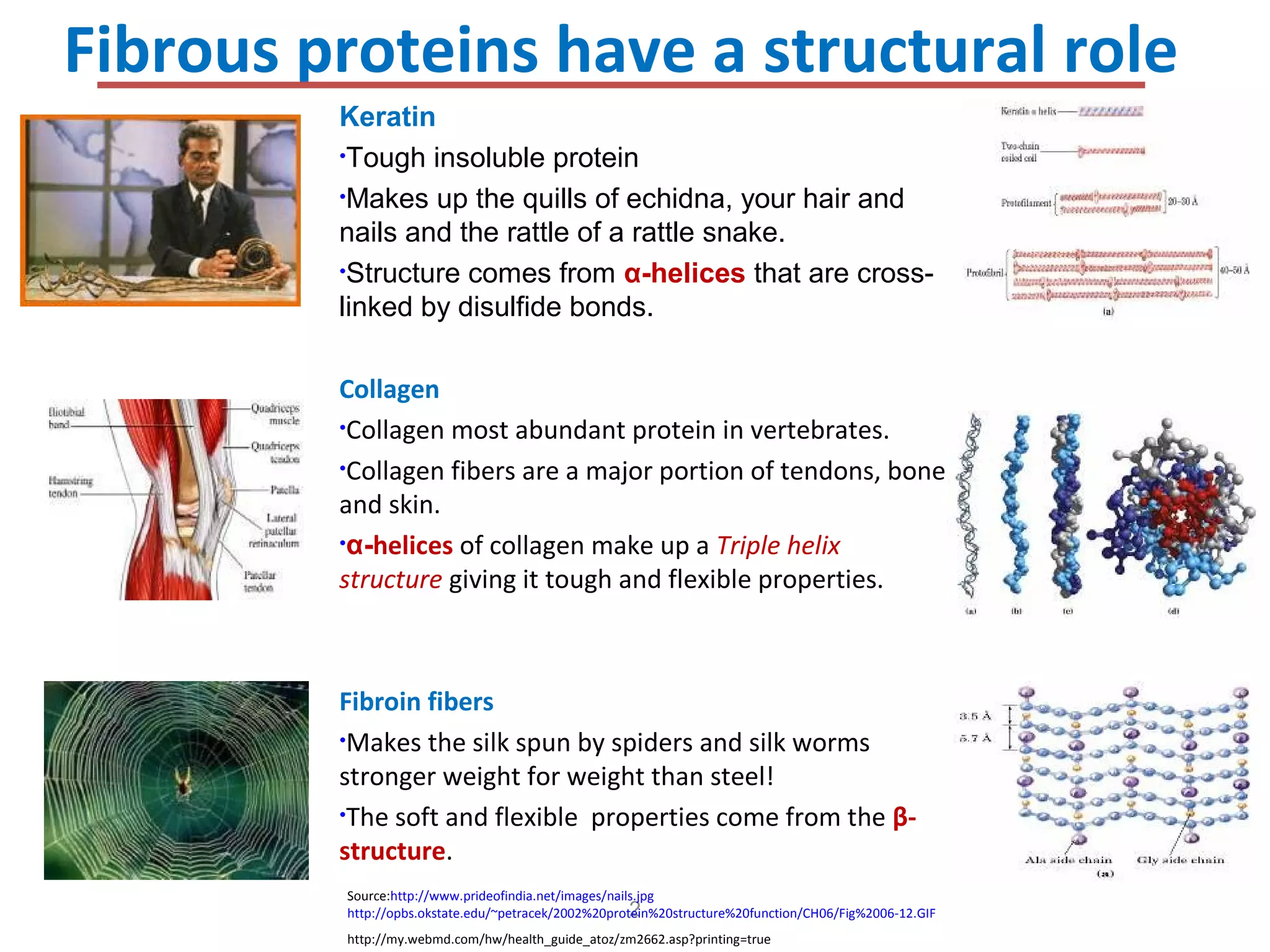
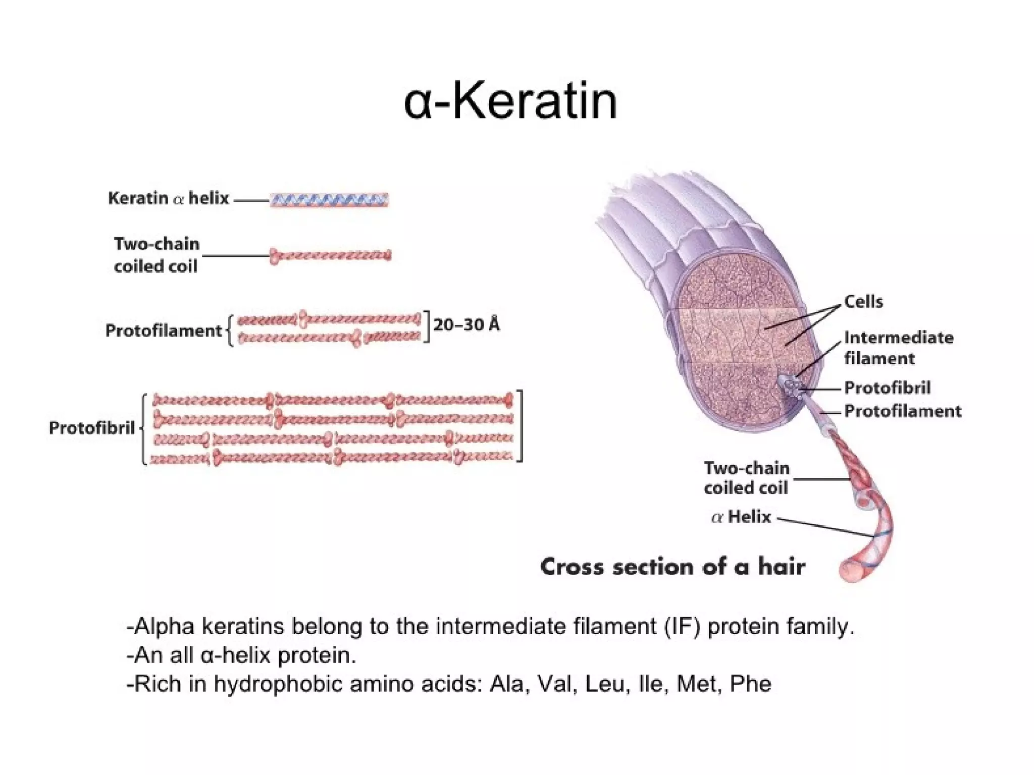
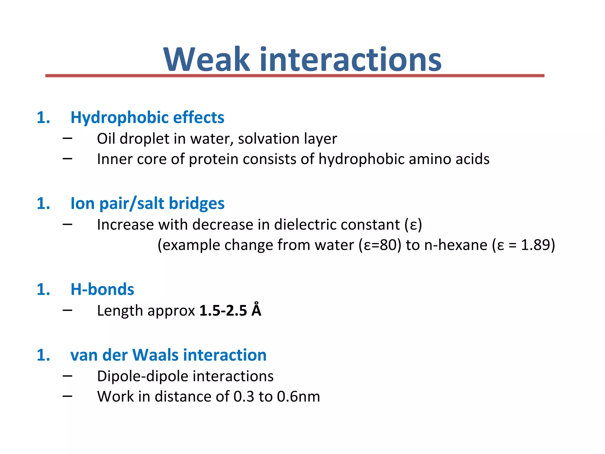
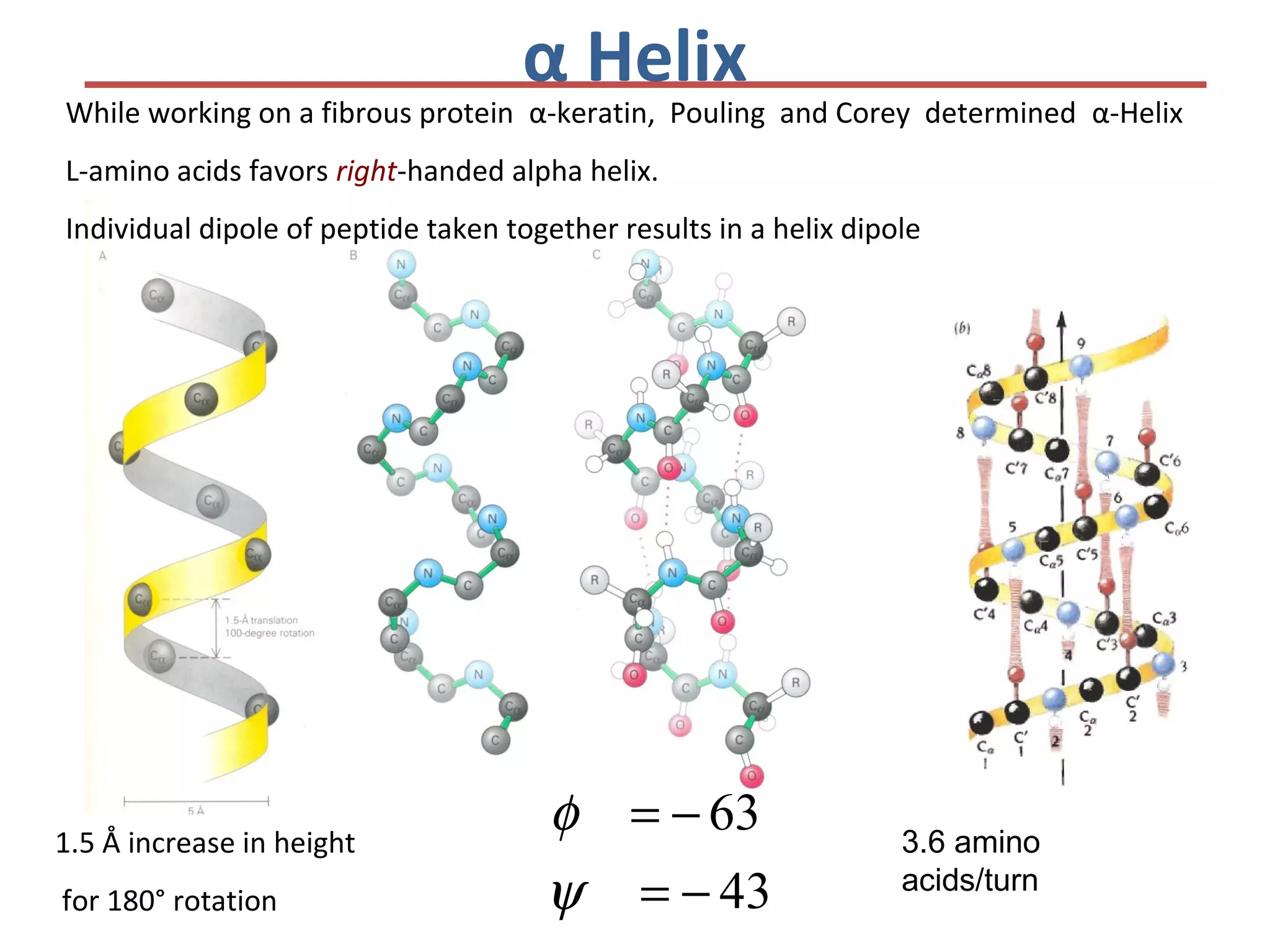
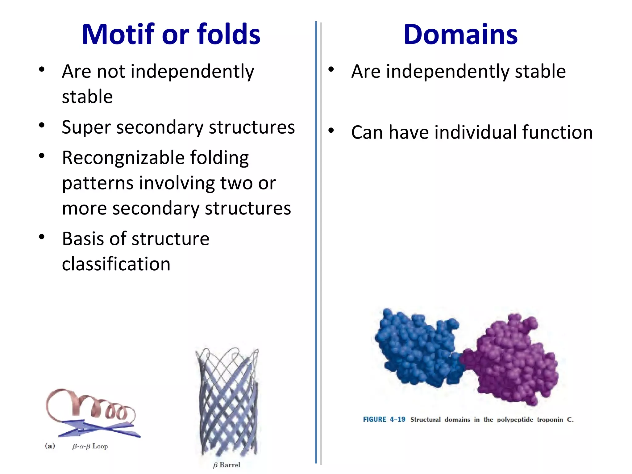
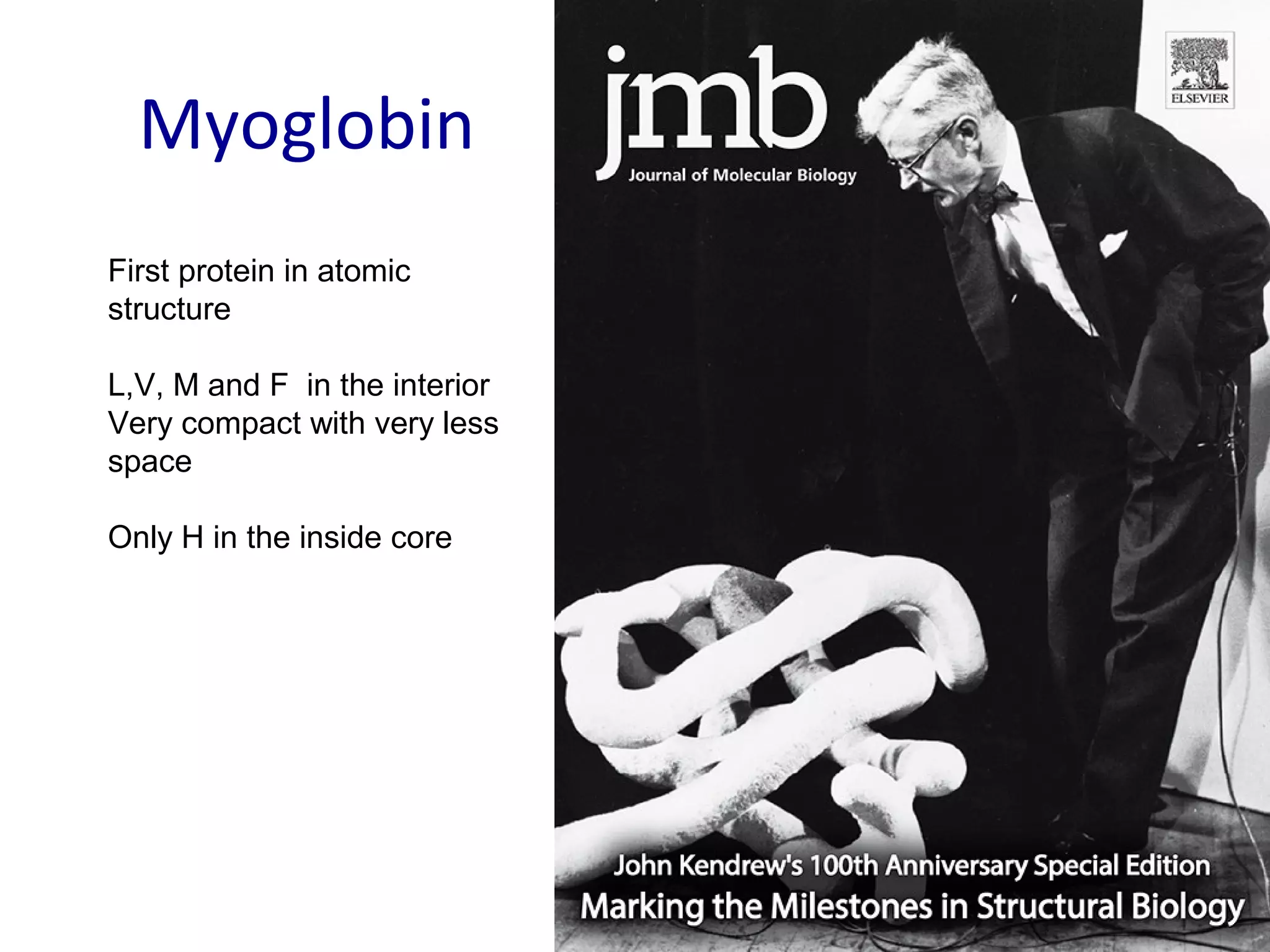
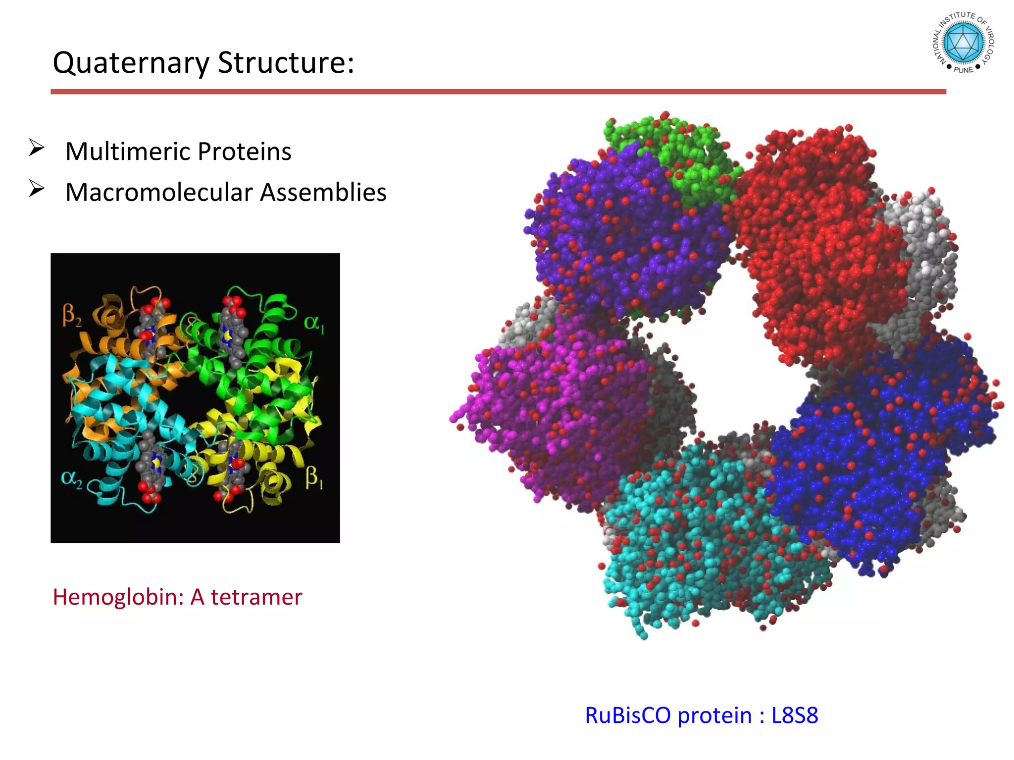
Presentation on Protein Structure for MSc class by Dr Anuj Kumar Scientist at National Institute of Virology, Indian Council of Medical Research (ICMR)
Also useful for Students preparing of CSIR/JRF NET and LS
2. Fibrous proteins have a structural role
2
Source:http://www.prideofindia.net/images/nails.jpg
http://opbs.okstate.edu/~petracek/2002%20protein%20structure%20function/CH06/Fig%2006-12.GIF
http://my.webmd.com/hw/health_guide_atoz/zm2662.asp?printing=true
Keratin
•Tough insoluble protein
•Makes up the quills of echidna, your hair and
nails and the rattle of a rattle snake.
•Structure comes from α-helices that are cross-
linked by disulfide bonds.
Collagen
•Collagen most abundant protein in vertebrates.
•Collagen fibers are a major portion of tendons, bone
and skin.
•α-helices of collagen make up a Triple helix
structure giving it tough and flexible properties.
Fibroin fibers
•Makes the silk spun by spiders and silk worms
stronger weight for weight than steel!
•The soft and flexible properties come from the β-
structure.
3.
4. Hierarchical nature of protein structure
Primary structure
(Amino acid sequence)
↓
Secondary structure
( α-helix, β-sheet )
↓
Tertiary structure
Three-dimensional structure formed by assembly of secondary structures
↓
Quaternary structure
Structure formed by more than one polypeptide chains
5. Protein Structure: Conformation
• The spatial arrangement of atoms in a protein or any part of
protein in called conformation (shape/structure)
• Conformation stabilized by weak interactions and disulphide
bond
– Difference between folded and unfolded approx 20-65
kJ/mol
– Weak interactions 0.4-30 kJ/mol (Peptide bond 200-460
kJ/mol)
• Proteins in any of their functional , folded conformations are
called native proteins
• In general, native conformation is lowest ΔG
6. Weak interactions
1. Hydrophobic effects
– Oil droplet in water, solvation layer
– Inner core of protein consists of hydrophobic amino acids
1. Ion pair/salt bridges
– Increase with decrease in dielectric constant (ε)
(example change from water (ε=80) to n-hexane (ε = 1.89)
1. H-bonds
– Length approx 1.5-2.5 Å
1. van der Waals interaction
– Dipole-dipole interactions
– Work in distance of 0.3 to 0.6nm
7. Hydrogen Bonding
• Involves three atoms:
– Donor electronegative atom (D)
(Nitrogen or Oxygen in proteins)
– Hydrogen bound to donor (H) may be from –N
atom –NH2
– Acceptor electronegative atom (A) in close
proximity may be Oxygen atom
Dδ-
– Hδ+
Aδ-
8. D-H Interaction
• Polarization due to electron withdrawal
from the hydrogen to D giving D partial negative
charge and the H a partial positive charge e.g.
H2O
• Proximity of the Acceptor A causes further charge
separation
D – H A
δ- δ+ δ-
9. D-H Interaction
• Polarization due to electron withdrawal from the
hydrogen to D giving D partial negative charge
and the H a partial positive charge
• Proximity of the Acceptor A causes further charge
separation
• Result:
– Closer approach of A to H
– Higher interaction energy than a simple van der Waals interaction
D – H A
δ- δ+ δ-
One liter Water=55.5M=55.5x 6.02x1023
=334.11x10²³
33,411,000,000,000,000,000,000,000 water molecules per liter!
13. X-Ray Crystallography
• Protein is crystallized
• Bombard with X-rays, record
scattering diffraction patterns
• Determine electron density map
from scattering and phase via
Fourier transform.
• Use electron density and
biochemical knowledge of the
protein to refine and determine a
model
14. An X-ray diffraction photograph of a crystal of sperm whale myoglobin.
The intensity of each diffraction maximum (the darkness of each spot) is a function of the crystal’s
electron density.
The distance between the spots is function of atomic distance
Bragg try to solve the structure of α-helix
X-ray diffraction photograph
15. Peptide Bond – Rigid and Planar
• A resonance in peptide bond (40% double bond
character)
• Partial negative charge on Oxygen and positive charge on
Nitrogen – Dipole
• All six atoms of the peptides are on single plane
• Three dihedral angle / torsion angle
– Φ angle between C-N-Cα -C
– ψ angle between N-Cα-C-N
– ω angle between Cα-N-C-Cα
• N-C (ω) cannot rotate freely
• Rotation allowed b/w N-Cα (Φ) and Cα-C(ψ)
16. Folding of the backbone chain: Phi (Φ)and Psi (ψ) Angles
Degrees of Freedom = 2
17. Ramachandran plot
• Phi (Φ) and Psi (ψ) rotate, allowing the
polypeptide to assume its various
conformations
• Some conformations of the polypeptide
backbone result in steric hindrance and
are disallowed
• Glycine has no side chain and is
therefore conformationally highly
flexible (it is often found in turns)
• Dr. Ramachandran used coordinated of
known di-, tri- and polypeptide of that
time including collagen and silk fibroin
Phi (Φ)
Psi (ψ)
20. Secondary structure
α-helix β-sheet
Secondary structures, α-helix and β-
sheet, have regular hydrogen-bonding
patterns.
Well defined and fixed pattern of H-bonding
Φ angle and ψ angle remain same
21. While working on a fibrous protein α-keratin, Pouling and Corey determined α-Helix
L-amino acids favors right-handed alpha helix.
Individual dipole of peptide taken together results in a helix dipole
1.5 Å increase in height
for 180° rotation 43
63
−=
−=
ψ
φ
α Helix
3.6 amino
acids/turn
22. Constraints affecting stability of helix
• The intrinsic propensity of aa
– V, T, I destabilize due to steric clashes
– S, D, N destabilize due to H-donor
– P destabilize due to fix phi angle (60 degrees) and
No H atom to donate for H-bonding
– Glycine highly flexible
• Aa at the end
23. Pleated appearance of Beta
sheet.
β strand
• Extended conformation, zigzag
structure
• Adjacent aa distance 3.5 A
• R groups in opposite direction
• Parallel and anti parallel
• Interstrand H-bonds
– in-line in antiparallel strands
– distorted in parallel strands
25. β Turns
• Four residues
• H-bonding between i and i+3
• Gly and Pro often occur in β turns
26. Motif or folds
• Are not independently
stable
• Super secondary structures
• Recongnizable folding
patterns involving two or
more secondary structures
• Basis of structure
classification
Domains
• Are independently stable
• Can have individual function
27. Tertiary
structure
Fold defines function
3D structure of HA protein of Influenza
A/H1N1; Known epitopes highlighted in
different colors
Visualization: DiscoveryStudio
Tertiary Structure
28. Myoglobin
First protein in atomic
structure
L,V, M and F in the interior
Very compact with very less
space
Only H in the inside core
29. Quaternary Structure
• non-linear
• 3 dimensional
• global, and across distinct
amino acid polymers
• formed by hydrogen bonding,
covalent bonding,
hydrophobic packing and
hydrophilic exposure
• favorable, functional
structures occur frequently
and have been categorized
30. Multimeric Proteins
Macromolecular Assemblies
Hemoglobin: A tetramer
Quaternary Structure:
RuBisCO protein : L8S8
34. Intrinsically Disordered Proteins
• Lack of hydrophobic core
• Large number of charged amino acids in core
• A third of all human proteins are intrinsically disordered
• Large number of viral proteins are intrinsically disordered
• Intrinsically Disordered Proteins are at the centre of protein-
protein interaction network
35. Protein denaturation and folding
Christian Anfinsen experiment
• Denaturation by temperature, pH,
chaotropic agent
• Ribonuclease A denaturation
• By urea and reducing agent
• After removal of urea and reducing agent
complete gain in activity
• Primary sequence have all the
information required to gain native
structure
36. Protein folding
• Levinthal’s paradox
– 100aa protein on
random folding
– 10-13
sec/conformation
– 1077
years
• Protein folding not
random process
• But a stepwise process
37. Homology modeling and threading
• Homology modeling = knowledge-based approach, given a sequence
database, use multiple sequence alignment on this database to
identify structurally conserved regions and construct structure
backbone and loops based on these regions, restore side-chains and
refine through energy minimization (apply to proteins that have high
sequence similarity to those in the database)
• Threading = knowledge-based approach, given a structure database of
interest (e.g. one that provides a limited set of possible structures per
given sequence for fold recognition, one that provides a one structure
per given limited set of possible sequences for inverse folding) use
scoring functions and correlations from this database to derive
structure that is in agreement (apply to proteins with moderate
sequence similarity to those in the database)
39. Hemoglobin: Background
• Protein in red blood cells
• Composed of four subunits, each
containing a heme group: a ring-like
structure with a central iron atom that
binds oxygen
• Picks up oxygen in lungs, releases it in
peripheral tissues (e.g. muscles)
40. • Sickle-cell anemia resulting from one residue
change in hemoglobin protein
• Replace highly polar (hydrophilic) glutamate
with nonpolar (hydrophobic) valine
42. Normal Trait
• Hemoglobin molecules exist as single,
isolated units in RBC, whether oxygen
bound or not
• Cells maintain basic disc shape, whether
transporting oxygen or not
43. Sickle-cell Trait
• Oxy-hemoglobin is isolated, but de-
oxyhemoglobin sticks together in
polymers, distorting RBC
• Some cells take on “sickle” shape
44. RBC Distortion
• Hydrophobic valine replaces hydrophilic glutamate
• Causes hemoglobin molecules to repel water and be
attracted to one another
• Leads to the formation of long hemoglobin filaments
• Filaments distort the shape of red blood cells
(analogy: icicle in a water balloon)
• Rigid structure of sickle cells blocks capillaries and
prevents red blood cells from delivering oxygen
45. Sickle-cell Trait
• Oxy-hemoglobin is isolated, but de-
oxyhemoglobin sticks together in
polymers, distorting RBC
• Some cells take on “sickle” shape
• When hemoglobin again binds oxygen,
again becomes isolated
• Cyclic alteration damages hemoglobin
and ultimately RBC itself