
Recommended
More Related Content
What's hot
What's hot (20)
Similar to SPRENGEL SHOULDER.pptx
Similar to SPRENGEL SHOULDER.pptx (20)
More from Salman Syed
More from Salman Syed (17)
Recently uploaded
Recently uploaded (20)
SPRENGEL SHOULDER.pptx
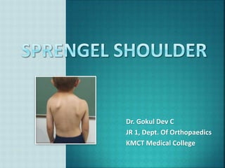
- 1. Dr. Gokul Dev C JR 1, Dept. Of Orthopaedics KMCT Medical College
- 2. Congenital Elevation Of Scapula Congenital High Scapula Scapula Elevata Earliest description : Eulenberg (1863) Named after Otto Gerhard Karl Sprengel (1852-1915) , a German surgeon who described four cases in 1891.
- 3. Rare - Most common congenital shoulder anomaly in children Typically congenital May not become apparent until grows older Girls > Boys (3:1) Unilateral > Bilateral Bilateral in 10–30% Right scapulae = left scapulae Sporadic (Rarely – run in families : AD Trait)
- 6. Develops embryologically with the upper limb Appears - 5th week of gestation along with arm bud (opposite to C4,C5,C6) Descends to the final anatomical position (T2-T8 vertebrae) by 12 weeks of gestation Develop from pleuripotent mesenchymal cells Cellular signaling pathways guide growth and development of scapula, surrounding muscles, bones, and nerves.
- 7. Results from failure of scapular descent. Usually during the 9-12 th week The scapula remains Hypoplastic High-riding Medially rotated With varying degrees of reduced scapulo-thoracic movement. Bone, Cartilage and Scapular muscles poorly developed and are replaced by fibrous bands.
- 8. The exact, underlying cause - unknown. ? Interruption of embryonic subclavian blood supply At level of subclavian, internal thoracic or suprascapular artery
- 9. Deformity is cardinal symptom - Painless Variable loss of ROM of Arm, Shoulder Blade and Cervical Spine Affected scapula appears Hypoplastic/Dysplastic and high riding into the C spine Increased width to height ratio Inferior pole is rotated medially with glenoid pointing inferiorly - thus restricting gleno-humeral abduction (≤ 90°) Convexity of upper part of scapula increased, curvature of clavicle shaft reduced – narrow scapulo-clavicular space – leads to brachial plexus injury Other associated abnormalities
- 10. • Omovertebral bar/bone • Omoclavicular bars • Klippel-Feil syndrome (35% with Sprengel deformity) • Kyphosis, Scoliosis • Spina bifida • Torticollis , Brevicollis • Scapular winging • Rib anomalies (fused or absent ribs) • Facial asymmetry • VACTERL association • Foot deformities
- 11. In about 50% of patients An abnormal bridge between the superior scapular angle and the cervical spine (spinous process/lamina/transverse process) Omovertebral Bar: fibrous/cartilaginous bridge Omovertebral Bone: trapezoid/rhomboidal bone
- 12. Brachial plexus palsy Rickets Osteomalacia Paralysis (long thoracic nerve – Winging of Scapula) Malunited scapular fractures Scoliosis
- 13. Examine both sides of patient Active movements of shoulder Passive movements Scapulo - humeral rhythm Scapular control Resisted isometric movements Functional assessment Special tests - DRST (Dynamic Rotatory Stability Test), Apley's scratch test, Rowe’s sign, Gagey hyperabduction test
- 14. X-ray : Shoulder AP Confirm diagnosis Omovertebral bone – may be seen Presence of associated abnormalities. MRI or CT Scan Identify omovertebral connection, Scapula dysplasia/ Position Used in preoperative planning Diagnosed prenatally - USG
- 15. The Radiographic RIGAULT CLASSIFICATION Grade I: Superomedial angle lower than T2 but above T4 transverse process Grade II: Superomedial angle located between C5 and T2 transverse process Grade III: Superomedial angle above C5 transverse process
- 18. Non operative – Observation + Physiotherapy Mild cases (no shoulder dysfunction) No severe cosmetic concerns Cavendish score of 1-2 Annual observation till skeletal maturity, to assess for progression of deformities Operative – Surgical fixation For conspicuous deformities with disability (abduction < 110-120) Severe cosmetic concerns Cavendish score of 3-4 To decrease deformity and improve shoulder function
- 19. Based on Cavendish classification Watch for spinal abnormalities prior Age of surgical intervention - often debated. Early intervention - maximal correction during further growth. But child may not bear extensive nature of intervention. Age of at least 3 years - the optimal time for intervention (3 to 6 yrs).
- 20. Putti’s Procedure The Woodward Procedure The Green Procedure Wikinson’s Osteotomy Mear’s Procedure
- 21. Surgical Principles Release the scapular tethers by resection of the superior angle (superomedial) of scapula and omovertebral bar. Scapula is derotated and relocated to a more caudal position by shift of either the origin or insertion of the scapular muscles. To relieve compression of brachial plexus and subclavian artery Morcellation of the clavicle (mid clavicular resection osteotomy ) is performed In older patients ( > 8 years)
- 22. To prevent brachial plexopathy when mobilizing the scapula The surgeon must take reference from contralateral scapular spine and not from inferior margin on affected side
- 23. Additional Procedures Bony Resection Extraperiosteal resection of proximal scapular prominence For cosmetic concerns May be done with other procedures or alone Clavicular Osteotomy / Morcellization. To reduce risk of neurovascular injury and to provide anterior release. Helps in additional scapular descent. Usually done in older children (>8 years) Done along with other procedures
- 24. Detatchment of scapular insertion of the para- scapular muscles (Rhomboids and Trapezius) Omovertebral bar resection Lowering the scapula and fixing its inferior angle to a rib at the corrected level Not done now Shrock Modified Putti’s Procedure Subperiosteal resection of the musculature Adding an osteotomy of supraspinous scapular region and the acromial base To facilitate scapular descent.
- 25. Most commonly used For correction of a moderate or severe Sprengel’s deformity Relocates scapula by detachment and caudal relocation of the midline origin of the para- scapular muscles (Trapezius and Rhomboids) The spine of scapula kept at the same level as that on the opposite side Clavicular osteotomy allows additional scapular descent. Cervical spine anomalies – Negative affect on outcome
- 27. Borges Modification of the Woodward Procedure The superomedial scapular prominence resected Better abduction and correction of glenoid tilt/ vara. Scapula anchored to lower dorsal vertebrae by a stout absorbable suture Good cosmetic and functional improvement (can improve abduction by 40-50 degrees). Better results The muscles are incised farther from scapula lowers risk of formation of scar keloid. A larger mobilization is possible. The postoperative scar is not as thick as with Green’s procedure.
- 29. Scapula is derotated by extraperiosteal detachment of scapular muscles at scapular insertion Resection of the supraspinous part of scapula and omovertebral bone (if present) Scapula is laid free Caudal relocation of scapula and fixation using scapular traction cables done The parascapular detached muscles are reattached. The traction wire is removed after 3 weeks Can improve abduction by 40-50 degrees
- 30. Leibovic Modification of The Green Technique - The reduced scapula is secured in a pocket developed in the latissimus dorsi muscle Belleman’s and Lamoureux Modification of The Green Technique - Serratus anterior muscle is not released - Immediate postoperative mobilization encouraged Andrault Modification of The Green Technique - Dis-insertion of supraspinatus - Clavicular osteotomy - A limited release of serratus anterior to facilitate the descent of scapula.
- 32. Vertical Scapular Osteotomy Lateral half of scapula is brought down caudally With concomitant Clavicular Osteotomy or Morcellization. Performed before movement of scapula The correction achieved is limited (compared to other procedures).
- 34. Subperiosteal release of muscle insertions on the superomedial aspect of scapula Extraperiosteal resection of the omovertebral bone Osteotomy of the supraspinatus fossa Release of long head of triceps and part of the origin of teres minor Inferomedial resection of scapula to 160° of shoulder abduction The gain in shoulder abduction is more as compared to others.
- 35. Straight incision over clavicle extending from 1.5 cm lateral to sternoclavicular joint to 1.5 cm medial to acromioclavicular joint. Expose clavicle subperiosteally. Divide the bone 2 cm from each end, remove it, and cut it into small pieces (morcellate). Replace the pieces in the periosteal tube and close the tube with interrupted sutures.
- 36. Brachial plexus injury Injury to blood vessels Regrowth of the superior pole of the scapula Hypertrophic scar / Keloid Winging of scapula due to incomplete reattachment of the serratus anterior
- 37. Not favorable – even after surgical management Literatures indicate – mobility of the shoulder may be improved, but the asymmetry almost always persists.
- 38. The arm is supported with a immobilizer postoperatively Gentle passive range of motion of the shoulder & scapula exercises are started. Suitable pain relief modality TENS, IFT, hot packs used to induce relaxation A sling is used after 3 weeks. Gradually, active ROM and strengthening exercises are instituted
- 39. Special attention is given to achieve early mobility of scapula and the shoulder abduction, elevation. Physiotherapy is continued for up to 6 months
- 40. 1. Apley’s System Of Orthopaedics & Fractures Tenth Edition. 2. Campbell's Operative Orthopaedics - 14th Edition 3. Essential Orthopedics Principles & Practice By Manish Kumar Varshney. 4. Pubmed: Kadavkolan et. Al. Sprengel's deformity of the shoulder: Current perspectives in management. Int J Shoulder Surg. 2011 Jan;5(1):1-8. doi: 10.4103/0973-6042.80459.