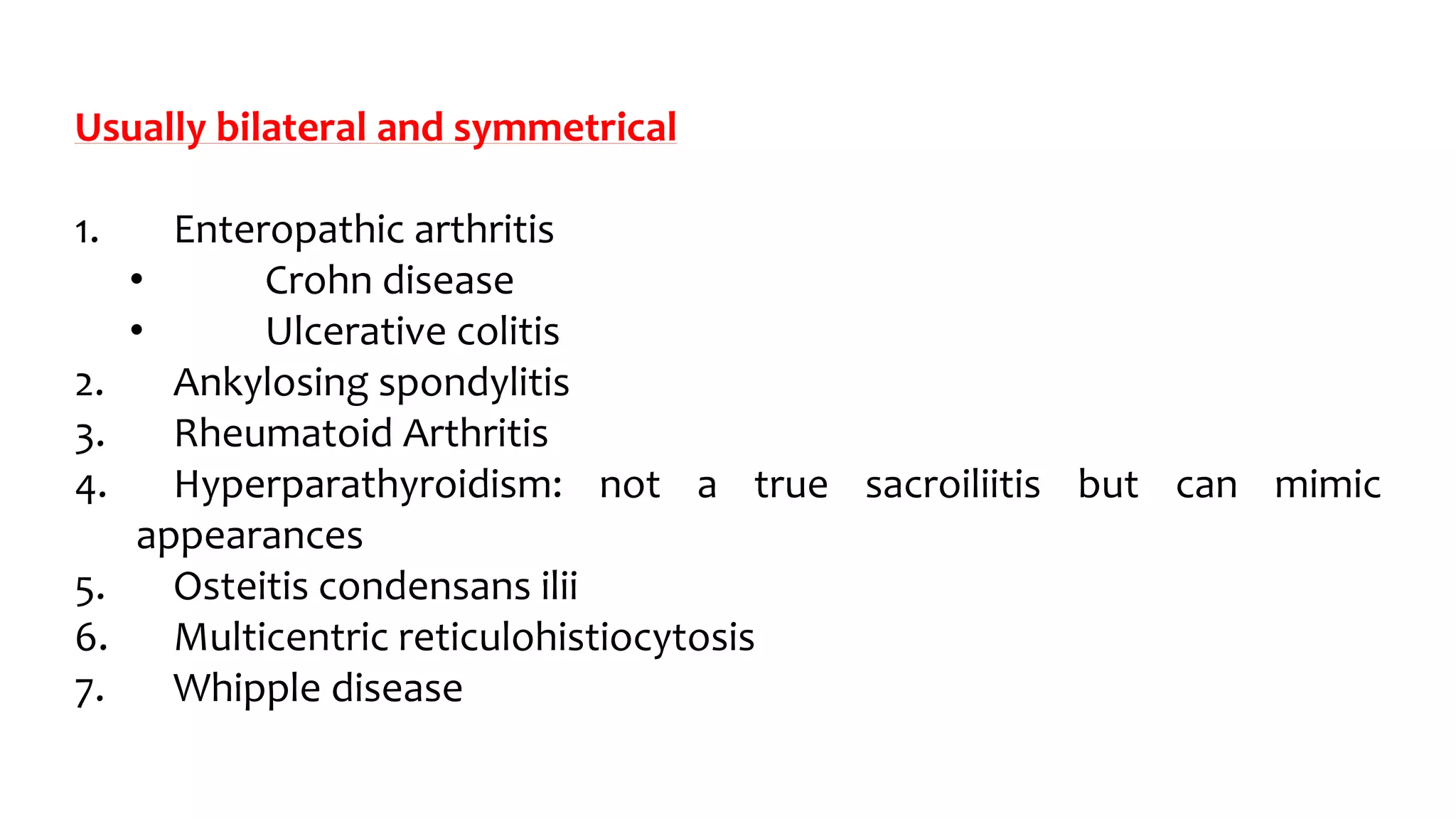
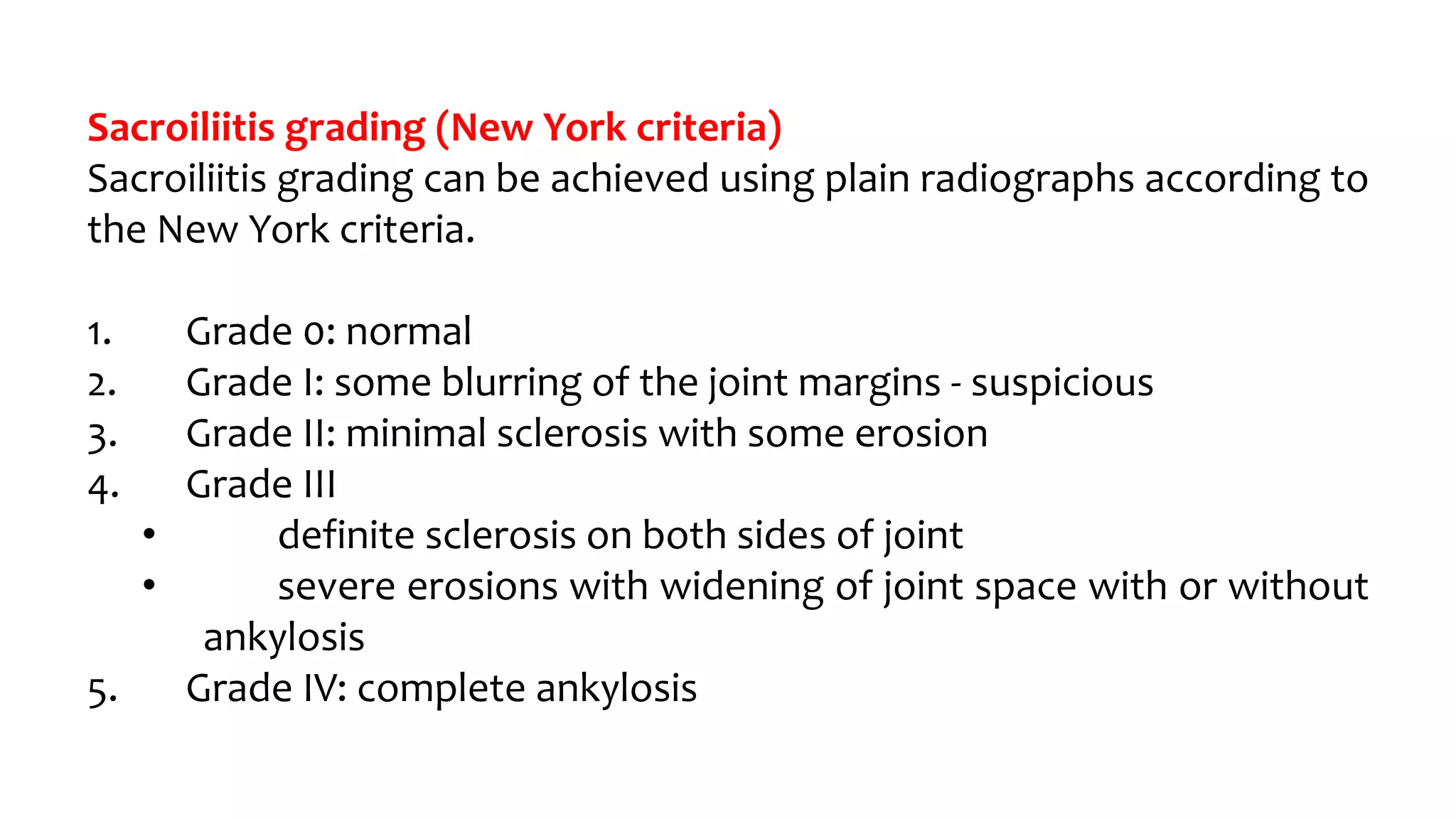
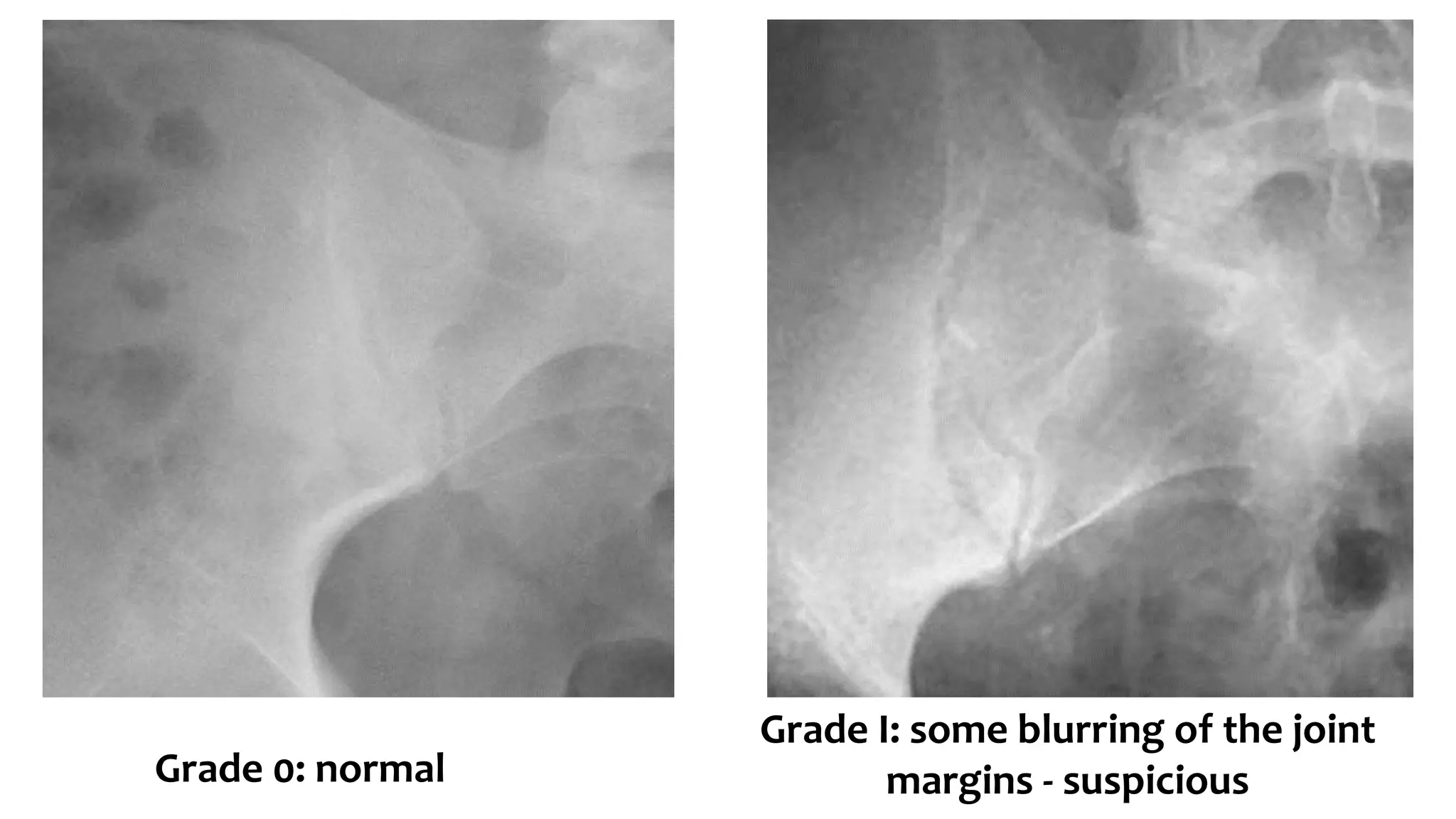
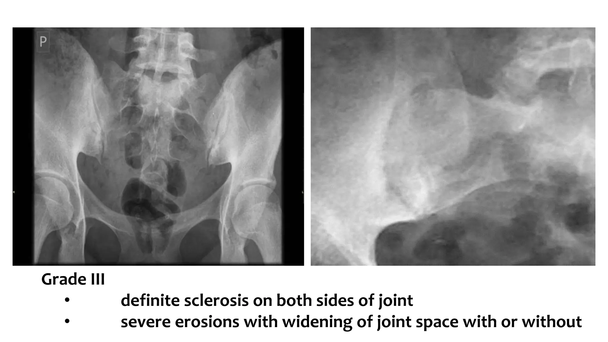
Sacroiliitis
Mohamed Ahmed Hefny, MD.
Usually bilateral and symmetrical
1. Enteropathic arthritis
• Crohn disease
• Ulcerative colitis
2. Ankylosing spondylitis
3. Rheumatoid Arthritis
4. Hyperparathyroidism: not a true sacroiliitis but can mimic
appearances
5. Osteitis condensans ilii
6. Multicentric reticulohistiocytosis
7. Whipple disease
Usually bilateral but asymmetrical
1. Gout
2. Psoriatic arthritis
3. Reactive arthritis (Reiter syndrome)
4. Osteoarthritis
5. Relapsing polychondritis
6. Behcet disease
7. Sacroiliitis circumscripta
Usually unilateral
1. Neoplastic destructive process
2. Infective
• pyogenic septic arthritis
• tuberculous sacroiliitis
• brucellosis
3. Paraplegia
4. SAPHO syndrome
Causes of sacroiliitis - symmetric vs asymmetric (mnemonic)
PAIR
Mnemonic
P: psoriatic arthritis
A: ankylosing spondylitis
I: inflammatory bowel disease related
R: reactive (e.g. Reiter syndrome)
P & R (letters are far apart) are the asymmetric causes and the inside
letters A & I (letters are close together) are the symmetric causes.
Sacroiliitis grading (New York criteria)
Sacroiliitis grading can be achieved using plain radiographs according to
the New York criteria.
1. Grade 0: normal
2. Grade I: some blurring of the joint margins - suspicious
3. Grade II: minimal sclerosis with some erosion
4. Grade III
• definite sclerosis on both sides of joint
• severe erosions with widening of joint space with or without
ankylosis
5. Grade IV: complete ankylosis
Grade 0: normal
Grade I: some blurring of the joint
margins - suspicious
Grade III
• definite sclerosis on both sides of joint
• severe erosions with widening of joint space with or without
Grade IV: complete ankylosis
Grade 1 (a) sacroiliitis was defined as focal erosions seen on
only one of either semicoronal or axial images.
Grade 2 (b) sacroiliitis was defined as erosions seen on 25 %
of consecutive semicoronal or axial images.
Grade 3 (c, d) sacroiliitis included bony erosions on C25 % of
consecutive semicoronal or axial images, or joint space
alteration, or partial ankylosis
Sacroiliitis

Sacroiliitis

  • 1.
  • 3.
    Usually bilateral andsymmetrical 1. Enteropathic arthritis • Crohn disease • Ulcerative colitis 2. Ankylosing spondylitis 3. Rheumatoid Arthritis 4. Hyperparathyroidism: not a true sacroiliitis but can mimic appearances 5. Osteitis condensans ilii 6. Multicentric reticulohistiocytosis 7. Whipple disease
  • 4.
    Usually bilateral butasymmetrical 1. Gout 2. Psoriatic arthritis 3. Reactive arthritis (Reiter syndrome) 4. Osteoarthritis 5. Relapsing polychondritis 6. Behcet disease 7. Sacroiliitis circumscripta
  • 5.
    Usually unilateral 1. Neoplasticdestructive process 2. Infective • pyogenic septic arthritis • tuberculous sacroiliitis • brucellosis 3. Paraplegia 4. SAPHO syndrome
  • 6.
    Causes of sacroiliitis- symmetric vs asymmetric (mnemonic) PAIR Mnemonic P: psoriatic arthritis A: ankylosing spondylitis I: inflammatory bowel disease related R: reactive (e.g. Reiter syndrome) P & R (letters are far apart) are the asymmetric causes and the inside letters A & I (letters are close together) are the symmetric causes.
  • 7.
    Sacroiliitis grading (NewYork criteria) Sacroiliitis grading can be achieved using plain radiographs according to the New York criteria. 1. Grade 0: normal 2. Grade I: some blurring of the joint margins - suspicious 3. Grade II: minimal sclerosis with some erosion 4. Grade III • definite sclerosis on both sides of joint • severe erosions with widening of joint space with or without ankylosis 5. Grade IV: complete ankylosis
  • 8.
    Grade 0: normal GradeI: some blurring of the joint margins - suspicious
  • 9.
    Grade III • definitesclerosis on both sides of joint • severe erosions with widening of joint space with or without
  • 10.
  • 11.
    Grade 1 (a)sacroiliitis was defined as focal erosions seen on only one of either semicoronal or axial images. Grade 2 (b) sacroiliitis was defined as erosions seen on 25 % of consecutive semicoronal or axial images. Grade 3 (c, d) sacroiliitis included bony erosions on C25 % of consecutive semicoronal or axial images, or joint space alteration, or partial ankylosis