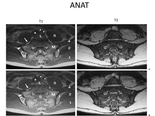
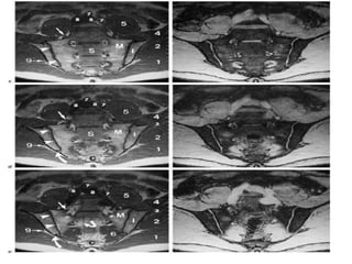
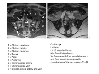
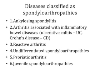
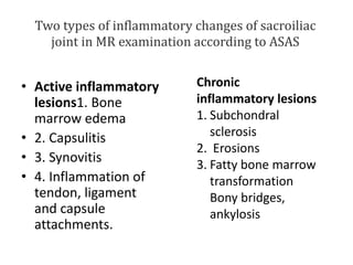
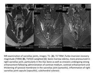
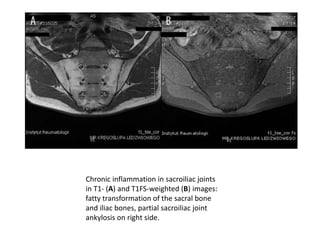
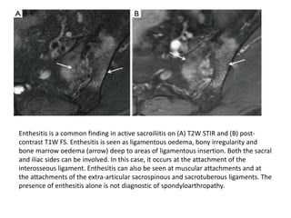
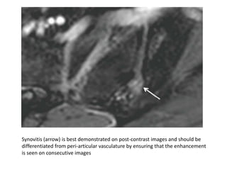
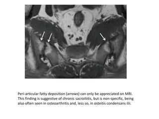
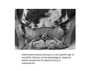
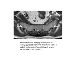
SlideShare a Scribd company logo
SI JOINT PATHOLOGIES
ANAT
T1 T2
1 = Gluteus maximus
2 = Gluteus medius
3 = Gluteus minimus
4 = Iliacus
5 = Psoas
6 = Piriformis
7 = Common iliac artery
8 = Common iliac vein
9 = Inferior gluteal artery and vein
C = Coccyx
I = Ilium
L = L5 vertebral body
M = Sacral lateral mass
S = Sacrum with four sacral elements
and four neural foramina with
visualization of the nerve roots S1–S4
Diseases classified as
spondyloarthropathies
• 1.Ankylosing spondylitis
• 2.Arthritis associated with inflammatory
bowel diseases (ulcerative colitis – UC,
Crohn’s disease – CD)
• 3.Reactive arthritis
• 4.Undifferentiated spondyloarthropathies
• 5.Psoriatic arthritis
• 6.Juvenile spondyloarthropathies
Radiological classification of sacroilitis
according to New York criteria
• Grade 0:- No changes/sacroiliac joint normal
• Grade 1:-Suspected changes (blurry margins of
sacroiliac joint gap)
• Grade 2:-Minimal changes (single erosions and
periarticular sclerosis involving small area of iliac
or sacral bone)
• Grade 3:-Advanced changes (distinct periarticular
sclerosis, numerous erosions with widening of
articular gap, possible partial ankylosis)
• Grade 4:-Total ankylosis
Grade 1,2,3,4
Two types of inflammatory changes of sacroiliac
joint in MR examination according to ASAS
• Active inflammatory
lesions1. Bone
marrow edema
• 2. Capsulitis
• 3. Synovitis
• 4. Inflammation of
tendon, ligament
and capsule
attachments.
Chronic
inflammatory lesions
1. Subchondral
sclerosis
2. Erosions
3. Fatty bone marrow
transformation
Bony bridges,
ankylosis
MR examination of sacroiliac joints, images: T1- (A), T2 TIRM ;Turbo inversion recovery
magnitude (TIRM) (B), T1FSCE-weighted (C): bone marrow edema, more pronounced in
right sacroiliac joint, particularly in the iliac bone as well as erosions undergoing strong
enhancement following administration of contrast medium, contrast enhancement and
thickening of synovial membrane in the sacroiliac joint (synovitis), inflammation of right
sacroiliac joint capsule (capsulitis), subchondral sclerosis
Chronic inflammation in sacroiliac joints
in T1- (A) and T1FS-weighted (B) images:
fatty transformation of the sacral bone
and iliac bones, partial sacroiliac joint
ankylosis on right side.
Active inflammatory lesions in MR examination
Bone marrow edema (osteomyelitis, osteitis, BME)
It is visible as an area of increased signal intensity in T2-weighted images and STIR/TIRM
sequences and as an area of decreased signal intensity in T1-weighted images that become
enhanced on administration of contrast medium. Bone marrow edema is not specific
for sacroilitis, as it also occurs in other inflammatory, traumatic or neoplastic pathologies.
Subchondral, periarticular BME specific for sacroilitis is located in iliac or sacral bones. It may
surround erosions . It is thought that symmetric lesions are characteristic for ankylosing
spondylitis, while unilateral, asymmetric location of bone marrow edema point to other forms of
SpA – most commonly psoriatic arthritis . literature data indicate that at early stage of ankylosing
spondylitis bone marrow edema is asymmetrical and unilateral.
Synovitis and capsulitis
Synovitis and capsulitis are diagnosed based on the presence of increased synovial signal intensity
in T2-weighted and STIR/TIRM sequences. They are characteristic for inflammation of sacroiliac
joints provided that there is an adjacent area of bone edema . Presence of articular exudate is
often observed in the course of synovitis and capsulitis, which may be differentiated from
synovitis in T1FS-weighted images following intravenous administration of contrast .
Inflammation of tendon, ligament and capsule insertions (enthesitis)
Enthesitis is visible in the form of increased signal intensity of bony or tendinous (either
ligamentous or fibrous layer of the capsule) part of enthesis on STIR;short inversion time
inversion recovery/TIRM or T1FSCE images .
• CHRONIC INFLAMMATORY CHANGES IN MR EXAMINATION
Subchondral sclerosis of bones that constitute the sacroiliac joint characterized
by low signal in T1-weighted and STIR sequences as well as lack of contrast
enhancement. Area of sclerosis specific for SpA has blurry margins and, as
disease progresses, becomes broader as opposed to osteoarthritis, in which it is
clearly demarcated and narrower .
Erosions in the subchondral layer of sacral or iliac bones, which may be active, i.e.
filled with inflamed tissue (areas of low signal in T1-weighted sequences and
hyperintense in STIR and T1CE sequences), or inactive (hypointense in all
sequences).
Subchondral fatty bone marrow transformation with high signal in T1-weighted
images, which becomes completely suppressed in fat-saturation sequences and
does not become enhanced following administration of contrast medium.
Ankylosis, characterized by low signal in all sequences, is sometimes surrounded
by high signal in T1-weighted images corresponding to fatty bone marrow
transformation.
„Shiny corners” of anterior vertebral bodies in the lumbar region; inflammation of
intervertebral joints L1–L5 with signs of bone marrow edema in T2 TIRM image (A)
enhanced after administration of contrast medium in T2FSCE-weighted image (B).
Visualizing >3 shiny corners indicates SpA with 81% specificity, while in young patients
<40 years old, specificity of this symptom raises to 97%
Sacroiliitis on MRI. (A) Bone marrow oedema (arrows) on T2W FS oblique coronal
images and (B) corresponding enhancement (arrowheads) on T1W FS post-contrast
axial images. This degree of bone marrow oedema is sufficient to diagnose sacroiliitis.
Capsulitis can be visible (A) anteriorly (arrow) or (B) posteriorly
(arrowhead) and may extend to involve the adjacent bone. The presence of
capsulitis alone is not diagnostic of spondyloarthropathy.
Enthesitis is a common finding in active sacroiliitis on (A) T2W STIR and (B) post-
contrast T1W FS. Enthesitis is seen as ligamentous oedema, bony irregularity and
bone marrow oedema (arrow) deep to areas of ligamentous insertion. Both the sacral
and iliac sides can be involved. In this case, it occurs at the attachment of the
interosseous ligament. Enthesitis can also be seen at muscular attachments and at
the attachments of the extra-articular sacrospinous and sacrotuberous ligaments. The
presence of enthesitis alone is not diagnostic of spondyloarthropathy.
Synovitis (arrow) is best demonstrated on post-contrast images and should be
differentiated from peri-articular vasculature by ensuring that the enhancement
is seen on consecutive images
Peri-articular fatty deposition (arrows) can only be appreciated on MRI.
This finding is suggestive of chronic sacroiliitis, but is non-specific, being
also often seen in osteoarthritis and, less so, in osteitis condensans ilii.
Subchondral sclerosis (arrows) is a non-specific sign of
sacroiliitis. Sclerosis can be physiological, related to
osteitis condensans ilii, physical stress or
osteoarthritis.
Ankylosis or bony bridging (arrows) can be
readily appreciated on MRI. One ideally wants to
make the diagnosis of sacroiliitis well before
ankylosis becomes apparent.
INFECTIVE SACROILIITIS
Three features of infective sacroiliitis are particularly
helpful in differentiating infective from inflammatory
sacroiliitis.
First, bone marrow oedema in infective sacroiliitis
tends to be more intense and there is more intra-
articular fluid (Figure 17A).
Second, inflammation in infective sacroiliitis spreads to
involve the peri-articular soft tissues, particularly the
iliacus and gluteal muscles (Figure 17B).
Third, peri-articular fluid collection or abscess is
practically pathognomonic of an infective sacroiliitis
infective sacroiliitis. (A) Excessive sacroiliac joint fluid
(red arrow); (B) inflammation of iliacus muscles (white
arrow); and (C) abscess formation (arrowhead)
supports the diagnosis of infective sacroiliitis.
Osteoarthritis. (A) Inferior osteophytosis (arrow) on radiograph and (B)
anterior marginal osteophytosis (arrowhead) seen on MRI as features of
osteoarthritis or instability. (C) Joint irregularity with subchondral
sclerosis is due to osteoarthritis rather than sacroiliitis on radiograph
Stress reaction. Focal patchy
oedema (arrow) at the mid-sacral
area close to the left sacroiliac
joint in a 28-year-old male
athlete. No fracture line is
evident. The patient recovered
with rest.
Reactive stress-related soft tissue changes
around the sacroiliac joint. Twenty-seven-
year-old professional football player.
Radiograph was unremarkable. (A) On MRI,
there is severe thickening of the anterior
capsule (arrow) on the right side; (B) more
proximally, similar though less severe
capsular thickening (arrowhead) with
osteoarthritis is present on the left side.
insufficiency fracture. MRI has a very high positive and negative
predictive value for insufficiency fracture. It will demonstrate either (A)
bone oedema (arrow) ± or (B) a discrete fracture line (arrowhead).
OSTEITIS CONDENSANS ILII is caused by bone deposition at
stress areas alongside the sacroiliac joint. It is usually seen in
childbearing women and is most likely related to the abnormal
high stresses incurred during pregnancy. Radiography and MRI
typically reveal bilateral, symmetrical, sharply circumscribed,
triangular-shaped areas of subchondral sclerosis, without
erosions or joint space widening, at the anteroinferior aspect of
the iliac bone alongside the sacroiliac joint.
Bone marrow oedema and fatty infiltration are recognized
additional MR features of osteitis condensans ilii .
Bone marrow oedema in osteitis condensans ilii tends to appear
in a continuous pattern as an arcuate line beneath the anterior
subchondral bone sclerosis in contrast to inflammatory
sacroiliitis
Osteitis condensans ilii (arrows). Well-defined
triangular-shaped symmetrical subchondral sclerosis,
without erosions or joint space widening at the
anterior aspect of the iliac bone on (A) radiograph
and (B) MR

More Related Content

Similar to SI JOINT PATHOLOGIES.pptx

Presentation2.pptx wrist joint.
Presentation2.pptx wrist joint.Presentation2.pptx wrist joint.
Presentation2.pptx wrist joint.
Abdellah Nazeer
 
Presentation2.pptx wrist joint.
Presentation2.pptx wrist joint.Presentation2.pptx wrist joint.
Presentation2.pptx wrist joint.
Abdellah Nazeer
 
Presentation1, radiological imaging of anterior knee pain.
Presentation1, radiological imaging of anterior knee pain.Presentation1, radiological imaging of anterior knee pain.
Presentation1, radiological imaging of anterior knee pain.
Abdellah Nazeer
 
Presentation1.pptx mri of elbow joint
Presentation1.pptx mri of elbow jointPresentation1.pptx mri of elbow joint
Presentation1.pptx mri of elbow joint
Abdellah Nazeer
 
Presentation1.pptx, radiological imaging of gout disease.
Presentation1.pptx, radiological imaging of gout disease.Presentation1.pptx, radiological imaging of gout disease.
Presentation1.pptx, radiological imaging of gout disease.
Abdellah Nazeer
 

Similar to SI JOINT PATHOLOGIES.pptx (20)

joints Radiological evaluation pdf.pdf
joints Radiological evaluation pdf.pdfjoints Radiological evaluation pdf.pdf
joints Radiological evaluation pdf.pdf
 
Transient Osteoporosis of Hip
Transient Osteoporosis of HipTransient Osteoporosis of Hip
Transient Osteoporosis of Hip
 
Mri knee bone
Mri knee boneMri knee bone
Mri knee bone
 
Rheumatoid arthritis and osteoarthritis
Rheumatoid arthritis and osteoarthritisRheumatoid arthritis and osteoarthritis
Rheumatoid arthritis and osteoarthritis
 
Seronegative spondyloarthropathies
Seronegative  spondyloarthropathiesSeronegative  spondyloarthropathies
Seronegative spondyloarthropathies
 
Presentation2.pptx wrist joint.
Presentation2.pptx wrist joint.Presentation2.pptx wrist joint.
Presentation2.pptx wrist joint.
 
Presentation2.pptx wrist joint.
Presentation2.pptx wrist joint.Presentation2.pptx wrist joint.
Presentation2.pptx wrist joint.
 
Charcot joint / Neuropathic Joint
Charcot joint / Neuropathic JointCharcot joint / Neuropathic Joint
Charcot joint / Neuropathic Joint
 
Arthritis and arthroplasty- dr. Mahmoud Abdel Kareem
Arthritis and arthroplasty- dr. Mahmoud Abdel KareemArthritis and arthroplasty- dr. Mahmoud Abdel Kareem
Arthritis and arthroplasty- dr. Mahmoud Abdel Kareem
 
Paraplegia and spinal cord syndromes
Paraplegia and spinal cord syndromesParaplegia and spinal cord syndromes
Paraplegia and spinal cord syndromes
 
Presentation1, radiological imaging of anterior knee pain.
Presentation1, radiological imaging of anterior knee pain.Presentation1, radiological imaging of anterior knee pain.
Presentation1, radiological imaging of anterior knee pain.
 
23204928
2320492823204928
23204928
 
Presentation1.pptx mri of elbow joint
Presentation1.pptx mri of elbow jointPresentation1.pptx mri of elbow joint
Presentation1.pptx mri of elbow joint
 
Swann2009
Swann2009Swann2009
Swann2009
 
Artrites
ArtritesArtrites
Artrites
 
Imaging of inflammatory arthritis ppt
Imaging of inflammatory arthritis pptImaging of inflammatory arthritis ppt
Imaging of inflammatory arthritis ppt
 
Presentation1, radiological imaging of ostepopikilosis
Presentation1, radiological imaging of ostepopikilosisPresentation1, radiological imaging of ostepopikilosis
Presentation1, radiological imaging of ostepopikilosis
 
Presentation1.pptx, radiological imaging of gout disease.
Presentation1.pptx, radiological imaging of gout disease.Presentation1.pptx, radiological imaging of gout disease.
Presentation1.pptx, radiological imaging of gout disease.
 
Imaging in arthritis
Imaging in arthritisImaging in arthritis
Imaging in arthritis
 
Presentation1, mri quiz cases.
Presentation1, mri quiz cases.Presentation1, mri quiz cases.
Presentation1, mri quiz cases.
 

More from ChintanBanugariya1 (6)

SPOTS of radiology for exam going students
SPOTS of radiology for exam going studentsSPOTS of radiology for exam going students
SPOTS of radiology for exam going students
 
liver.pptx
liver.pptxliver.pptx
liver.pptx
 
Bronchiectasis.pptx
Bronchiectasis.pptxBronchiectasis.pptx
Bronchiectasis.pptx
 
spinaltumors-copy-160925185617 (1).pptx
spinaltumors-copy-160925185617 (1).pptxspinaltumors-copy-160925185617 (1).pptx
spinaltumors-copy-160925185617 (1).pptx
 
nikhilknee-150625213218-lva1-app6891.pptx
nikhilknee-150625213218-lva1-app6891.pptxnikhilknee-150625213218-lva1-app6891.pptx
nikhilknee-150625213218-lva1-app6891.pptx
 
Fibroepithelial polyps.pptx
Fibroepithelial polyps.pptxFibroepithelial polyps.pptx
Fibroepithelial polyps.pptx
 

Recently uploaded

678020731-Sumas-y-Restas-Para-Colorear.pdf
678020731-Sumas-y-Restas-Para-Colorear.pdf678020731-Sumas-y-Restas-Para-Colorear.pdf
678020731-Sumas-y-Restas-Para-Colorear.pdf
CarlosHernanMontoyab2
 
The Roman Empire A Historical Colossus.pdf
The Roman Empire A Historical Colossus.pdfThe Roman Empire A Historical Colossus.pdf
The Roman Empire A Historical Colossus.pdf
kaushalkr1407
 
plant breeding methods in asexually or clonally propagated crops
plant breeding methods in asexually or clonally propagated cropsplant breeding methods in asexually or clonally propagated crops
plant breeding methods in asexually or clonally propagated crops
parmarsneha2
 

Recently uploaded (20)

Matatag-Curriculum and the 21st Century Skills Presentation.pptx
Matatag-Curriculum and the 21st Century Skills Presentation.pptxMatatag-Curriculum and the 21st Century Skills Presentation.pptx
Matatag-Curriculum and the 21st Century Skills Presentation.pptx
 
How to Split Bills in the Odoo 17 POS Module
How to Split Bills in the Odoo 17 POS ModuleHow to Split Bills in the Odoo 17 POS Module
How to Split Bills in the Odoo 17 POS Module
 
678020731-Sumas-y-Restas-Para-Colorear.pdf
678020731-Sumas-y-Restas-Para-Colorear.pdf678020731-Sumas-y-Restas-Para-Colorear.pdf
678020731-Sumas-y-Restas-Para-Colorear.pdf
 
Phrasal Verbs.XXXXXXXXXXXXXXXXXXXXXXXXXX
Phrasal Verbs.XXXXXXXXXXXXXXXXXXXXXXXXXXPhrasal Verbs.XXXXXXXXXXXXXXXXXXXXXXXXXX
Phrasal Verbs.XXXXXXXXXXXXXXXXXXXXXXXXXX
 
Sectors of the Indian Economy - Class 10 Study Notes pdf
Sectors of the Indian Economy - Class 10 Study Notes pdfSectors of the Indian Economy - Class 10 Study Notes pdf
Sectors of the Indian Economy - Class 10 Study Notes pdf
 
aaaaaaaaaaaaaaaaaaaaaaaaaaaaaaaaaaaaaaaaaaaaaaaaaaaaaaa
aaaaaaaaaaaaaaaaaaaaaaaaaaaaaaaaaaaaaaaaaaaaaaaaaaaaaaaaaaaaaaaaaaaaaaaaaaaaaaaaaaaaaaaaaaaaaaaaaaaaaaaaaaaaaa
aaaaaaaaaaaaaaaaaaaaaaaaaaaaaaaaaaaaaaaaaaaaaaaaaaaaaaa
 
Instructions for Submissions thorugh G- Classroom.pptx
Instructions for Submissions thorugh G- Classroom.pptxInstructions for Submissions thorugh G- Classroom.pptx
Instructions for Submissions thorugh G- Classroom.pptx
 
Basic_QTL_Marker-assisted_Selection_Sourabh.ppt
Basic_QTL_Marker-assisted_Selection_Sourabh.pptBasic_QTL_Marker-assisted_Selection_Sourabh.ppt
Basic_QTL_Marker-assisted_Selection_Sourabh.ppt
 
The Challenger.pdf DNHS Official Publication
The Challenger.pdf DNHS Official PublicationThe Challenger.pdf DNHS Official Publication
The Challenger.pdf DNHS Official Publication
 
The Roman Empire A Historical Colossus.pdf
The Roman Empire A Historical Colossus.pdfThe Roman Empire A Historical Colossus.pdf
The Roman Empire A Historical Colossus.pdf
 
MARUTI SUZUKI- A Successful Joint Venture in India.pptx
MARUTI SUZUKI- A Successful Joint Venture in India.pptxMARUTI SUZUKI- A Successful Joint Venture in India.pptx
MARUTI SUZUKI- A Successful Joint Venture in India.pptx
 
Overview on Edible Vaccine: Pros & Cons with Mechanism
Overview on Edible Vaccine: Pros & Cons with MechanismOverview on Edible Vaccine: Pros & Cons with Mechanism
Overview on Edible Vaccine: Pros & Cons with Mechanism
 
Introduction to Quality Improvement Essentials
Introduction to Quality Improvement EssentialsIntroduction to Quality Improvement Essentials
Introduction to Quality Improvement Essentials
 
Basic Civil Engineering Notes of Chapter-6, Topic- Ecosystem, Biodiversity G...
Basic Civil Engineering Notes of Chapter-6,  Topic- Ecosystem, Biodiversity G...Basic Civil Engineering Notes of Chapter-6,  Topic- Ecosystem, Biodiversity G...
Basic Civil Engineering Notes of Chapter-6, Topic- Ecosystem, Biodiversity G...
 
The approach at University of Liverpool.pptx
The approach at University of Liverpool.pptxThe approach at University of Liverpool.pptx
The approach at University of Liverpool.pptx
 
Jose-Rizal-and-Philippine-Nationalism-National-Symbol-2.pptx
Jose-Rizal-and-Philippine-Nationalism-National-Symbol-2.pptxJose-Rizal-and-Philippine-Nationalism-National-Symbol-2.pptx
Jose-Rizal-and-Philippine-Nationalism-National-Symbol-2.pptx
 
UNIT – IV_PCI Complaints: Complaints and evaluation of complaints, Handling o...
UNIT – IV_PCI Complaints: Complaints and evaluation of complaints, Handling o...UNIT – IV_PCI Complaints: Complaints and evaluation of complaints, Handling o...
UNIT – IV_PCI Complaints: Complaints and evaluation of complaints, Handling o...
 
How to Create Map Views in the Odoo 17 ERP
How to Create Map Views in the Odoo 17 ERPHow to Create Map Views in the Odoo 17 ERP
How to Create Map Views in the Odoo 17 ERP
 
plant breeding methods in asexually or clonally propagated crops
plant breeding methods in asexually or clonally propagated cropsplant breeding methods in asexually or clonally propagated crops
plant breeding methods in asexually or clonally propagated crops
 
Basic phrases for greeting and assisting costumers
Basic phrases for greeting and assisting costumersBasic phrases for greeting and assisting costumers
Basic phrases for greeting and assisting costumers
 

SI JOINT PATHOLOGIES.pptx

  • 3.
  • 4. 1 = Gluteus maximus 2 = Gluteus medius 3 = Gluteus minimus 4 = Iliacus 5 = Psoas 6 = Piriformis 7 = Common iliac artery 8 = Common iliac vein 9 = Inferior gluteal artery and vein C = Coccyx I = Ilium L = L5 vertebral body M = Sacral lateral mass S = Sacrum with four sacral elements and four neural foramina with visualization of the nerve roots S1–S4
  • 5. Diseases classified as spondyloarthropathies • 1.Ankylosing spondylitis • 2.Arthritis associated with inflammatory bowel diseases (ulcerative colitis – UC, Crohn’s disease – CD) • 3.Reactive arthritis • 4.Undifferentiated spondyloarthropathies • 5.Psoriatic arthritis • 6.Juvenile spondyloarthropathies
  • 6.
  • 7.
  • 8. Radiological classification of sacroilitis according to New York criteria • Grade 0:- No changes/sacroiliac joint normal • Grade 1:-Suspected changes (blurry margins of sacroiliac joint gap) • Grade 2:-Minimal changes (single erosions and periarticular sclerosis involving small area of iliac or sacral bone) • Grade 3:-Advanced changes (distinct periarticular sclerosis, numerous erosions with widening of articular gap, possible partial ankylosis) • Grade 4:-Total ankylosis
  • 10. Two types of inflammatory changes of sacroiliac joint in MR examination according to ASAS • Active inflammatory lesions1. Bone marrow edema • 2. Capsulitis • 3. Synovitis • 4. Inflammation of tendon, ligament and capsule attachments. Chronic inflammatory lesions 1. Subchondral sclerosis 2. Erosions 3. Fatty bone marrow transformation Bony bridges, ankylosis
  • 11. MR examination of sacroiliac joints, images: T1- (A), T2 TIRM ;Turbo inversion recovery magnitude (TIRM) (B), T1FSCE-weighted (C): bone marrow edema, more pronounced in right sacroiliac joint, particularly in the iliac bone as well as erosions undergoing strong enhancement following administration of contrast medium, contrast enhancement and thickening of synovial membrane in the sacroiliac joint (synovitis), inflammation of right sacroiliac joint capsule (capsulitis), subchondral sclerosis
  • 12. Chronic inflammation in sacroiliac joints in T1- (A) and T1FS-weighted (B) images: fatty transformation of the sacral bone and iliac bones, partial sacroiliac joint ankylosis on right side.
  • 13. Active inflammatory lesions in MR examination Bone marrow edema (osteomyelitis, osteitis, BME) It is visible as an area of increased signal intensity in T2-weighted images and STIR/TIRM sequences and as an area of decreased signal intensity in T1-weighted images that become enhanced on administration of contrast medium. Bone marrow edema is not specific for sacroilitis, as it also occurs in other inflammatory, traumatic or neoplastic pathologies. Subchondral, periarticular BME specific for sacroilitis is located in iliac or sacral bones. It may surround erosions . It is thought that symmetric lesions are characteristic for ankylosing spondylitis, while unilateral, asymmetric location of bone marrow edema point to other forms of SpA – most commonly psoriatic arthritis . literature data indicate that at early stage of ankylosing spondylitis bone marrow edema is asymmetrical and unilateral. Synovitis and capsulitis Synovitis and capsulitis are diagnosed based on the presence of increased synovial signal intensity in T2-weighted and STIR/TIRM sequences. They are characteristic for inflammation of sacroiliac joints provided that there is an adjacent area of bone edema . Presence of articular exudate is often observed in the course of synovitis and capsulitis, which may be differentiated from synovitis in T1FS-weighted images following intravenous administration of contrast . Inflammation of tendon, ligament and capsule insertions (enthesitis) Enthesitis is visible in the form of increased signal intensity of bony or tendinous (either ligamentous or fibrous layer of the capsule) part of enthesis on STIR;short inversion time inversion recovery/TIRM or T1FSCE images .
  • 14. • CHRONIC INFLAMMATORY CHANGES IN MR EXAMINATION Subchondral sclerosis of bones that constitute the sacroiliac joint characterized by low signal in T1-weighted and STIR sequences as well as lack of contrast enhancement. Area of sclerosis specific for SpA has blurry margins and, as disease progresses, becomes broader as opposed to osteoarthritis, in which it is clearly demarcated and narrower . Erosions in the subchondral layer of sacral or iliac bones, which may be active, i.e. filled with inflamed tissue (areas of low signal in T1-weighted sequences and hyperintense in STIR and T1CE sequences), or inactive (hypointense in all sequences). Subchondral fatty bone marrow transformation with high signal in T1-weighted images, which becomes completely suppressed in fat-saturation sequences and does not become enhanced following administration of contrast medium. Ankylosis, characterized by low signal in all sequences, is sometimes surrounded by high signal in T1-weighted images corresponding to fatty bone marrow transformation.
  • 15. „Shiny corners” of anterior vertebral bodies in the lumbar region; inflammation of intervertebral joints L1–L5 with signs of bone marrow edema in T2 TIRM image (A) enhanced after administration of contrast medium in T2FSCE-weighted image (B). Visualizing >3 shiny corners indicates SpA with 81% specificity, while in young patients <40 years old, specificity of this symptom raises to 97%
  • 16. Sacroiliitis on MRI. (A) Bone marrow oedema (arrows) on T2W FS oblique coronal images and (B) corresponding enhancement (arrowheads) on T1W FS post-contrast axial images. This degree of bone marrow oedema is sufficient to diagnose sacroiliitis.
  • 17. Capsulitis can be visible (A) anteriorly (arrow) or (B) posteriorly (arrowhead) and may extend to involve the adjacent bone. The presence of capsulitis alone is not diagnostic of spondyloarthropathy.
  • 18. Enthesitis is a common finding in active sacroiliitis on (A) T2W STIR and (B) post- contrast T1W FS. Enthesitis is seen as ligamentous oedema, bony irregularity and bone marrow oedema (arrow) deep to areas of ligamentous insertion. Both the sacral and iliac sides can be involved. In this case, it occurs at the attachment of the interosseous ligament. Enthesitis can also be seen at muscular attachments and at the attachments of the extra-articular sacrospinous and sacrotuberous ligaments. The presence of enthesitis alone is not diagnostic of spondyloarthropathy.
  • 19. Synovitis (arrow) is best demonstrated on post-contrast images and should be differentiated from peri-articular vasculature by ensuring that the enhancement is seen on consecutive images
  • 20. Peri-articular fatty deposition (arrows) can only be appreciated on MRI. This finding is suggestive of chronic sacroiliitis, but is non-specific, being also often seen in osteoarthritis and, less so, in osteitis condensans ilii.
  • 21. Subchondral sclerosis (arrows) is a non-specific sign of sacroiliitis. Sclerosis can be physiological, related to osteitis condensans ilii, physical stress or osteoarthritis.
  • 22. Ankylosis or bony bridging (arrows) can be readily appreciated on MRI. One ideally wants to make the diagnosis of sacroiliitis well before ankylosis becomes apparent.
  • 23. INFECTIVE SACROILIITIS Three features of infective sacroiliitis are particularly helpful in differentiating infective from inflammatory sacroiliitis. First, bone marrow oedema in infective sacroiliitis tends to be more intense and there is more intra- articular fluid (Figure 17A). Second, inflammation in infective sacroiliitis spreads to involve the peri-articular soft tissues, particularly the iliacus and gluteal muscles (Figure 17B). Third, peri-articular fluid collection or abscess is practically pathognomonic of an infective sacroiliitis
  • 24. infective sacroiliitis. (A) Excessive sacroiliac joint fluid (red arrow); (B) inflammation of iliacus muscles (white arrow); and (C) abscess formation (arrowhead) supports the diagnosis of infective sacroiliitis.
  • 25. Osteoarthritis. (A) Inferior osteophytosis (arrow) on radiograph and (B) anterior marginal osteophytosis (arrowhead) seen on MRI as features of osteoarthritis or instability. (C) Joint irregularity with subchondral sclerosis is due to osteoarthritis rather than sacroiliitis on radiograph
  • 26. Stress reaction. Focal patchy oedema (arrow) at the mid-sacral area close to the left sacroiliac joint in a 28-year-old male athlete. No fracture line is evident. The patient recovered with rest. Reactive stress-related soft tissue changes around the sacroiliac joint. Twenty-seven- year-old professional football player. Radiograph was unremarkable. (A) On MRI, there is severe thickening of the anterior capsule (arrow) on the right side; (B) more proximally, similar though less severe capsular thickening (arrowhead) with osteoarthritis is present on the left side.
  • 27. insufficiency fracture. MRI has a very high positive and negative predictive value for insufficiency fracture. It will demonstrate either (A) bone oedema (arrow) ± or (B) a discrete fracture line (arrowhead).
  • 28. OSTEITIS CONDENSANS ILII is caused by bone deposition at stress areas alongside the sacroiliac joint. It is usually seen in childbearing women and is most likely related to the abnormal high stresses incurred during pregnancy. Radiography and MRI typically reveal bilateral, symmetrical, sharply circumscribed, triangular-shaped areas of subchondral sclerosis, without erosions or joint space widening, at the anteroinferior aspect of the iliac bone alongside the sacroiliac joint. Bone marrow oedema and fatty infiltration are recognized additional MR features of osteitis condensans ilii . Bone marrow oedema in osteitis condensans ilii tends to appear in a continuous pattern as an arcuate line beneath the anterior subchondral bone sclerosis in contrast to inflammatory sacroiliitis
  • 29. Osteitis condensans ilii (arrows). Well-defined triangular-shaped symmetrical subchondral sclerosis, without erosions or joint space widening at the anterior aspect of the iliac bone on (A) radiograph and (B) MR