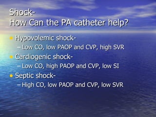
Shock Loring W. Rue, III, MD, FACS Professor and Vice Chairman Department of Surgery University of Alabama at Birmingham
Shock What is it? How to deal with it
Shock- Surgical Legacy G.J. Guthrie was the first to use the term shock in the context of physiologic instability Surgeon for Duke of Wellington in Peninsula campaign against Napoleon
Shock- Surgical Legacy George Crile Developed experimental models of hemorrhagic shock First to describe the decrease in CVP and cardiac output associated with hemorrhage An Experimental Research into Surgical Shock  (1899)
Shock- Surgical Legacy Alfred Blalock Demonstrated the hypovolemia associated with hemorrhage resulted in: HYPOTENSION METABOLIC ACIDOSIS IMPAIRED OXYGEN DELIVERY TO TISSUES Physiologic derangements corrected by volume resuscitation
What is Shock? Inadequate provision of oxygen to peripheral tissues resulting from either insufficient perfusion or abnormal extraction of oxygen Results in anaerobic metabolic pathways
Shock- Pathophysiology In the setting of adequate oxygen: Glucose metabolized via citric acid cycle yielding a net of 36 ATP In the setting of inadequate oxygen: No citric acid cycle-lactate generated yielding 2 ATP
Shock- Anaerobic Metabolism Results in significant intracellular energy deficits: Cell membrane repair mechanisms altered Electrolyte disturbances occur Rupture of endoplasmic reticulum Mitochondrial dysfunction occurs Rupture of lysosomes
Shock and Reperfusion Injury Resuscitation and tissue reperfusion in this setting results in free radical generation Can lead to cell death and organ dysfunction
Shock and Metabolic Acidosis: Not A Good Thing Cardiac Effects: Blood flow is redistributed from endocardium to epicardium with potential myocardial dysfunction Poor response to systemic catecholamines
Shock and Metabolic Acidosis: Not A Good Thing Pulmonary Effects Impairment of central respiratory drive CNS Effects Autoregulatory mechanisms of cerebral blood flow overridden with MAP < 60
Shock and Metabolic Acidosis: Not A Good Thing Renal Effects Afferent arteriolar vasoconstriction with RBF redistributed from cortex to medulla GFR decreases: oliguria / anuria Gastrointestinal Effects Gut ischemia > mucosal necrosis > bacterial translocation
Shock-  Systemic Compensatory Mechanisms Intravascular volume Baroreceptor activity Mechanoreceptor activity   CNS   Response Pituitary releases -ADH -ACTH Na/Water retention Cortisol released Sympathetic nervous stimulation Epinepherine Norepinepherine Renin/Angio/Aldo Enhanced cardiac function Peripheral vasoconstriction Na/Water retention Peripheral vasoconstriction
Shock- Clinical Features Hypotension Tachypnea Oliguria / Anuria Impaired Mental Status Depending upon cause, may also demonstrate Narrowing of pulse pressure Tachycardia
Shock- Management Priorities Airway adequacy Breathing restoration Circulatory support-  method dependent upon cause of shock
Shock - Resuscitation Endpoints Classic Restoration of blood pressure Normalization of heart rate and urine output Appropriate mental status Improved All of the above plus Normalization of serum lactate levels Resolution of base deficit
Shock- Judging the Adequacy of Resuscitation Monitor resuscitation with serial ABGs (attention to base deficit) and serum lactate levels.
Shock Causality Hypovolemic Cardiac Tamponade Tension Pneumothorax Cardiogenic Neurogenic Septic
Approach to the Trauma Patient - Appropriate for Any Patient in Shock Triage Primary Survey Resuscitation Secondary Survey Monitoring and Re-evaluation Definitive Care
Initial Assessment of the Trauma Patient– Primary Survey A – AIRWAY with C-SPINE CONTROL B – BREATHING C – CIRCULATION with BLEEDING CONTROL D – DISABILITY, NEUROLOGIC STATUS E – EXPOSURE and ENVIRONMENT Life threatening problems identified and managed simultaneously
Hypovolemic Shock Must treat the source of hypovolemia and arrest ongoing fluid losses The usual suspects: Thorax (CXR or CT) Abdomen (CT or Ultrasound) Pelvis (CT or plain film) Retroperitoneal (CT) Extremities – fractures or open wounds (plain films) All can be evaluated by operative intervention
Initial Assessment –  Radiographs Cervical spine Chest Pelvis
Hypovolemic Shock Categorized by severity as Class I-IV
Hypovolemic Shock Mainstay of therapy- Volume restoration Crystalloids Colloids Blood Plasma Blood Substitutes
Hypovolemic Shock –  Basic Management Principles-1 Immediate and rapid administration of 2 liters warm Lactated Ringers through large bore peripheral IVs Check for response to therapy If no response, either: Not enough “treatment given” Incorrect treatment
Hypovolemic Shock –  Basic Management Principles-2 Consequently, repeat fluid bolus Re-assess If no response, either: Massive exsanguinating hemorrhage Other causes: think Cardiogenic or Neurogenic in the trauma patient
Tension Pneumothorax
Tension Pneumothorax Progressive entry of air into pleural space Collapse of ipsilateral lung Mediastinal shift Compromised venous return to heart Hypotension / decreased cardiac output Cardiovascular collapse Needle decompression / chest tube
Tension Pneumothorax Hypotension Absent breath sounds Neck vein distension Hyperresonant thoracic percussion note Contralateral tracheal shift Subcutaneous emphysema Mechanism of injury Clinical diagnosis
Cardiogenic Shock The heart fails to deliver an adequate amount of blood to the body resulting in hypoperfusion of peripheral tissues The heart cannot generate a sufficient stroke volume and cardiac output Can be the result of impaired diastolic or systolic function or a combination of the two
Cardiogenic Shock Most often due to myocardial ischemia or infarction or resultant complications of papillary muscle rupture or ventricular septal rupture In trauma setting, think cardiac contusion in blunt force trauma and cardiac tamponade in penetrating trauma.
Cardiogenic Shock- Basic Management Principles For pure “pump” failure, options include: Pharmacologic agents Re-perfusion strategies Lytic agents Stents Coronary bypass Mechanical assists (Intra-aortic balloon) For tamponade physiology- surgery
Cardiac Tamponade
Penetrating Cardiac Injury
Penetrating Cardiac Injuries Etiology usually knives or bullets on the streets, but In the hospital lines, catheters
Penetrating Cardiac Injuries Relative role of tamponade vs. severe hemorrhage determined by : Size of pericardial rent Rate of bleeding from cardiac wound Chamber of heart involved STAB WOUNDS – TAMPONADE GSW - BLEED
Penetrating Cardiac Injuries Tamponade Physiology: Blood accumulates in pericardium Stroke volume decreases Right atrial pressure increases Right ventricle distends Ventricular septum shifts to left Left ventricle filling compromised Cardiac output decreases, hypotension ensues
Penetrating Cardiac Injuries Pre-hospital : Get patient to hospital ASAP Mattox / Feliciano study of 100 consecutive patients – if external CPR  > 3 minutes- 100% mortality Emergency department : Hemodynamics stable- ECHO +/- subxyphoid window Hemodynamics unstable – ED thoracotomy / OR *
Neurogenic Shock A form of distributive shock caused by the sudden loss of CNS signals to vascular smooth muscle following spinal cord injury Results in an immediate decrease in peripheral vascular resistance and hypotension May be associated with normal heart rate or even bradycardia
Neurogenic Shock Basic Management Principles Adequate oxygenation Spine immobilization Restore vasomotor tone after insuring adequate volume status Alpha agonist pharmacologic agents Often a role for early use of flow directed pulmonary artery catheter
Septic Shock A type of distributive shock related to loss of vasomotor tone in response to severe systemic infection or inflammatory response Results from release of multiple cellular mediators which stimulate a neurohormonal systemic response and influence vascular resistance
Septic Shock Inadequate treatment can lead to ARDS and multiple organ failure Mortality approaches 50%
Septic Shock Basic Management Principles Remove the source of infection or nidus for unlimited inflammatory response Antimicrobial therapy directed against offending organisms Support organ function Alpha agonists to maintain vasomotor tone Aggressive use of flow directed pulmonary artery catheters
Shock A Guide to Resuscitation Flow Directed Pulmonary Artery Catheter AKA the Swan- Ganz catheter, introduced into clinical practice in 1970 Integrates cardiopulmonary physiologic data of the patient Aides in determining appropriate theraputic interventions when cause of shock unknown Allows resuscitation to physiologic endpoints
Shock Flow Directed PA Catheters
Shock The Role of the PA Catheter Most often used in conjunction with standard intravascular monitoring devices (central venous line, arterial line) , EKG and pulse oximetry Provide both measured and calculated physiologic indices
Shock The Role of the PA Catheter, etc Measured physiologic indices: Mean arterial pressure (a-line) Heart rate (EKG, a-line) Central venous pressure (CVL, PA catheter) Pulmonary artery occlusion pressure, AKA the “wedge pressure” (PA catheter) Oxygen saturation (pulse oximetry, ABG)
Shock The Role of the PA Catheter, etc The measured indices enable  calculation of other parameters: Systemic Vascular Resistance [(MAP-CVP) x 80] / CO Stroke Volume  CO / HR Usually “indexed” by patient BSA (SV / BSA)
Shock The Role of the PA Catheter, etc Invasive hemodynamic monitoring can provide insight into the principal determinants of CO and hence tissue perfusion: Preload :  (CVP, better yet PAOP) Afterload : (SVR and MAP) Contractility : (SI)
Shock- How Can the PA catheter help? Hypovolemic shock- Low CO, low PAOP and CVP, high SVR Cardiogenic shock- Low CO, high PAOP and CVP, low SI Septic shock- High CO, low PAOP and CVP, low SVR
Shock The Role of the PA Catheter, etc Therapy can be directed where intervention needed: Inadequate Preload  = Hypovolemia Volume- either crystalloids or colloids depending upon patient Hb and coagulation parameters Excess Preload  = Congestive Heart Failure Diuretics +/- Inotropes
Shock The Role of the PA Catheter, etc Therapy can be directed where intervention needed: Inadequate afterload  = neurogenic or septic shock Alpha agonists  AKA Pressors (Norepinephrine or Neosynephreine) Excessive afterload  = Hypertensive CVD Vasodilators ( Sodium Nitroprusside, Nitroglycerine)
Shock The Role of the PA Catheter, etc Therapy can be directed where intervention needed: Inadequate contractility  = cardiogenic shock Beta agonists AKA Inotropes Epinepherine Dobutamine (good if increased afterload as well)
Shock- Pitfalls in Management Assuming blood pressure equates to cardiac output Extremes of age Hypothermia Athletes Pregnancy Medications / Pacemakers
Points for the Student Understand the rationale for the pitfalls in shock management as listed on slide What is the optimal and most efficient diagnostic modalities for the patient with presumed hypovolemic shock? Understand the clinical features of the 6 major causes of shock. Trivia question: Who is depicted on the slide “Penetrating Cardiac Injury” ?
Patient Scenario - 1 42 yo woman ejected from car following collision with a tree. Pre-hospital data: HR 110  BP  88/46  RR  26 Confused, skin cold and clammy Is she in shock? If so what are potential etiologies? How do we assess and treat?
Patient Scenario - 2 42 yo woman involved in MVC arrives in ED with full spine protection and supplement oxygen HR  120  BP  80 /46  RR 32 Is she in shock? If so what are potential etiologies? How do we assess and treat?
Patient Scenario - 2 After 2000cc warm lactated Ringers: HR  90  BP  110 / 80  RR  22 How has the patient responded? What are the diagnostic implications?
Patient Scenario - 2 What if the same patient, after 2000cc warm lactated Ringers: HR  130  BP  80 / 60  RR  40 Clinical interpretation?
Patient Scenario - 3 18 yo man stabbed in the left chest with large kitchen knife BP  80 / 60  HR  130  RR  30 Is he in shock ? Diagnostic possibilities ? How do you quickly evaluate and treat ?
Patient Scenario - 4 56 yo man now 6 days after undergoing a left hemicolectomy for cancer. Develops oliguria and altered sensorium. Transferred to the ICU- PA catheter placed Several hours later, patient is hypotensive BP 80 / 60  HR  140 What are the diagnostic possibilities ?  What would the PA catheter data look like for the various diagnostic possibilities?

S

  • 1.
    Shock Loring W.Rue, III, MD, FACS Professor and Vice Chairman Department of Surgery University of Alabama at Birmingham
  • 2.
    Shock What isit? How to deal with it
  • 3.
    Shock- Surgical LegacyG.J. Guthrie was the first to use the term shock in the context of physiologic instability Surgeon for Duke of Wellington in Peninsula campaign against Napoleon
  • 4.
    Shock- Surgical LegacyGeorge Crile Developed experimental models of hemorrhagic shock First to describe the decrease in CVP and cardiac output associated with hemorrhage An Experimental Research into Surgical Shock (1899)
  • 5.
    Shock- Surgical LegacyAlfred Blalock Demonstrated the hypovolemia associated with hemorrhage resulted in: HYPOTENSION METABOLIC ACIDOSIS IMPAIRED OXYGEN DELIVERY TO TISSUES Physiologic derangements corrected by volume resuscitation
  • 6.
    What is Shock?Inadequate provision of oxygen to peripheral tissues resulting from either insufficient perfusion or abnormal extraction of oxygen Results in anaerobic metabolic pathways
  • 7.
    Shock- Pathophysiology Inthe setting of adequate oxygen: Glucose metabolized via citric acid cycle yielding a net of 36 ATP In the setting of inadequate oxygen: No citric acid cycle-lactate generated yielding 2 ATP
  • 8.
    Shock- Anaerobic MetabolismResults in significant intracellular energy deficits: Cell membrane repair mechanisms altered Electrolyte disturbances occur Rupture of endoplasmic reticulum Mitochondrial dysfunction occurs Rupture of lysosomes
  • 9.
    Shock and ReperfusionInjury Resuscitation and tissue reperfusion in this setting results in free radical generation Can lead to cell death and organ dysfunction
  • 10.
    Shock and MetabolicAcidosis: Not A Good Thing Cardiac Effects: Blood flow is redistributed from endocardium to epicardium with potential myocardial dysfunction Poor response to systemic catecholamines
  • 11.
    Shock and MetabolicAcidosis: Not A Good Thing Pulmonary Effects Impairment of central respiratory drive CNS Effects Autoregulatory mechanisms of cerebral blood flow overridden with MAP < 60
  • 12.
    Shock and MetabolicAcidosis: Not A Good Thing Renal Effects Afferent arteriolar vasoconstriction with RBF redistributed from cortex to medulla GFR decreases: oliguria / anuria Gastrointestinal Effects Gut ischemia > mucosal necrosis > bacterial translocation
  • 13.
    Shock- SystemicCompensatory Mechanisms Intravascular volume Baroreceptor activity Mechanoreceptor activity CNS Response Pituitary releases -ADH -ACTH Na/Water retention Cortisol released Sympathetic nervous stimulation Epinepherine Norepinepherine Renin/Angio/Aldo Enhanced cardiac function Peripheral vasoconstriction Na/Water retention Peripheral vasoconstriction
  • 14.
    Shock- Clinical FeaturesHypotension Tachypnea Oliguria / Anuria Impaired Mental Status Depending upon cause, may also demonstrate Narrowing of pulse pressure Tachycardia
  • 15.
    Shock- Management PrioritiesAirway adequacy Breathing restoration Circulatory support- method dependent upon cause of shock
  • 16.
    Shock - ResuscitationEndpoints Classic Restoration of blood pressure Normalization of heart rate and urine output Appropriate mental status Improved All of the above plus Normalization of serum lactate levels Resolution of base deficit
  • 17.
    Shock- Judging theAdequacy of Resuscitation Monitor resuscitation with serial ABGs (attention to base deficit) and serum lactate levels.
  • 18.
    Shock Causality HypovolemicCardiac Tamponade Tension Pneumothorax Cardiogenic Neurogenic Septic
  • 19.
    Approach to theTrauma Patient - Appropriate for Any Patient in Shock Triage Primary Survey Resuscitation Secondary Survey Monitoring and Re-evaluation Definitive Care
  • 20.
    Initial Assessment ofthe Trauma Patient– Primary Survey A – AIRWAY with C-SPINE CONTROL B – BREATHING C – CIRCULATION with BLEEDING CONTROL D – DISABILITY, NEUROLOGIC STATUS E – EXPOSURE and ENVIRONMENT Life threatening problems identified and managed simultaneously
  • 21.
    Hypovolemic Shock Musttreat the source of hypovolemia and arrest ongoing fluid losses The usual suspects: Thorax (CXR or CT) Abdomen (CT or Ultrasound) Pelvis (CT or plain film) Retroperitoneal (CT) Extremities – fractures or open wounds (plain films) All can be evaluated by operative intervention
  • 22.
    Initial Assessment – Radiographs Cervical spine Chest Pelvis
  • 23.
    Hypovolemic Shock Categorizedby severity as Class I-IV
  • 24.
    Hypovolemic Shock Mainstayof therapy- Volume restoration Crystalloids Colloids Blood Plasma Blood Substitutes
  • 25.
    Hypovolemic Shock – Basic Management Principles-1 Immediate and rapid administration of 2 liters warm Lactated Ringers through large bore peripheral IVs Check for response to therapy If no response, either: Not enough “treatment given” Incorrect treatment
  • 26.
    Hypovolemic Shock – Basic Management Principles-2 Consequently, repeat fluid bolus Re-assess If no response, either: Massive exsanguinating hemorrhage Other causes: think Cardiogenic or Neurogenic in the trauma patient
  • 27.
  • 28.
    Tension Pneumothorax Progressiveentry of air into pleural space Collapse of ipsilateral lung Mediastinal shift Compromised venous return to heart Hypotension / decreased cardiac output Cardiovascular collapse Needle decompression / chest tube
  • 29.
    Tension Pneumothorax HypotensionAbsent breath sounds Neck vein distension Hyperresonant thoracic percussion note Contralateral tracheal shift Subcutaneous emphysema Mechanism of injury Clinical diagnosis
  • 30.
    Cardiogenic Shock Theheart fails to deliver an adequate amount of blood to the body resulting in hypoperfusion of peripheral tissues The heart cannot generate a sufficient stroke volume and cardiac output Can be the result of impaired diastolic or systolic function or a combination of the two
  • 31.
    Cardiogenic Shock Mostoften due to myocardial ischemia or infarction or resultant complications of papillary muscle rupture or ventricular septal rupture In trauma setting, think cardiac contusion in blunt force trauma and cardiac tamponade in penetrating trauma.
  • 32.
    Cardiogenic Shock- BasicManagement Principles For pure “pump” failure, options include: Pharmacologic agents Re-perfusion strategies Lytic agents Stents Coronary bypass Mechanical assists (Intra-aortic balloon) For tamponade physiology- surgery
  • 33.
  • 34.
  • 35.
    Penetrating Cardiac InjuriesEtiology usually knives or bullets on the streets, but In the hospital lines, catheters
  • 36.
    Penetrating Cardiac InjuriesRelative role of tamponade vs. severe hemorrhage determined by : Size of pericardial rent Rate of bleeding from cardiac wound Chamber of heart involved STAB WOUNDS – TAMPONADE GSW - BLEED
  • 37.
    Penetrating Cardiac InjuriesTamponade Physiology: Blood accumulates in pericardium Stroke volume decreases Right atrial pressure increases Right ventricle distends Ventricular septum shifts to left Left ventricle filling compromised Cardiac output decreases, hypotension ensues
  • 38.
    Penetrating Cardiac InjuriesPre-hospital : Get patient to hospital ASAP Mattox / Feliciano study of 100 consecutive patients – if external CPR > 3 minutes- 100% mortality Emergency department : Hemodynamics stable- ECHO +/- subxyphoid window Hemodynamics unstable – ED thoracotomy / OR *
  • 39.
    Neurogenic Shock Aform of distributive shock caused by the sudden loss of CNS signals to vascular smooth muscle following spinal cord injury Results in an immediate decrease in peripheral vascular resistance and hypotension May be associated with normal heart rate or even bradycardia
  • 40.
    Neurogenic Shock BasicManagement Principles Adequate oxygenation Spine immobilization Restore vasomotor tone after insuring adequate volume status Alpha agonist pharmacologic agents Often a role for early use of flow directed pulmonary artery catheter
  • 41.
    Septic Shock Atype of distributive shock related to loss of vasomotor tone in response to severe systemic infection or inflammatory response Results from release of multiple cellular mediators which stimulate a neurohormonal systemic response and influence vascular resistance
  • 42.
    Septic Shock Inadequatetreatment can lead to ARDS and multiple organ failure Mortality approaches 50%
  • 43.
    Septic Shock BasicManagement Principles Remove the source of infection or nidus for unlimited inflammatory response Antimicrobial therapy directed against offending organisms Support organ function Alpha agonists to maintain vasomotor tone Aggressive use of flow directed pulmonary artery catheters
  • 44.
    Shock A Guideto Resuscitation Flow Directed Pulmonary Artery Catheter AKA the Swan- Ganz catheter, introduced into clinical practice in 1970 Integrates cardiopulmonary physiologic data of the patient Aides in determining appropriate theraputic interventions when cause of shock unknown Allows resuscitation to physiologic endpoints
  • 45.
    Shock Flow DirectedPA Catheters
  • 46.
    Shock The Roleof the PA Catheter Most often used in conjunction with standard intravascular monitoring devices (central venous line, arterial line) , EKG and pulse oximetry Provide both measured and calculated physiologic indices
  • 47.
    Shock The Roleof the PA Catheter, etc Measured physiologic indices: Mean arterial pressure (a-line) Heart rate (EKG, a-line) Central venous pressure (CVL, PA catheter) Pulmonary artery occlusion pressure, AKA the “wedge pressure” (PA catheter) Oxygen saturation (pulse oximetry, ABG)
  • 48.
    Shock The Roleof the PA Catheter, etc The measured indices enable calculation of other parameters: Systemic Vascular Resistance [(MAP-CVP) x 80] / CO Stroke Volume CO / HR Usually “indexed” by patient BSA (SV / BSA)
  • 49.
    Shock The Roleof the PA Catheter, etc Invasive hemodynamic monitoring can provide insight into the principal determinants of CO and hence tissue perfusion: Preload : (CVP, better yet PAOP) Afterload : (SVR and MAP) Contractility : (SI)
  • 50.
    Shock- How Canthe PA catheter help? Hypovolemic shock- Low CO, low PAOP and CVP, high SVR Cardiogenic shock- Low CO, high PAOP and CVP, low SI Septic shock- High CO, low PAOP and CVP, low SVR
  • 51.
    Shock The Roleof the PA Catheter, etc Therapy can be directed where intervention needed: Inadequate Preload = Hypovolemia Volume- either crystalloids or colloids depending upon patient Hb and coagulation parameters Excess Preload = Congestive Heart Failure Diuretics +/- Inotropes
  • 52.
    Shock The Roleof the PA Catheter, etc Therapy can be directed where intervention needed: Inadequate afterload = neurogenic or septic shock Alpha agonists AKA Pressors (Norepinephrine or Neosynephreine) Excessive afterload = Hypertensive CVD Vasodilators ( Sodium Nitroprusside, Nitroglycerine)
  • 53.
    Shock The Roleof the PA Catheter, etc Therapy can be directed where intervention needed: Inadequate contractility = cardiogenic shock Beta agonists AKA Inotropes Epinepherine Dobutamine (good if increased afterload as well)
  • 54.
    Shock- Pitfalls inManagement Assuming blood pressure equates to cardiac output Extremes of age Hypothermia Athletes Pregnancy Medications / Pacemakers
  • 55.
    Points for theStudent Understand the rationale for the pitfalls in shock management as listed on slide What is the optimal and most efficient diagnostic modalities for the patient with presumed hypovolemic shock? Understand the clinical features of the 6 major causes of shock. Trivia question: Who is depicted on the slide “Penetrating Cardiac Injury” ?
  • 56.
    Patient Scenario -1 42 yo woman ejected from car following collision with a tree. Pre-hospital data: HR 110 BP 88/46 RR 26 Confused, skin cold and clammy Is she in shock? If so what are potential etiologies? How do we assess and treat?
  • 57.
    Patient Scenario -2 42 yo woman involved in MVC arrives in ED with full spine protection and supplement oxygen HR 120 BP 80 /46 RR 32 Is she in shock? If so what are potential etiologies? How do we assess and treat?
  • 58.
    Patient Scenario -2 After 2000cc warm lactated Ringers: HR 90 BP 110 / 80 RR 22 How has the patient responded? What are the diagnostic implications?
  • 59.
    Patient Scenario -2 What if the same patient, after 2000cc warm lactated Ringers: HR 130 BP 80 / 60 RR 40 Clinical interpretation?
  • 60.
    Patient Scenario -3 18 yo man stabbed in the left chest with large kitchen knife BP 80 / 60 HR 130 RR 30 Is he in shock ? Diagnostic possibilities ? How do you quickly evaluate and treat ?
  • 61.
    Patient Scenario -4 56 yo man now 6 days after undergoing a left hemicolectomy for cancer. Develops oliguria and altered sensorium. Transferred to the ICU- PA catheter placed Several hours later, patient is hypotensive BP 80 / 60 HR 140 What are the diagnostic possibilities ? What would the PA catheter data look like for the various diagnostic possibilities?