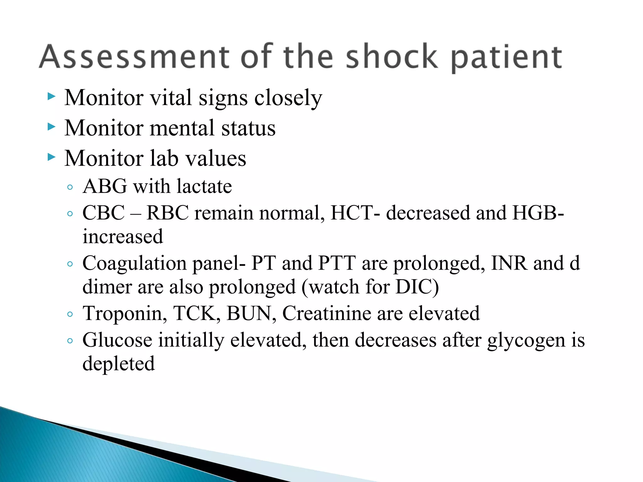
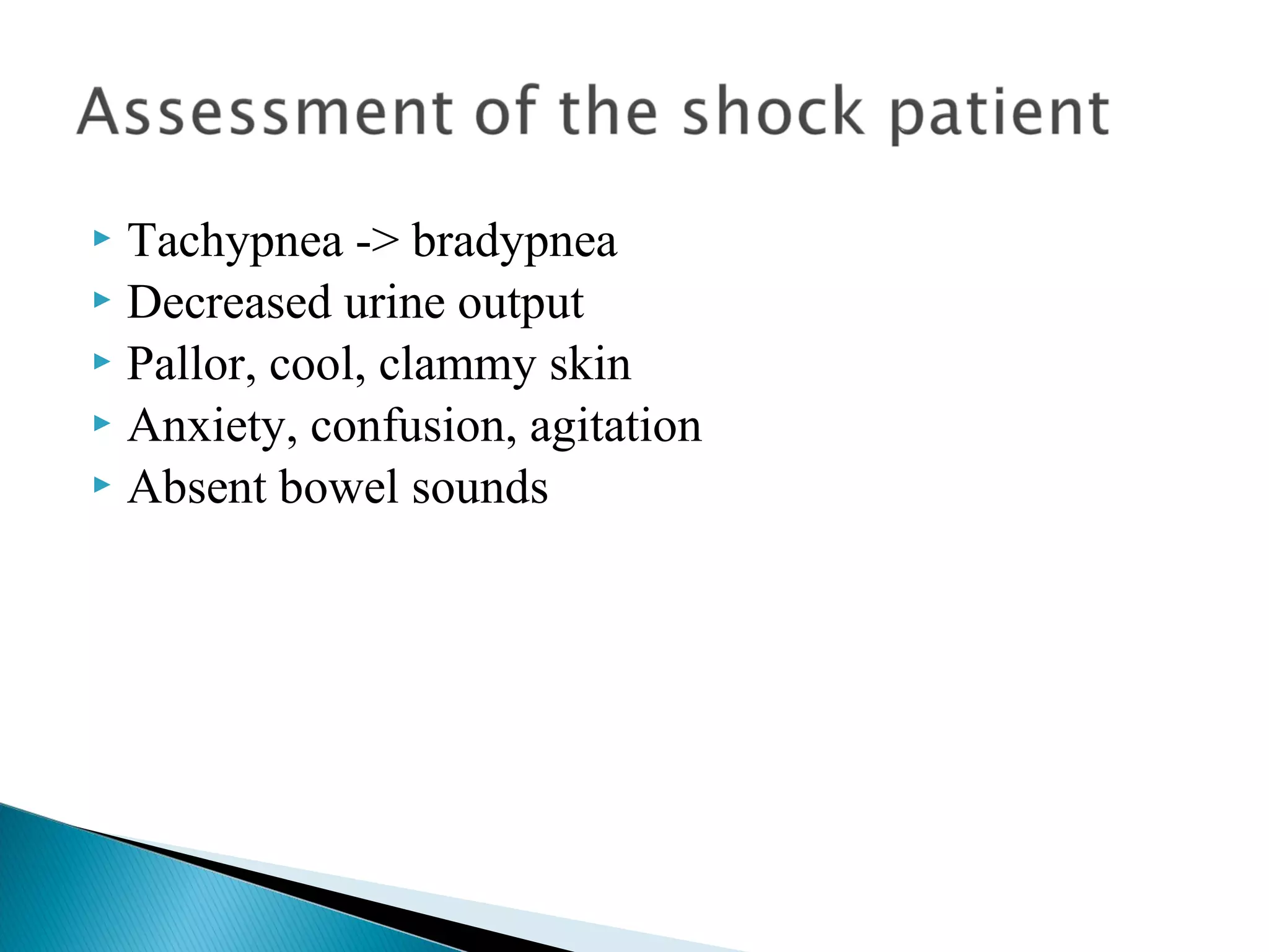
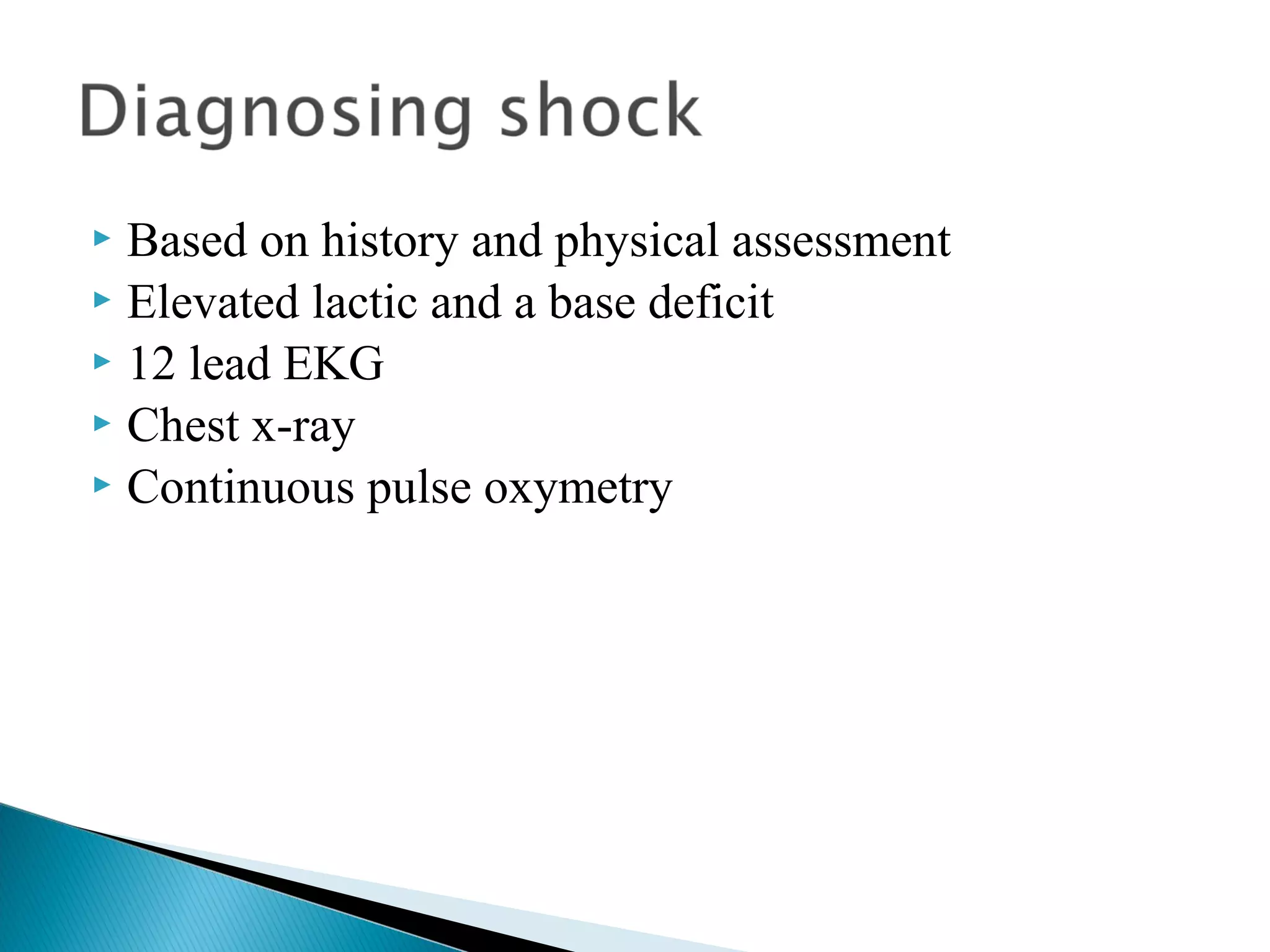
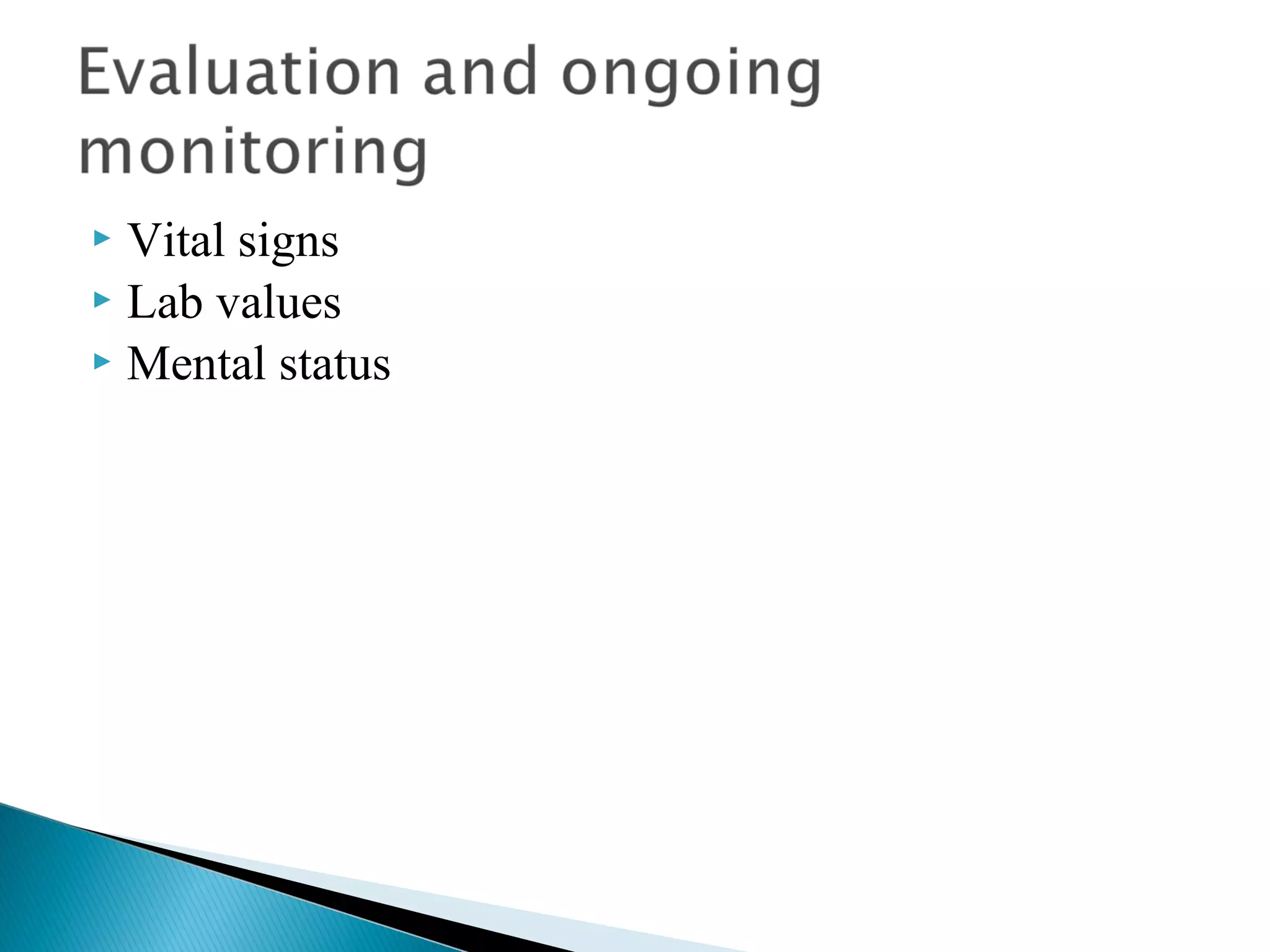
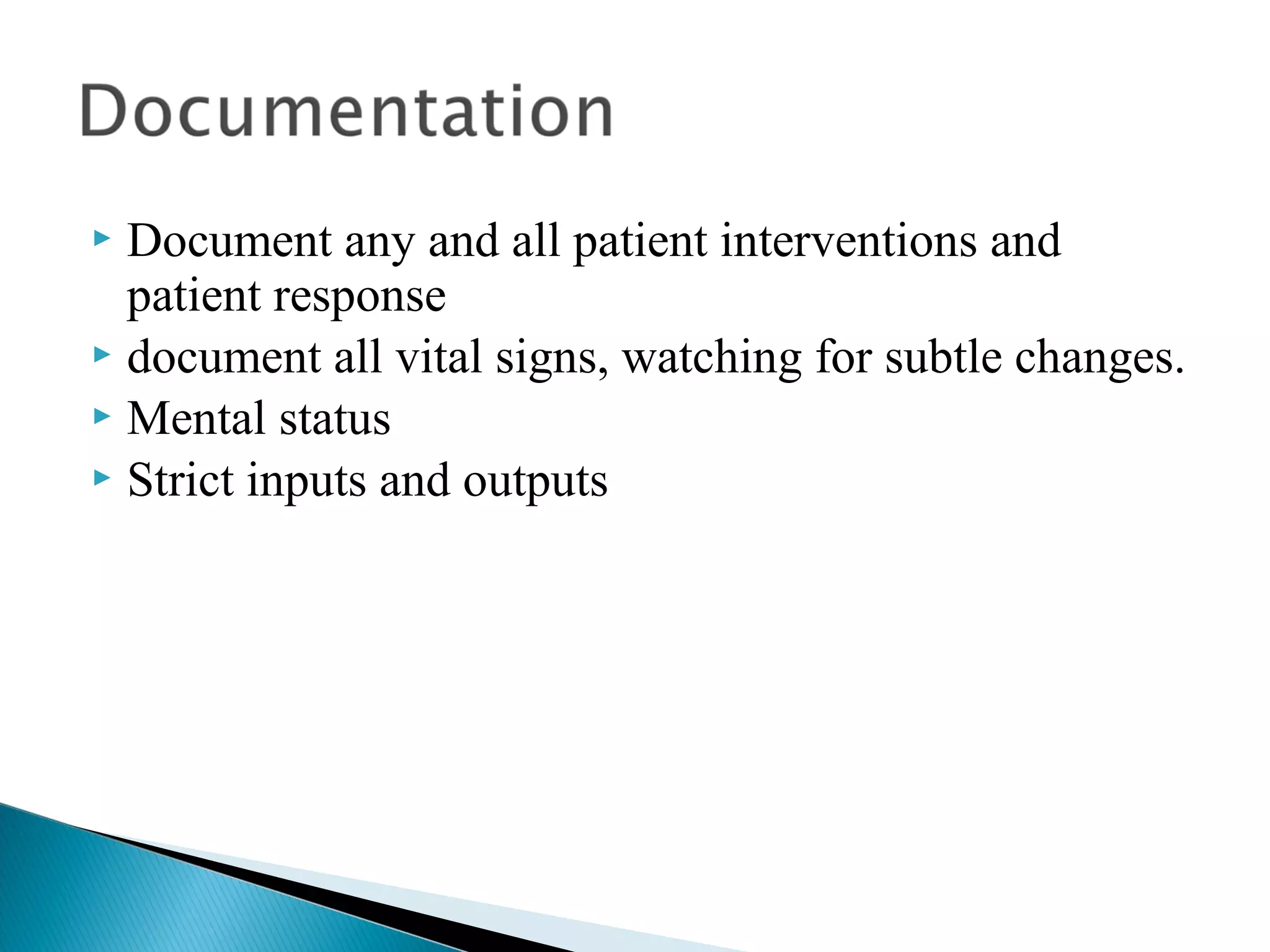
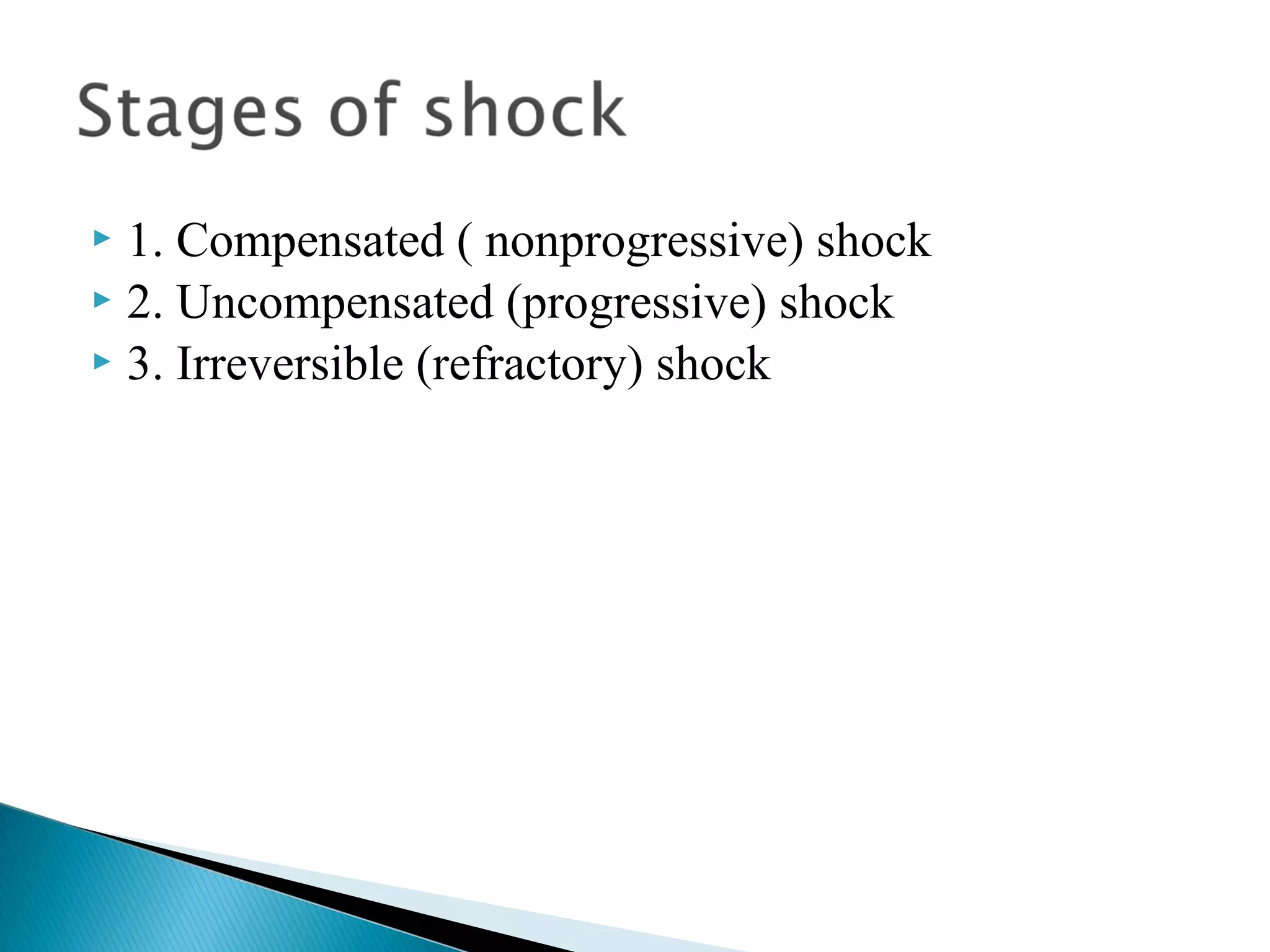
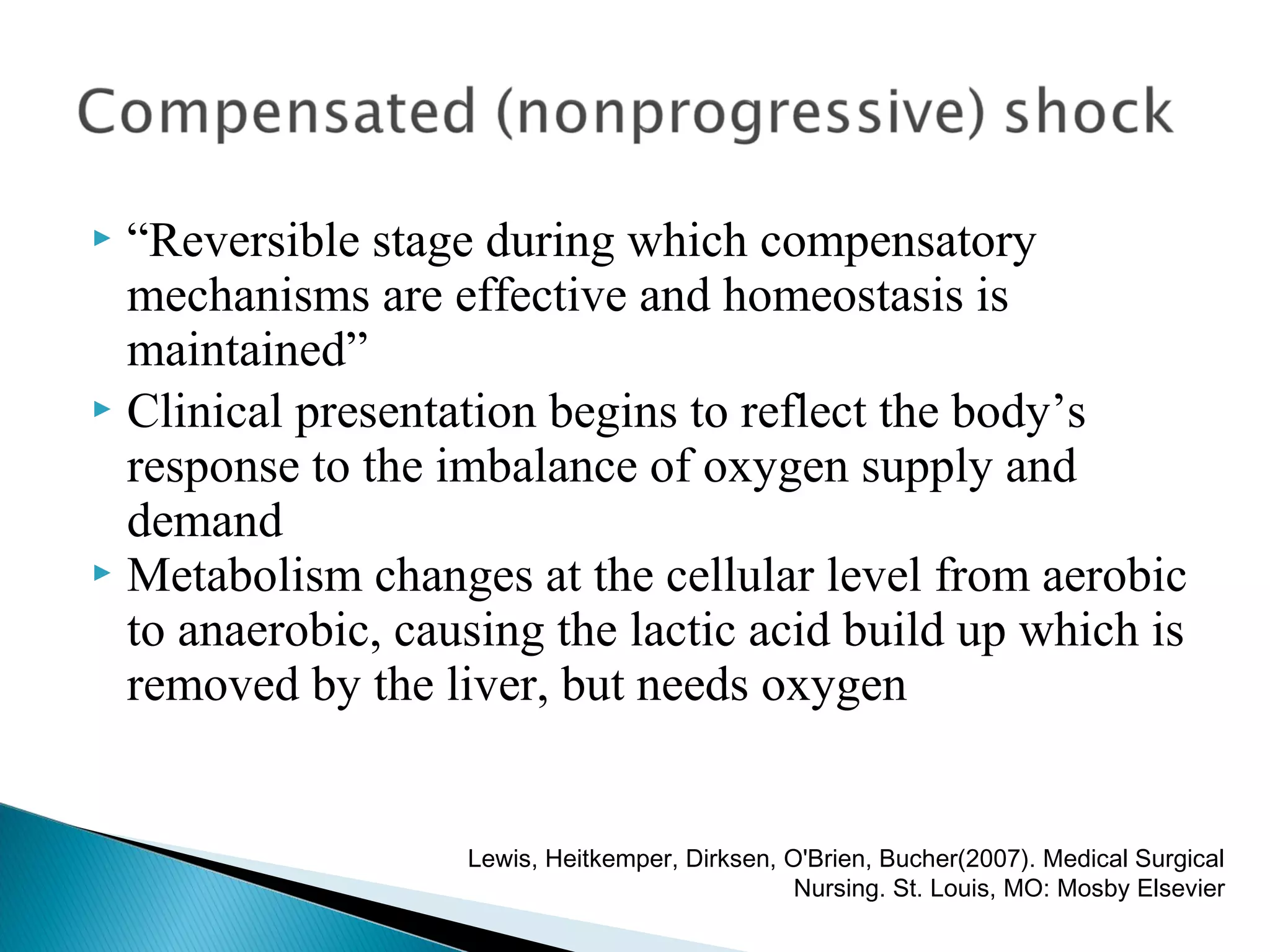
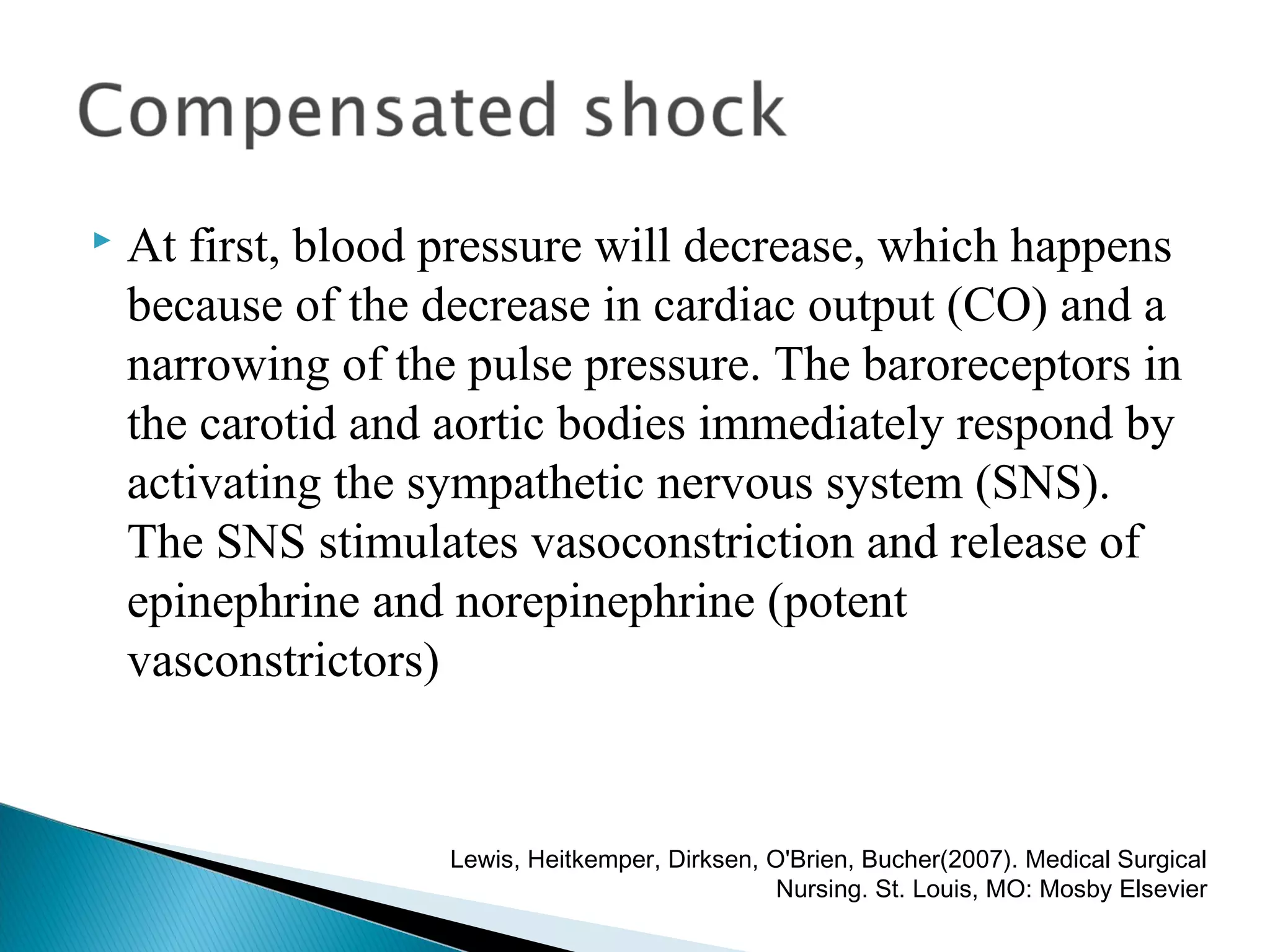
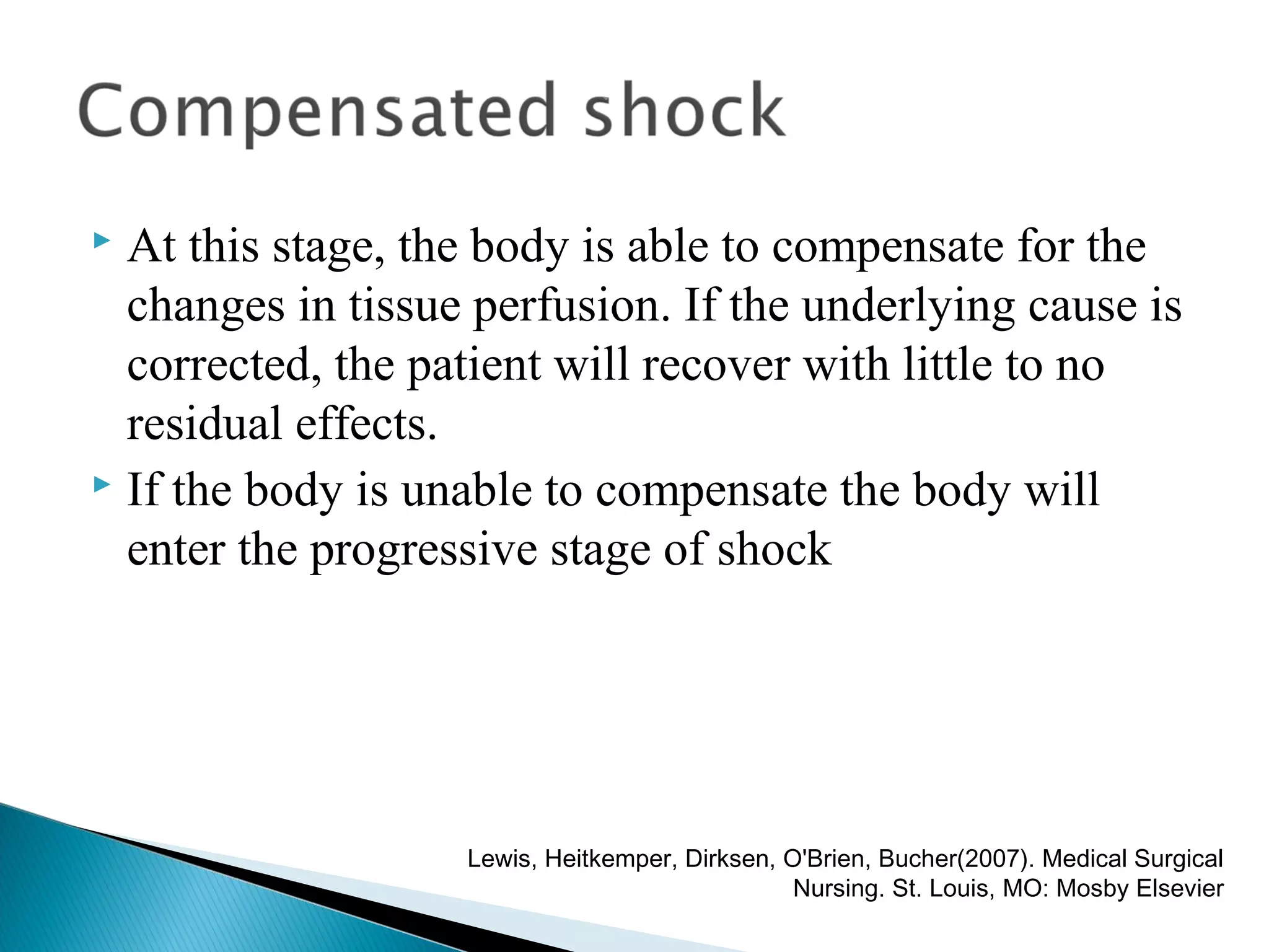
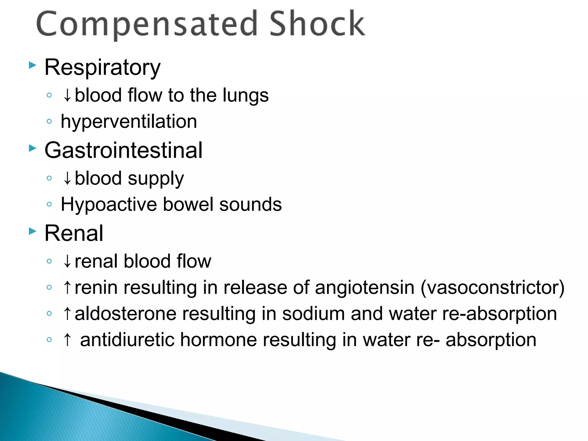
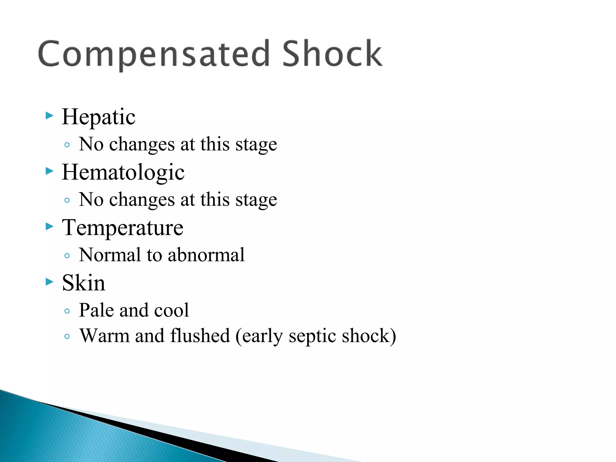
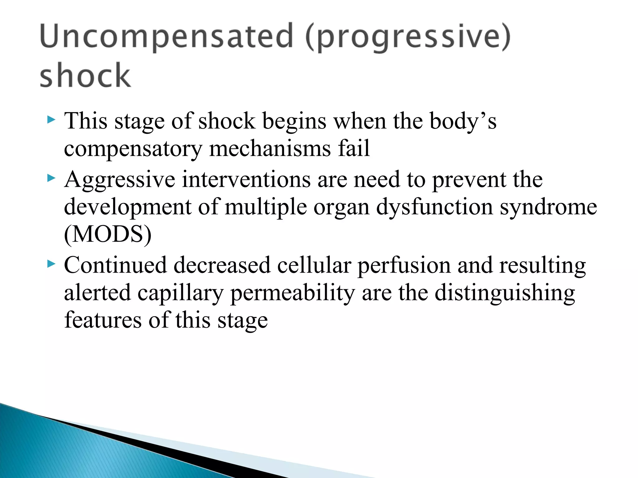
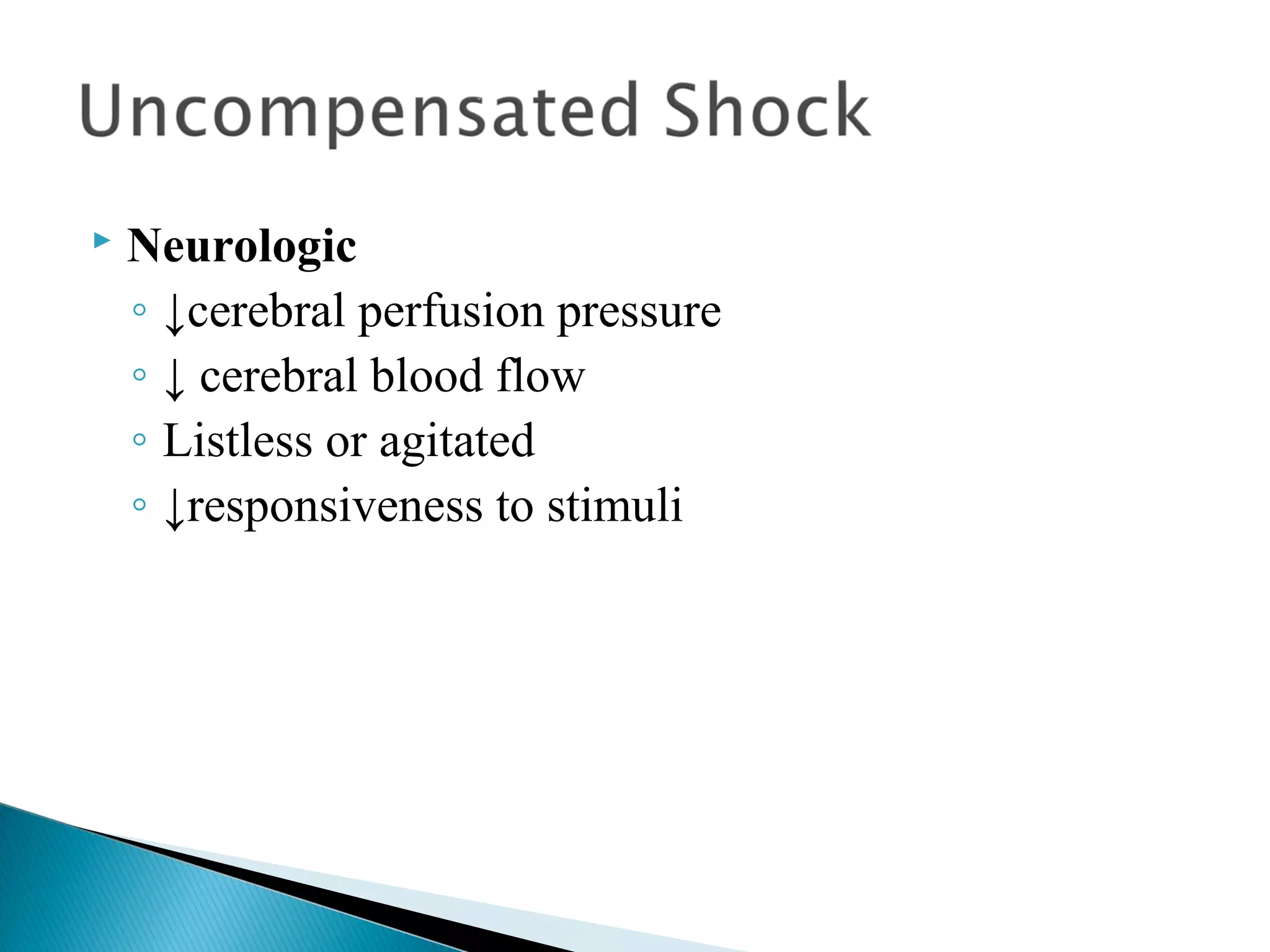
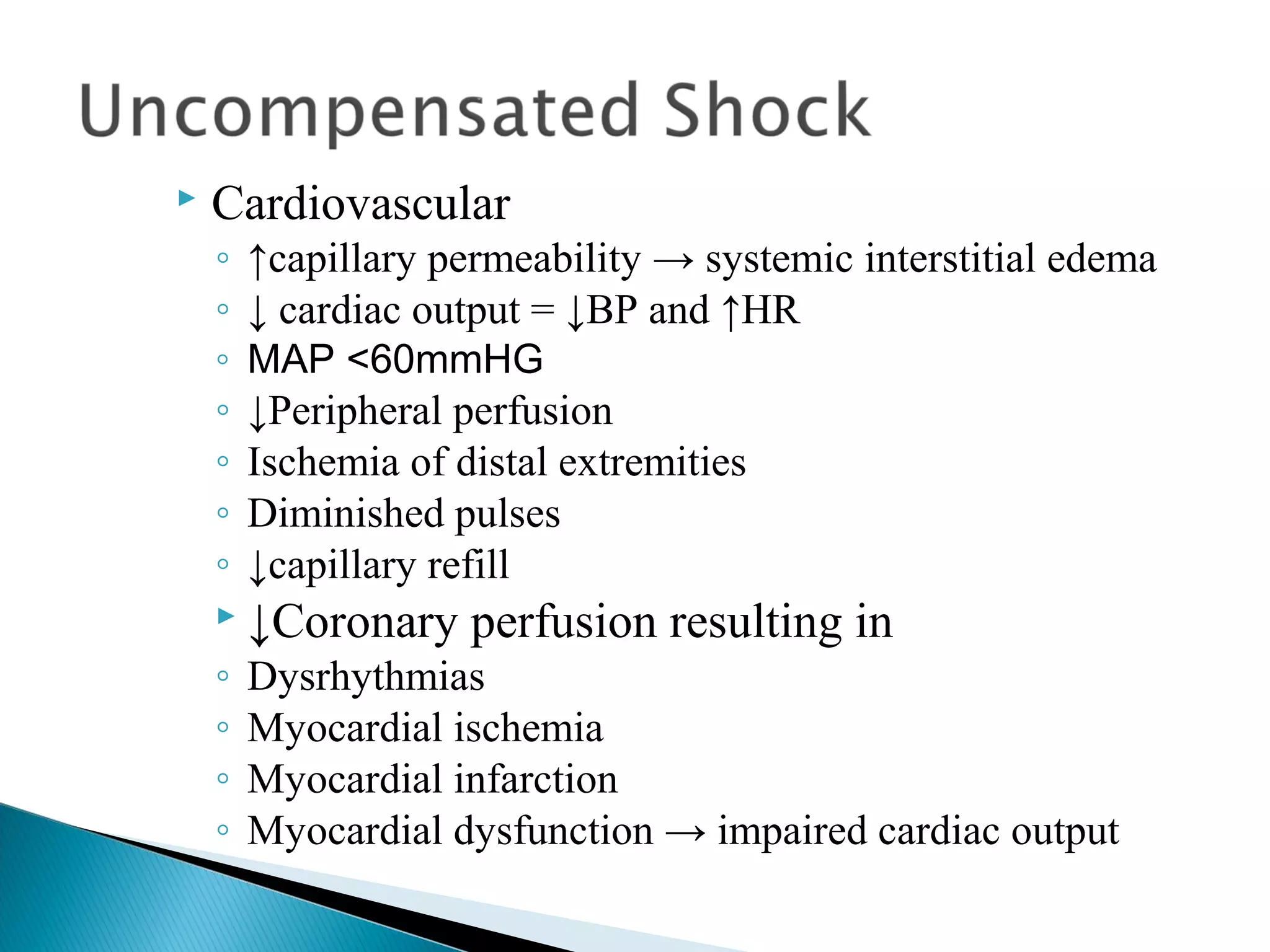
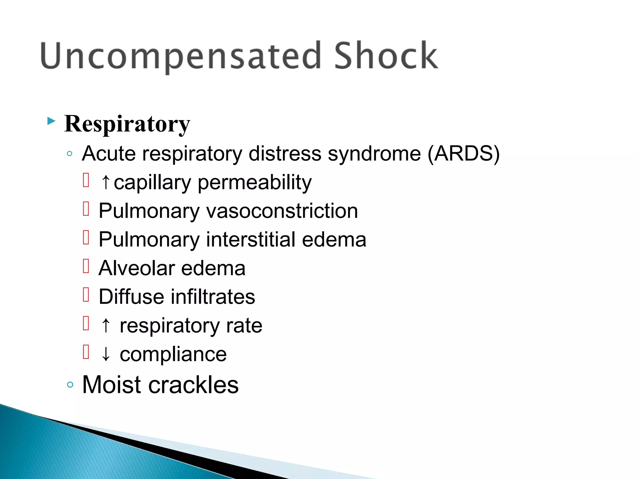
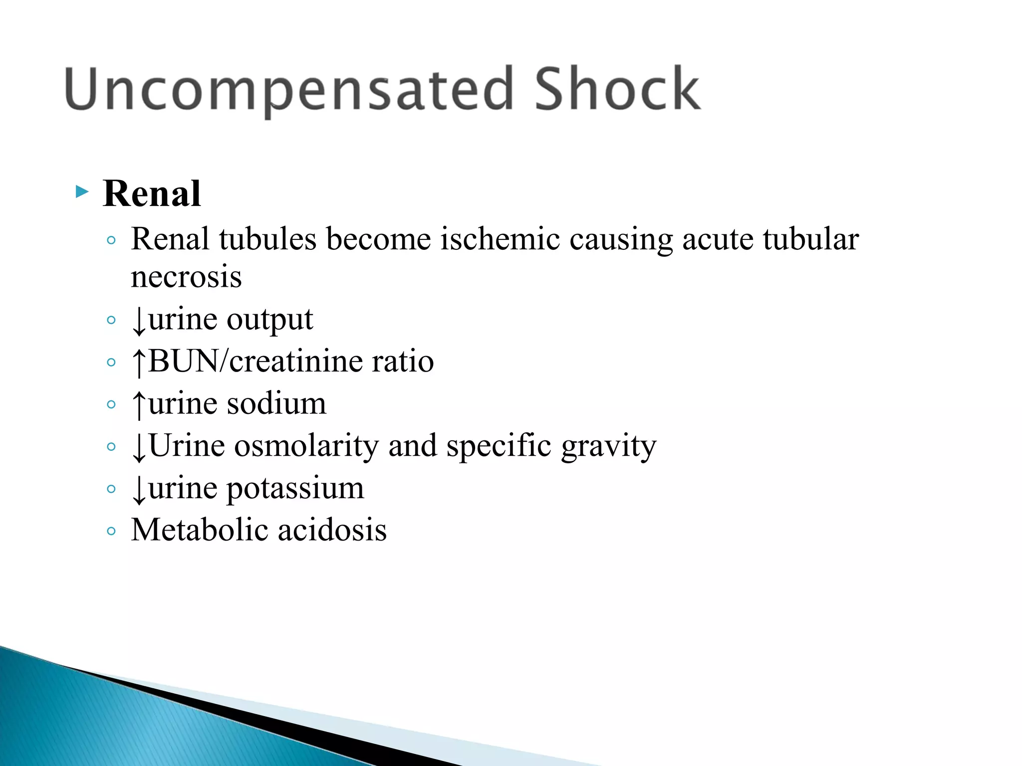
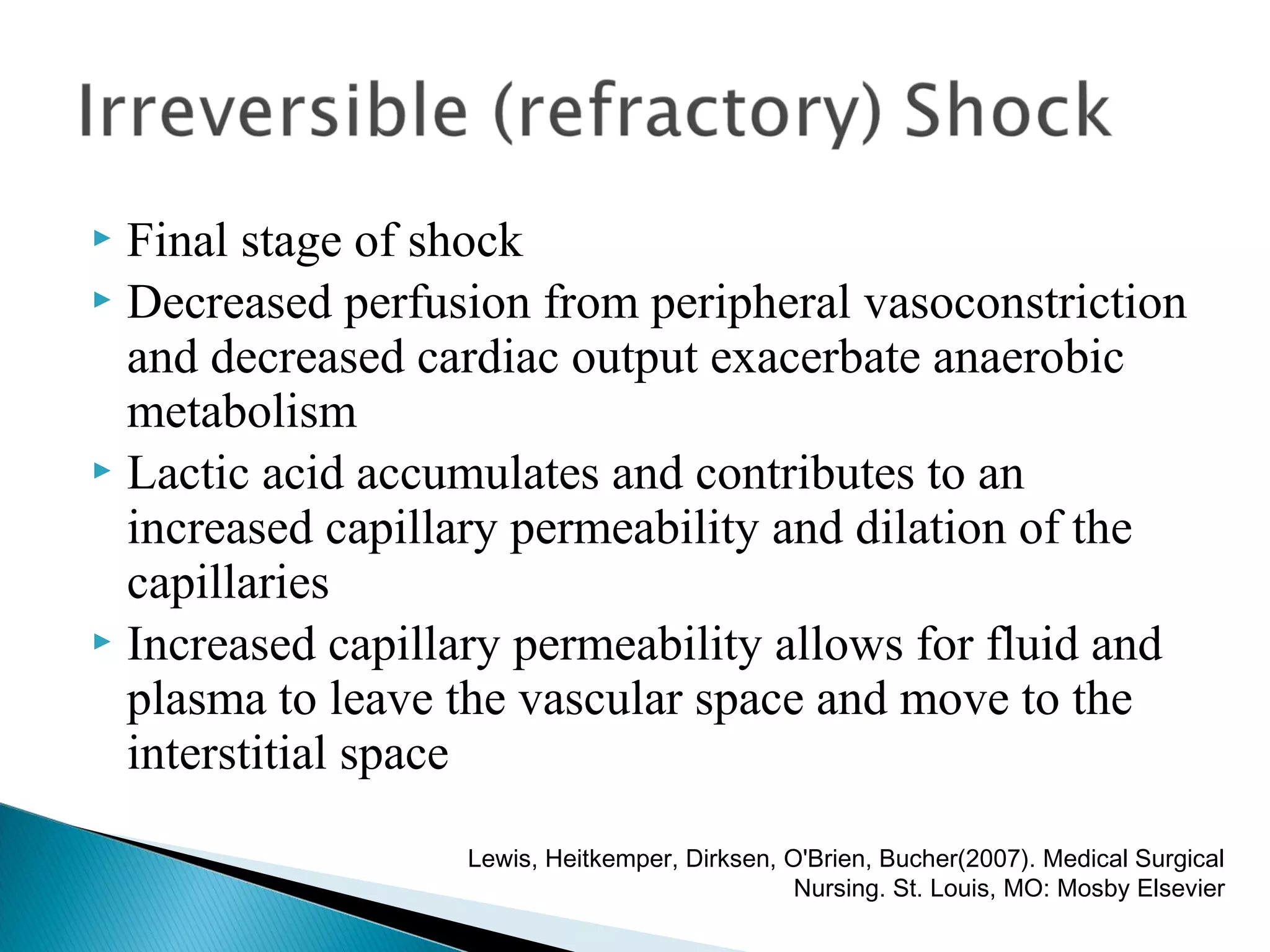
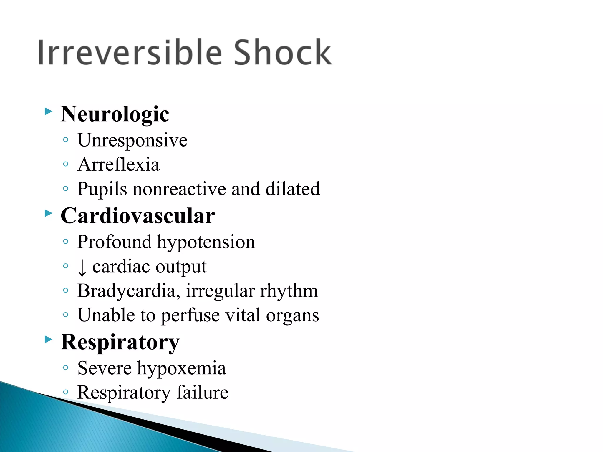
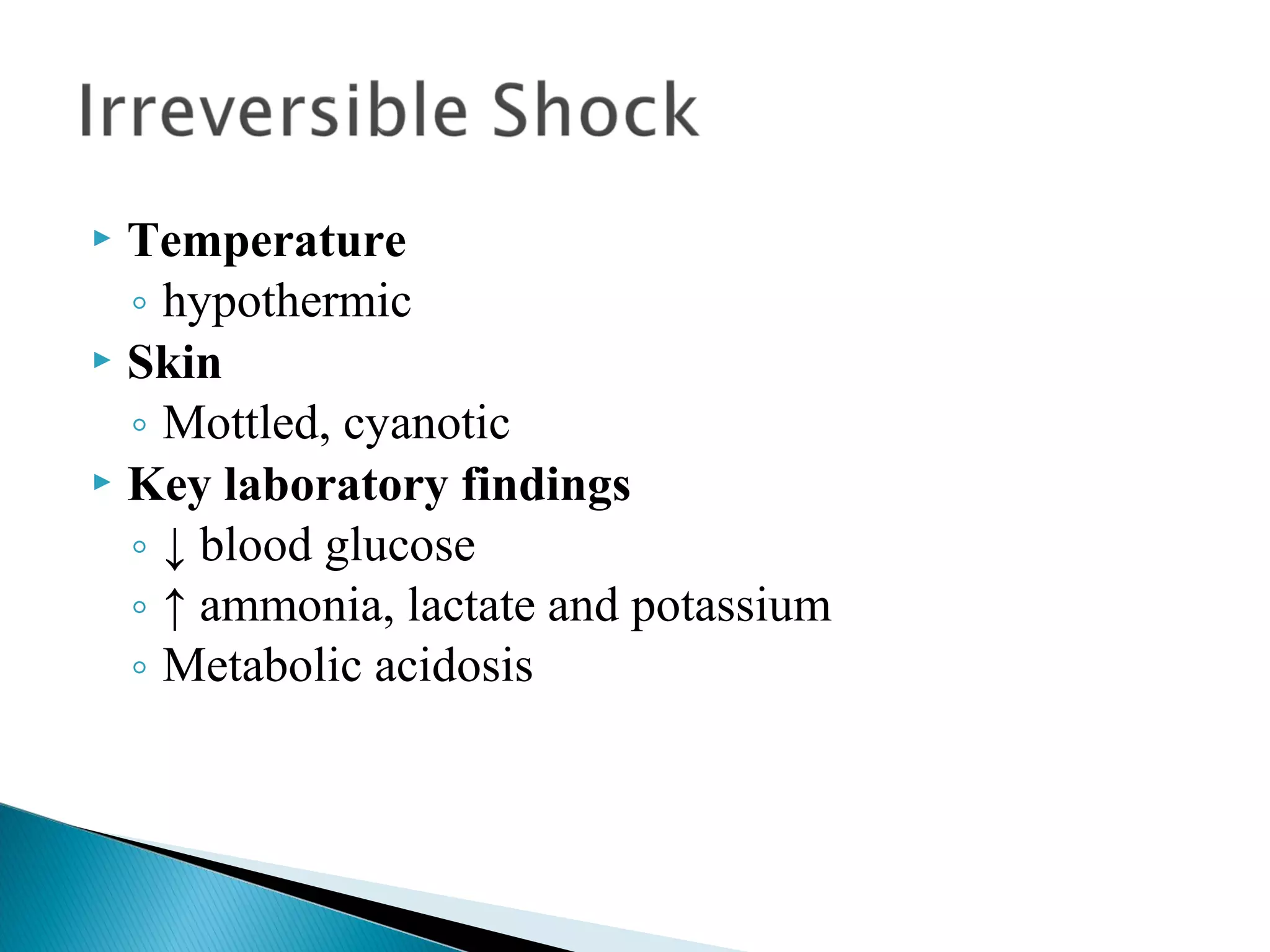
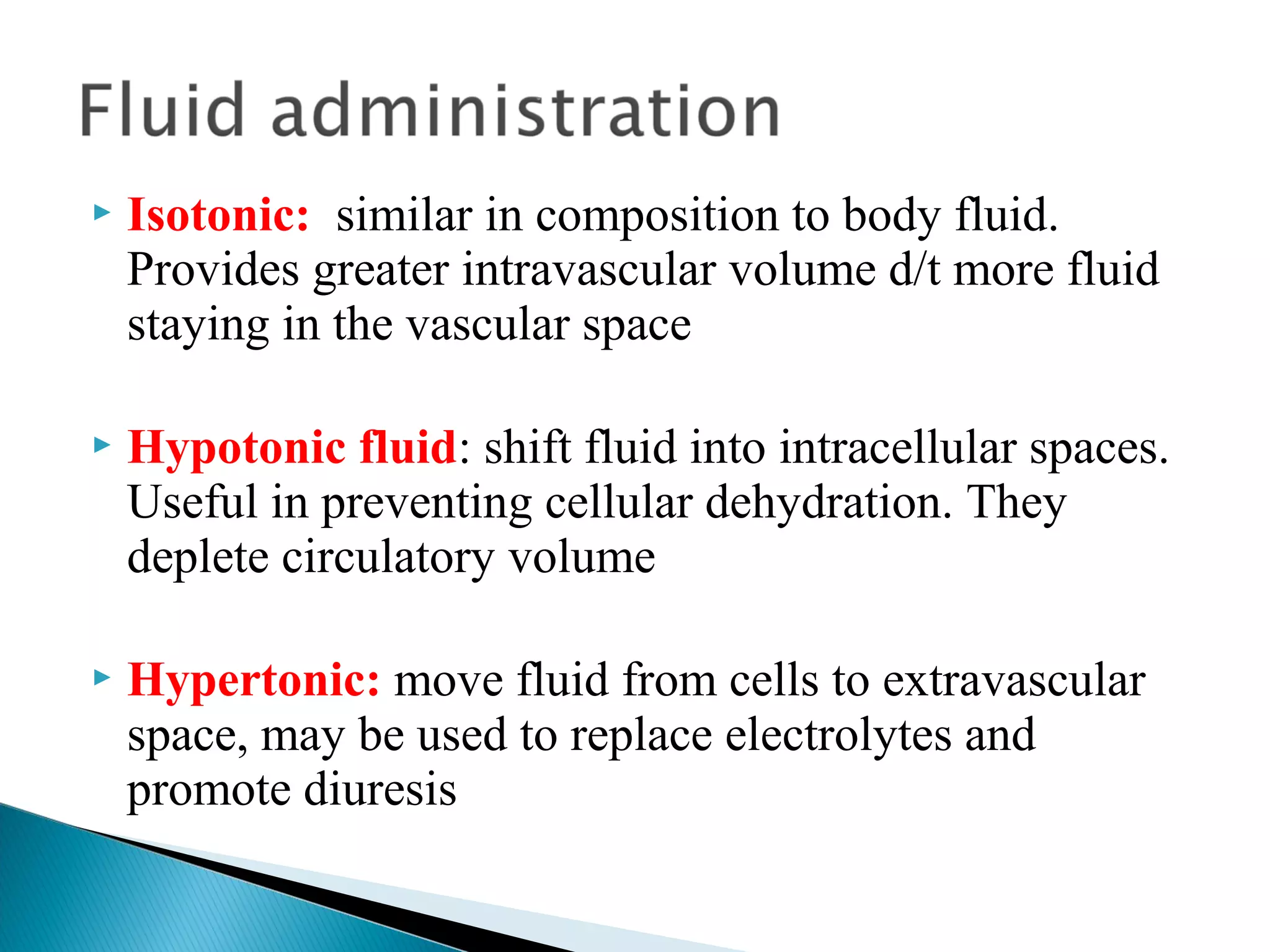
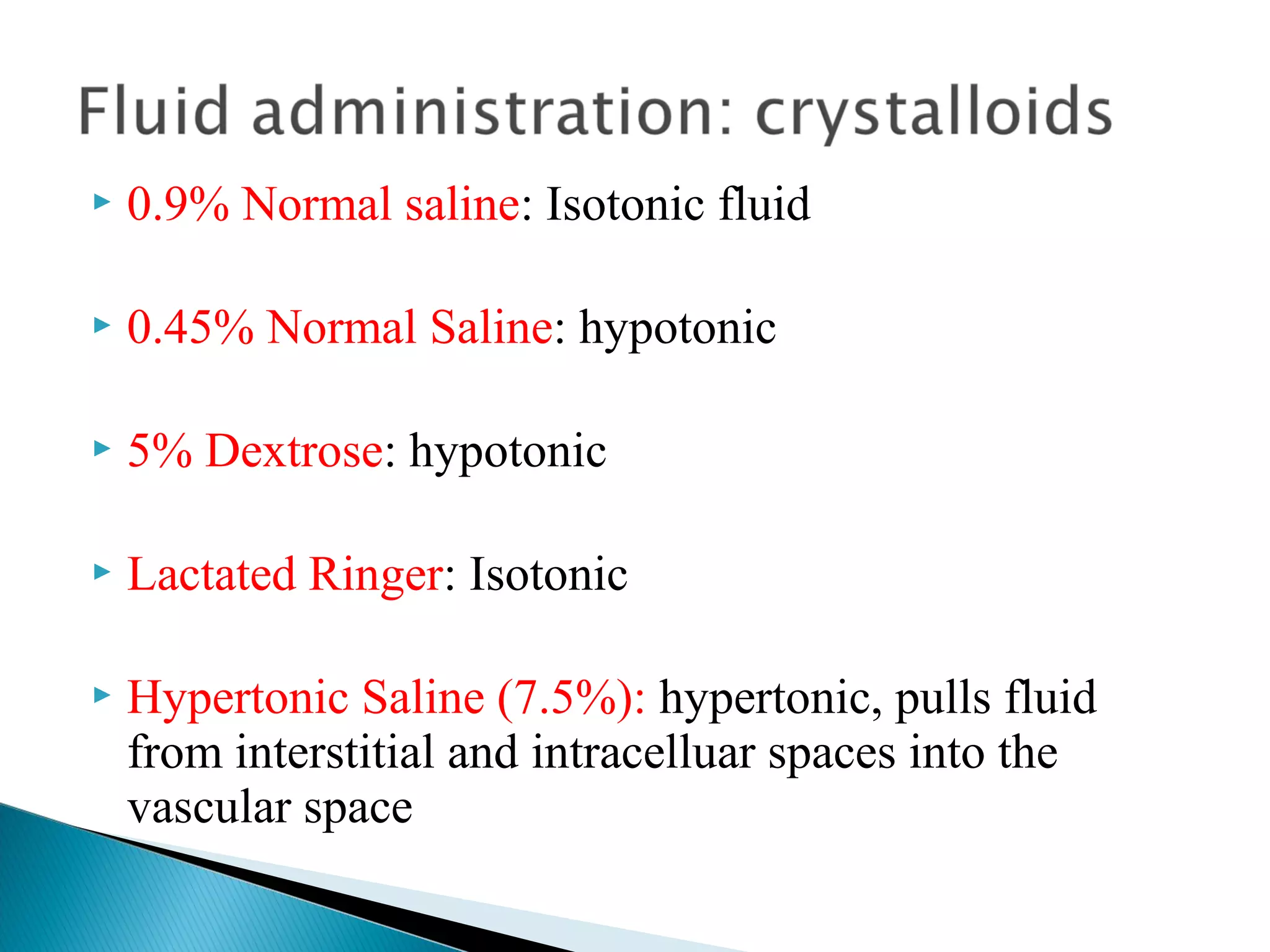
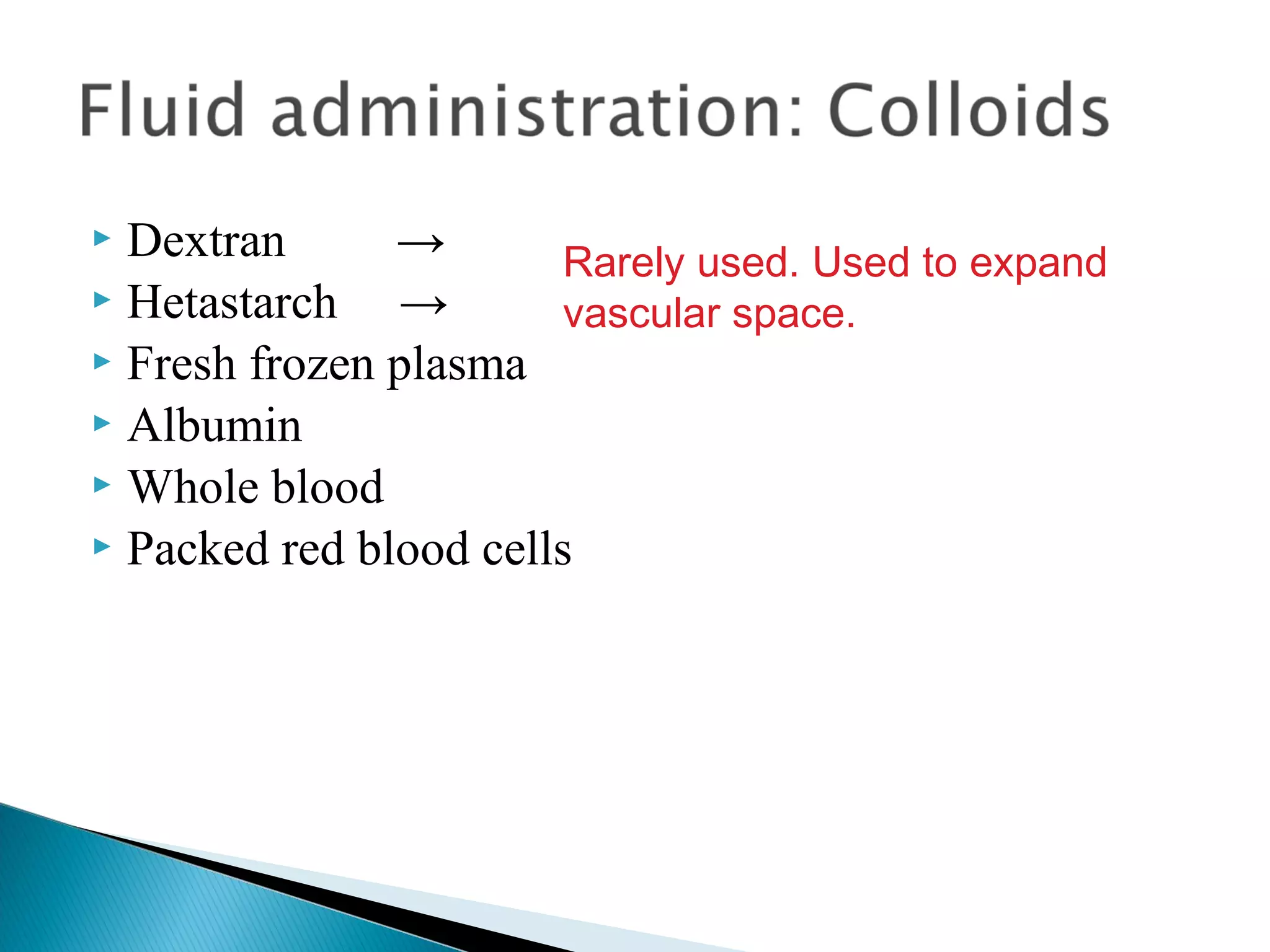
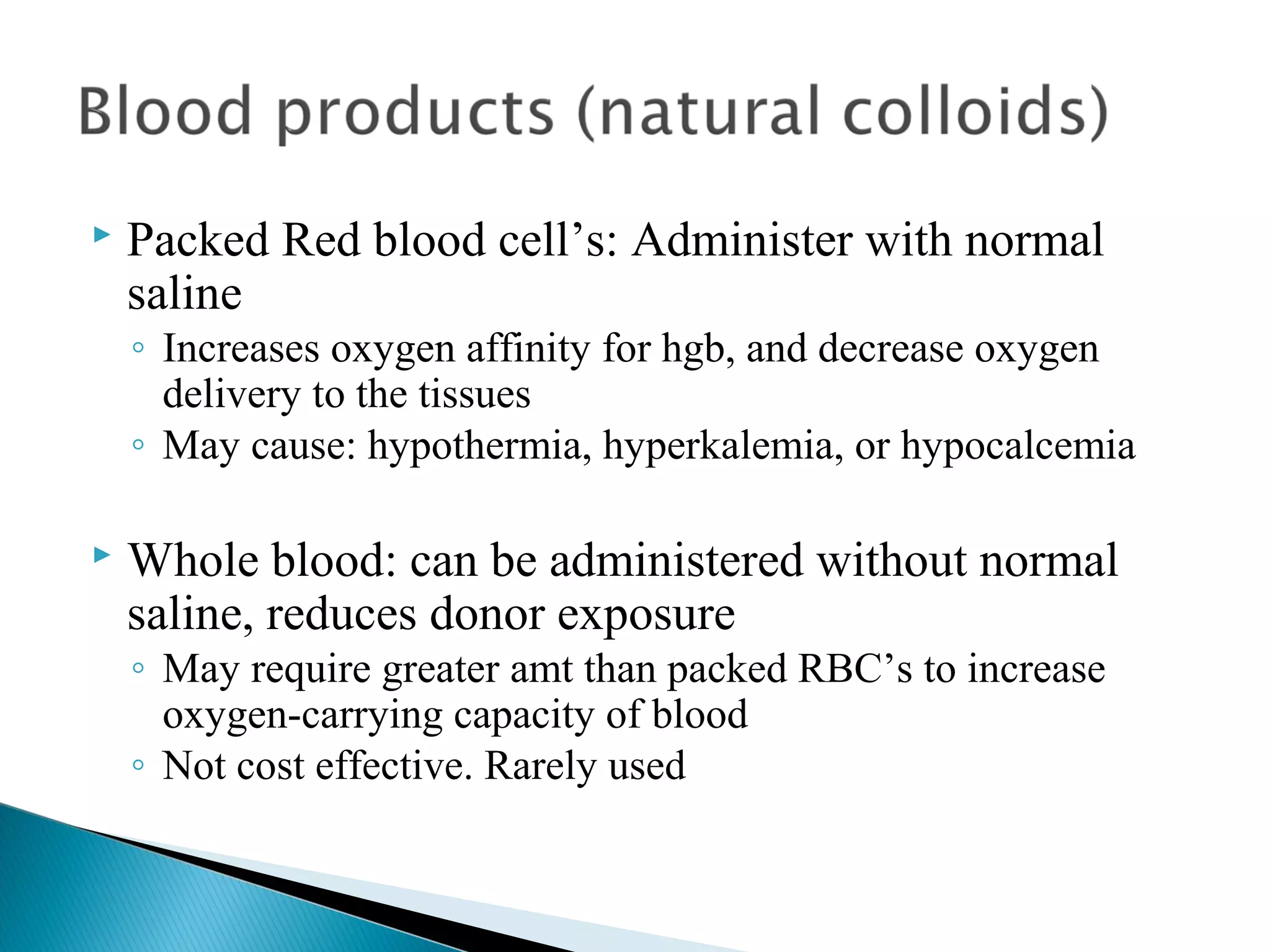
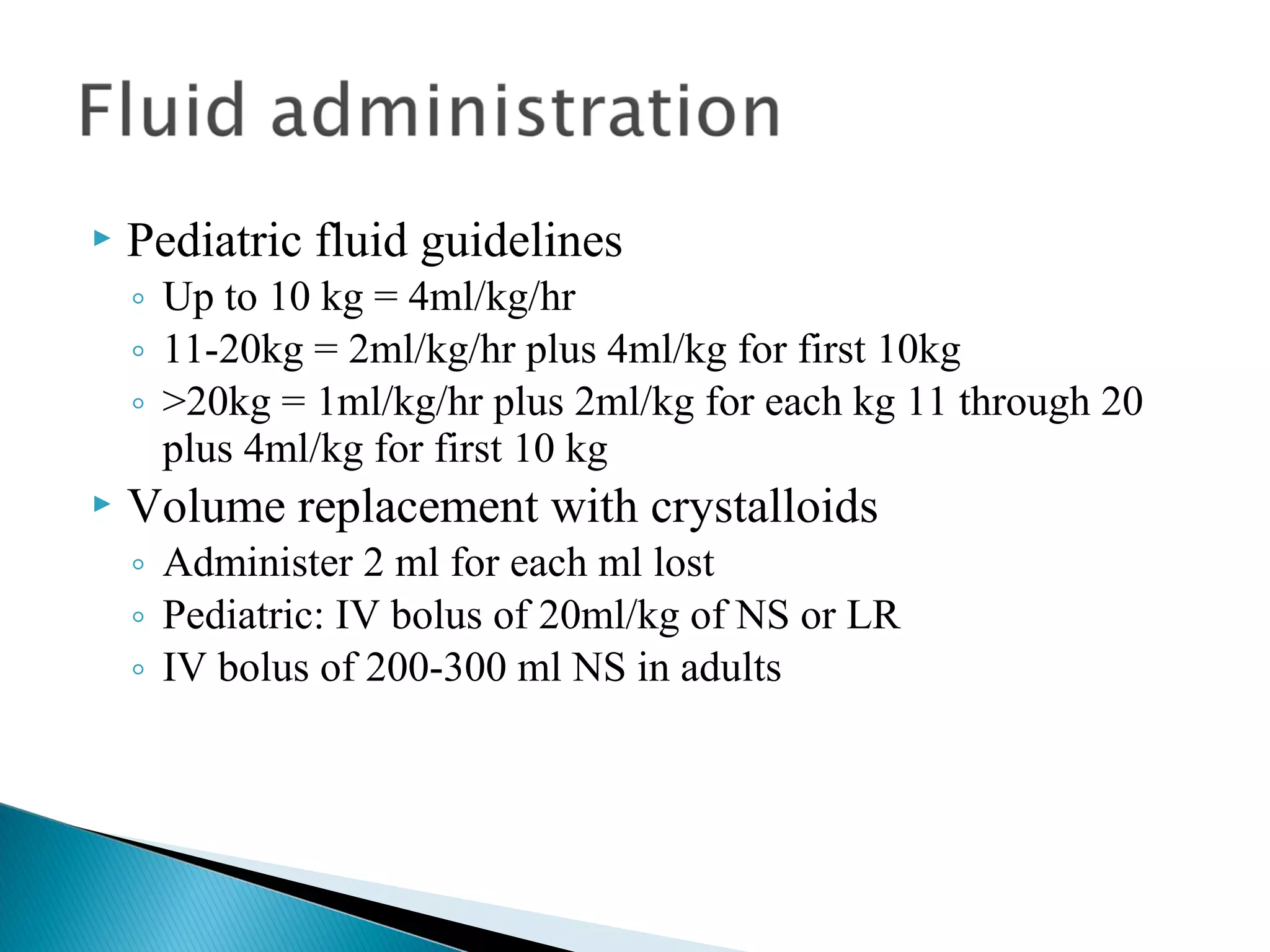
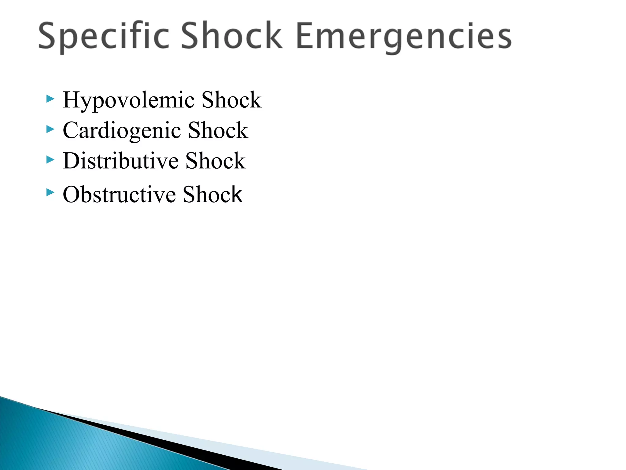
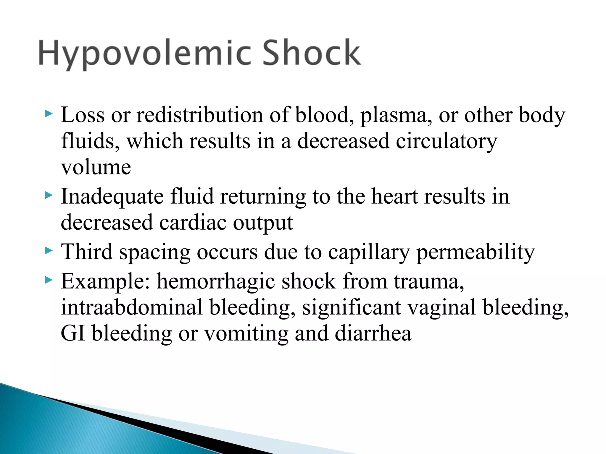
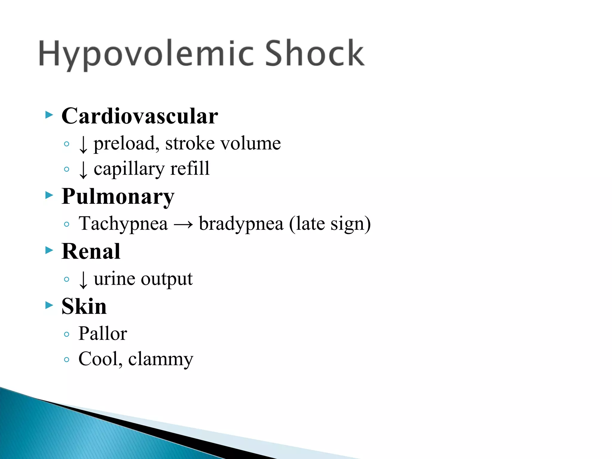
The document provides guidance on the nursing management of shock. It discusses assessing the type and phase of shock, providing emergency nursing care, monitoring the patient closely, making a diagnosis based on history and assessments, treating with fluid resuscitation and blood products, and monitoring the patient's response. It also covers age-related considerations and the three phases of shock: compensated, uncompensated, and irreversible.
Cardiogenic shock is a condition of diminished cardiac output that severely impairs cardiac perfusion. In this condition in which the heart suddenly can't pump enough blood to meet the body's needs.
Definition of heart failure - causes and types of heart failure - pathophysiology and risky factors for heart failure - Diagnosis clinical manifestations and investigations and classification of heart failure- treatment of chronic heart failure
Also Acute heart failure causes - clinical picture and treatment
Acute scrotum is a general term referring to an emergency condition affecting the contents or the wall of the scrotum.
There are a number of conditions that present acutely, predominantly with pain and/or swelling
A careful and detailed history and examination, and in some cases, investigations allow differentiation between these diagnoses. A prompt diagnosis is essential as the patient may require urgent surgical intervention
Testicular torsion refers to twisting of the spermatic cord, causing ischaemia of the testicle.
Testicular torsion results from inadequate fixation of the testis to the tunica vaginalis producing ischemia from reduced arterial inflow and venous outflow obstruction.
The prevalence of testicular torsion in adult patients hospitalized with acute scrotal pain is approximately 25 to 50 percent
Tom Selleck Health: A Comprehensive Look at the Iconic Actor’s Wellness Journeygreendigital
Tom Selleck, an enduring figure in Hollywood. has captivated audiences for decades with his rugged charm, iconic moustache. and memorable roles in television and film. From his breakout role as Thomas Magnum in Magnum P.I. to his current portrayal of Frank Reagan in Blue Bloods. Selleck's career has spanned over 50 years. But beyond his professional achievements. fans have often been curious about Tom Selleck Health. especially as he has aged in the public eye.
Follow us on: Pinterest
Introduction
Many have been interested in Tom Selleck health. not only because of his enduring presence on screen but also because of the challenges. and lifestyle choices he has faced and made over the years. This article delves into the various aspects of Tom Selleck health. exploring his fitness regimen, diet, mental health. and the challenges he has encountered as he ages. We'll look at how he maintains his well-being. the health issues he has faced, and his approach to ageing .
Early Life and Career
Childhood and Athletic Beginnings
Tom Selleck was born on January 29, 1945, in Detroit, Michigan, and grew up in Sherman Oaks, California. From an early age, he was involved in sports, particularly basketball. which played a significant role in his physical development. His athletic pursuits continued into college. where he attended the University of Southern California (USC) on a basketball scholarship. This early involvement in sports laid a strong foundation for his physical health and disciplined lifestyle.
Transition to Acting
Selleck's transition from an athlete to an actor came with its physical demands. His first significant role in "Magnum P.I." required him to perform various stunts and maintain a fit appearance. This role, which he played from 1980 to 1988. necessitated a rigorous fitness routine to meet the show's demands. setting the stage for his long-term commitment to health and wellness.
Fitness Regimen
Workout Routine
Tom Selleck health and fitness regimen has evolved. adapting to his changing roles and age. During his "Magnum, P.I." days. Selleck's workouts were intense and focused on building and maintaining muscle mass. His routine included weightlifting, cardiovascular exercises. and specific training for the stunts he performed on the show.
Selleck adjusted his fitness routine as he aged to suit his body's needs. Today, his workouts focus on maintaining flexibility, strength, and cardiovascular health. He incorporates low-impact exercises such as swimming, walking, and light weightlifting. This balanced approach helps him stay fit without putting undue strain on his joints and muscles.
Importance of Flexibility and Mobility
In recent years, Selleck has emphasized the importance of flexibility and mobility in his fitness regimen. Understanding the natural decline in muscle mass and joint flexibility with age. he includes stretching and yoga in his routine. These practices help prevent injuries, improve posture, and maintain mobilit
New Directions in Targeted Therapeutic Approaches for Older Adults With Mantl...i3 Health
i3 Health is pleased to make the speaker slides from this activity available for use as a non-accredited self-study or teaching resource.
This slide deck presented by Dr. Kami Maddocks, Professor-Clinical in the Division of Hematology and
Associate Division Director for Ambulatory Operations
The Ohio State University Comprehensive Cancer Center, will provide insight into new directions in targeted therapeutic approaches for older adults with mantle cell lymphoma.
STATEMENT OF NEED
Mantle cell lymphoma (MCL) is a rare, aggressive B-cell non-Hodgkin lymphoma (NHL) accounting for 5% to 7% of all lymphomas. Its prognosis ranges from indolent disease that does not require treatment for years to very aggressive disease, which is associated with poor survival (Silkenstedt et al, 2021). Typically, MCL is diagnosed at advanced stage and in older patients who cannot tolerate intensive therapy (NCCN, 2022). Although recent advances have slightly increased remission rates, recurrence and relapse remain very common, leading to a median overall survival between 3 and 6 years (LLS, 2021). Though there are several effective options, progress is still needed towards establishing an accepted frontline approach for MCL (Castellino et al, 2022). Treatment selection and management of MCL are complicated by the heterogeneity of prognosis, advanced age and comorbidities of patients, and lack of an established standard approach for treatment, making it vital that clinicians be familiar with the latest research and advances in this area. In this activity chaired by Michael Wang, MD, Professor in the Department of Lymphoma & Myeloma at MD Anderson Cancer Center, expert faculty will discuss prognostic factors informing treatment, the promising results of recent trials in new therapeutic approaches, and the implications of treatment resistance in therapeutic selection for MCL.
Target Audience
Hematology/oncology fellows, attending faculty, and other health care professionals involved in the treatment of patients with mantle cell lymphoma (MCL).
Learning Objectives
1.) Identify clinical and biological prognostic factors that can guide treatment decision making for older adults with MCL
2.) Evaluate emerging data on targeted therapeutic approaches for treatment-naive and relapsed/refractory MCL and their applicability to older adults
3.) Assess mechanisms of resistance to targeted therapies for MCL and their implications for treatment selection
Title: Sense of Smell
Presenter: Dr. Faiza, Assistant Professor of Physiology
Qualifications:
MBBS (Best Graduate, AIMC Lahore)
FCPS Physiology
ICMT, CHPE, DHPE (STMU)
MPH (GC University, Faisalabad)
MBA (Virtual University of Pakistan)
Learning Objectives:
Describe the primary categories of smells and the concept of odor blindness.
Explain the structure and location of the olfactory membrane and mucosa, including the types and roles of cells involved in olfaction.
Describe the pathway and mechanisms of olfactory signal transmission from the olfactory receptors to the brain.
Illustrate the biochemical cascade triggered by odorant binding to olfactory receptors, including the role of G-proteins and second messengers in generating an action potential.
Identify different types of olfactory disorders such as anosmia, hyposmia, hyperosmia, and dysosmia, including their potential causes.
Key Topics:
Olfactory Genes:
3% of the human genome accounts for olfactory genes.
400 genes for odorant receptors.
Olfactory Membrane:
Located in the superior part of the nasal cavity.
Medially: Folds downward along the superior septum.
Laterally: Folds over the superior turbinate and upper surface of the middle turbinate.
Total surface area: 5-10 square centimeters.
Olfactory Mucosa:
Olfactory Cells: Bipolar nerve cells derived from the CNS (100 million), with 4-25 olfactory cilia per cell.
Sustentacular Cells: Produce mucus and maintain ionic and molecular environment.
Basal Cells: Replace worn-out olfactory cells with an average lifespan of 1-2 months.
Bowman’s Gland: Secretes mucus.
Stimulation of Olfactory Cells:
Odorant dissolves in mucus and attaches to receptors on olfactory cilia.
Involves a cascade effect through G-proteins and second messengers, leading to depolarization and action potential generation in the olfactory nerve.
Quality of a Good Odorant:
Small (3-20 Carbon atoms), volatile, water-soluble, and lipid-soluble.
Facilitated by odorant-binding proteins in mucus.
Membrane Potential and Action Potential:
Resting membrane potential: -55mV.
Action potential frequency in the olfactory nerve increases with odorant strength.
Adaptation Towards the Sense of Smell:
Rapid adaptation within the first second, with further slow adaptation.
Psychological adaptation greater than receptor adaptation, involving feedback inhibition from the central nervous system.
Primary Sensations of Smell:
Camphoraceous, Musky, Floral, Pepperminty, Ethereal, Pungent, Putrid.
Odor Detection Threshold:
Examples: Hydrogen sulfide (0.0005 ppm), Methyl-mercaptan (0.002 ppm).
Some toxic substances are odorless at lethal concentrations.
Characteristics of Smell:
Odor blindness for single substances due to lack of appropriate receptor protein.
Behavioral and emotional influences of smell.
Transmission of Olfactory Signals:
From olfactory cells to glomeruli in the olfactory bulb, involving lateral inhibition.
Primitive, less old, and new olfactory systems with different path
Title: Sense of Taste
Presenter: Dr. Faiza, Assistant Professor of Physiology
Qualifications:
MBBS (Best Graduate, AIMC Lahore)
FCPS Physiology
ICMT, CHPE, DHPE (STMU)
MPH (GC University, Faisalabad)
MBA (Virtual University of Pakistan)
Learning Objectives:
Describe the structure and function of taste buds.
Describe the relationship between the taste threshold and taste index of common substances.
Explain the chemical basis and signal transduction of taste perception for each type of primary taste sensation.
Recognize different abnormalities of taste perception and their causes.
Key Topics:
Significance of Taste Sensation:
Differentiation between pleasant and harmful food
Influence on behavior
Selection of food based on metabolic needs
Receptors of Taste:
Taste buds on the tongue
Influence of sense of smell, texture of food, and pain stimulation (e.g., by pepper)
Primary and Secondary Taste Sensations:
Primary taste sensations: Sweet, Sour, Salty, Bitter, Umami
Chemical basis and signal transduction mechanisms for each taste
Taste Threshold and Index:
Taste threshold values for Sweet (sucrose), Salty (NaCl), Sour (HCl), and Bitter (Quinine)
Taste index relationship: Inversely proportional to taste threshold
Taste Blindness:
Inability to taste certain substances, particularly thiourea compounds
Example: Phenylthiocarbamide
Structure and Function of Taste Buds:
Composition: Epithelial cells, Sustentacular/Supporting cells, Taste cells, Basal cells
Features: Taste pores, Taste hairs/microvilli, and Taste nerve fibers
Location of Taste Buds:
Found in papillae of the tongue (Fungiform, Circumvallate, Foliate)
Also present on the palate, tonsillar pillars, epiglottis, and proximal esophagus
Mechanism of Taste Stimulation:
Interaction of taste substances with receptors on microvilli
Signal transduction pathways for Umami, Sweet, Bitter, Sour, and Salty tastes
Taste Sensitivity and Adaptation:
Decrease in sensitivity with age
Rapid adaptation of taste sensation
Role of Saliva in Taste:
Dissolution of tastants to reach receptors
Washing away the stimulus
Taste Preferences and Aversions:
Mechanisms behind taste preference and aversion
Influence of receptors and neural pathways
Impact of Sensory Nerve Damage:
Degeneration of taste buds if the sensory nerve fiber is cut
Abnormalities of Taste Detection:
Conditions: Ageusia, Hypogeusia, Dysgeusia (parageusia)
Causes: Nerve damage, neurological disorders, infections, poor oral hygiene, adverse drug effects, deficiencies, aging, tobacco use, altered neurotransmitter levels
Neurotransmitters and Taste Threshold:
Effects of serotonin (5-HT) and norepinephrine (NE) on taste sensitivity
Supertasters:
25% of the population with heightened sensitivity to taste, especially bitterness
Increased number of fungiform papillae
Knee anatomy and clinical tests 2024.pdfvimalpl1234
This includes all relevant anatomy and clinical tests compiled from standard textbooks, Campbell,netter etc..It is comprehensive and best suited for orthopaedicians and orthopaedic residents.
These lecture slides, by Dr Sidra Arshad, offer a quick overview of physiological basis of a normal electrocardiogram.
Learning objectives:
1. Define an electrocardiogram (ECG) and electrocardiography
2. Describe how dipoles generated by the heart produce the waveforms of the ECG
3. Describe the components of a normal electrocardiogram of a typical bipolar leads (limb II)
4. Differentiate between intervals and segments
5. Enlist some common indications for obtaining an ECG
Study Resources:
1. Chapter 11, Guyton and Hall Textbook of Medical Physiology, 14th edition
2. Chapter 9, Human Physiology - From Cells to Systems, Lauralee Sherwood, 9th edition
3. Chapter 29, Ganong’s Review of Medical Physiology, 26th edition
4. Electrocardiogram, StatPearls - https://www.ncbi.nlm.nih.gov/books/NBK549803/
5. ECG in Medical Practice by ABM Abdullah, 4th edition
6. ECG Basics, http://www.nataliescasebook.com/tag/e-c-g-basics
The prostate is an exocrine gland of the male mammalian reproductive system
It is a walnut-sized gland that forms part of the male reproductive system and is located in front of the rectum and just below the urinary bladder
Function is to store and secrete a clear, slightly alkaline fluid that constitutes 10-30% of the volume of the seminal fluid that along with the spermatozoa, constitutes semen
A healthy human prostate measures (4cm-vertical, by 3cm-horizontal, 2cm ant-post ).
It surrounds the urethra just below the urinary bladder. It has anterior, median, posterior and two lateral lobes
It’s work is regulated by androgens which are responsible for male sex characteristics
Generalised disease of the prostate due to hormonal derangement which leads to non malignant enlargement of the gland (increase in the number of epithelial cells and stromal tissue)to cause compression of the urethra leading to symptoms (LUTS
Couples presenting to the infertility clinic- Do they really have infertility...Sujoy Dasgupta
Dr Sujoy Dasgupta presented the study on "Couples presenting to the infertility clinic- Do they really have infertility? – The unexplored stories of non-consummation" in the 13th Congress of the Asia Pacific Initiative on Reproduction (ASPIRE 2024) at Manila on 24 May, 2024.
Lung Cancer: Artificial Intelligence, Synergetics, Complex System Analysis, S...Oleg Kshivets
RESULTS: Overall life span (LS) was 2252.1±1742.5 days and cumulative 5-year survival (5YS) reached 73.2%, 10 years – 64.8%, 20 years – 42.5%. 513 LCP lived more than 5 years (LS=3124.6±1525.6 days), 148 LCP – more than 10 years (LS=5054.4±1504.1 days).199 LCP died because of LC (LS=562.7±374.5 days). 5YS of LCP after bi/lobectomies was significantly superior in comparison with LCP after pneumonectomies (78.1% vs.63.7%, P=0.00001 by log-rank test). AT significantly improved 5YS (66.3% vs. 34.8%) (P=0.00000 by log-rank test) only for LCP with N1-2. Cox modeling displayed that 5YS of LCP significantly depended on: phase transition (PT) early-invasive LC in terms of synergetics, PT N0—N12, cell ratio factors (ratio between cancer cells- CC and blood cells subpopulations), G1-3, histology, glucose, AT, blood cell circuit, prothrombin index, heparin tolerance, recalcification time (P=0.000-0.038). Neural networks, genetic algorithm selection and bootstrap simulation revealed relationships between 5YS and PT early-invasive LC (rank=1), PT N0—N12 (rank=2), thrombocytes/CC (3), erythrocytes/CC (4), eosinophils/CC (5), healthy cells/CC (6), lymphocytes/CC (7), segmented neutrophils/CC (8), stick neutrophils/CC (9), monocytes/CC (10); leucocytes/CC (11). Correct prediction of 5YS was 100% by neural networks computing (area under ROC curve=1.0; error=0.0).
CONCLUSIONS: 5YS of LCP after radical procedures significantly depended on: 1) PT early-invasive cancer; 2) PT N0--N12; 3) cell ratio factors; 4) blood cell circuit; 5) biochemical factors; 6) hemostasis system; 7) AT; 8) LC characteristics; 9) LC cell dynamics; 10) surgery type: lobectomy/pneumonectomy; 11) anthropometric data. Optimal diagnosis and treatment strategies for LC are: 1) screening and early detection of LC; 2) availability of experienced thoracic surgeons because of complexity of radical procedures; 3) aggressive en block surgery and adequate lymph node dissection for completeness; 4) precise prediction; 5) adjuvant chemoimmunoradiotherapy for LCP with unfavorable prognosis.
Pulmonary Thromboembolism - etilogy, types, medical- Surgical and nursing man...VarunMahajani
Disruption of blood supply to lung alveoli due to blockage of one or more pulmonary blood vessels is called as Pulmonary thromboembolism. In this presentation we will discuss its causes, types and its management in depth.
Pulmonary Thromboembolism - etilogy, types, medical- Surgical and nursing man...
Management of shock
1.
2. Assess and identify the type and phase of shock in a
presenting patient
Manage the emergency nursing care of the patient
with shock
3. Assessment
Analysis
Planning and implementation/intervention
Evaluation and ongoing monitoring
Documentation of interventions and patient response
Age-related considerations (pediatric/geriatric)
4. Inadequate tissue perfusion
Multiple causes, but the pathophysiology is usually
the same
Life threatening
Imbalance between the supply of and demand of
oxygen and nutrients
5. Monitor vital signs closely
Monitor mental status
Monitor lab values
◦ ABG with lactate
◦ CBC – RBC remain normal, HCT- decreased and HGB-
increased
◦ Coagulation panel- PT and PTT are prolonged, INR and d
dimer are also prolonged (watch for DIC)
◦ Troponin, TCK, BUN, Creatinine are elevated
◦ Glucose initially elevated, then decreases after glycogen is
depleted
10. Document any and all patient interventions and
patient response
document all vital signs, watching for subtle changes.
Mental status
Strict inputs and outputs
11. Pediatric
◦ Increases cardiac output by increasing heart rate
◦ Sustains arterial pressure despite significant volume loss
◦ Loses 25% of circulating volume before signs of shock
occur
Geriatric
◦ Shock progression is rapid
◦ Reduced compensatory mechanisms
◦ Preexisting disease states contribute to co-morbidities
13. “Reversible stage during which compensatory
mechanisms are effective and homeostasis is
maintained”
Clinical presentation begins to reflect the body’s
response to the imbalance of oxygen supply and
demand
Metabolism changes at the cellular level from aerobic
to anaerobic, causing the lactic acid build up which is
removed by the liver, but needs oxygen
Lewis, Heitkemper, Dirksen, O'Brien, Bucher(2007). Medical Surgical
Nursing. St. Louis, MO: Mosby Elsevier
14. At first, blood pressure will decrease, which happens
because of the decrease in cardiac output (CO) and a
narrowing of the pulse pressure. The baroreceptors in
the carotid and aortic bodies immediately respond by
activating the sympathetic nervous system (SNS).
The SNS stimulates vasoconstriction and release of
epinephrine and norepinephrine (potent
vasconstrictors)
Lewis, Heitkemper, Dirksen, O'Brien, Bucher(2007). Medical Surgical
Nursing. St. Louis, MO: Mosby Elsevier
15. Blood flow to the vital organs, such as the heart and
brain, are maintained, while blood flow to non-vital
organs, the kidneys, liver, skin, GI tract and the
lungs, is shunted.
Decreased blood flow to the kidneys activates the
renin-angiotensin system.
Renin is released, which activates angiotensinogen to
produce angiotensin I, which is then converted to
antiotesnsin II.
Angiotensin II causes vasoconstriction in both the
arteries and venous system
Lewis, Heitkemper, Dirksen, O'Brien, Bucher(2007). Medical Surgical
Nursing. St. Louis, MO: Mosby Elsevier
16. At this stage, the body is able to compensate for the
changes in tissue perfusion. If the underlying cause is
corrected, the patient will recover with little to no
residual effects.
If the body is unable to compensate the body will
enter the progressive stage of shock
Lewis, Heitkemper, Dirksen, O'Brien, Bucher(2007). Medical Surgical
Nursing. St. Louis, MO: Mosby Elsevier
17. Neurologic
◦ Alert and oriented to person, place and time
◦ Restless, apprehensive, confused
◦ Change in level of consciousness
Cardiovascular
◦ Release of epinephrine/norepinephrine which
promotes vasoconstriction
◦ ↑contractility
◦ ↑heart rate
◦ Coronary artery dilation
◦ Narrow pulse pressure
◦ BP remains adequate to perfuse vital organs
18. Respiratory
◦ ↓blood flow to the lungs
◦ hyperventilation
Gastrointestinal
◦ ↓blood supply
◦ Hypoactive bowel sounds
Renal
◦ ↓renal blood flow
◦ ↑renin resulting in release of angiotensin (vasoconstrictor)
◦ ↑aldosterone resulting in sodium and water re-absorption
◦ ↑ antidiuretic hormone resulting in water re- absorption
19. Hepatic
◦ No changes at this stage
Hematologic
◦ No changes at this stage
Temperature
◦ Normal to abnormal
Skin
◦ Pale and cool
◦ Warm and flushed (early septic shock)
21. This stage of shock begins when the body’s
compensatory mechanisms fail
Aggressive interventions are need to prevent the
development of multiple organ dysfunction syndrome
(MODS)
Continued decreased cellular perfusion and resulting
alerted capillary permeability are the distinguishing
features of this stage
22. Altered capillary permiability allows leakage of fluid
and protein out of the vascular space into the
surrounding interstitial space causing a decrease in
circulating volume and an increase in systemic
interstitial edema.
This fluid leak from the vascular space also affects
the solid organs, liver, spleen, GI tract, lungs, and
peripheral tissues by further decreasing oxygen
perfusion
Lewis, Heitkemper, Dirksen, O'Brien, Bucher(2007). Medical Surgical
Nursing. St. Louis, MO: Mosby Elsevier
23. Neurologic
◦ ↓cerebral perfusion pressure
◦ ↓ cerebral blood flow
◦ Listless or agitated
◦ ↓responsiveness to stimuli
26. Gastrointestinal
◦ Vasoconstriction and decreased perfusion lead to
ischemic gut (stomach, small and large intestines,
gallbladder and pancreas)
◦ Erosive ulcers
◦ GI bleeding
◦ Translocation of GI bacteria
◦ Impaired absorption of nutrients
27. Renal
◦ Renal tubules become ischemic causing acute tubular
necrosis
◦ ↓urine output
◦ ↑BUN/creatinine ratio
◦ ↑urine sodium
◦ ↓Urine osmolarity and specific gravity
◦ ↓urine potassium
◦ Metabolic acidosis
28. Hepatic
◦ Failure to metabolize drugs and waste products
◦ Jaundice
◦ Increase in lactate and ammonia
Hematologic
◦ DIC
◦ Thrombin clots in microcirculation
◦ Consumption of clots in microcirculation
29. Temperature
◦ Hypothermia
◦ Sepsis: hyper or hypothermia
Skin
◦ Cold and clammy
Key laboratory findings
◦ ↑ liver enzymes: ALT, AST, GGT
◦ ↑ bleeding times
◦ thrombocytopenia
30. Final stage of shock
Decreased perfusion from peripheral vasoconstriction
and decreased cardiac output exacerbate anaerobic
metabolism
Lactic acid accumulates and contributes to an
increased capillary permeability and dilation of the
capillaries
Increased capillary permeability allows for fluid and
plasma to leave the vascular space and move to the
interstitial space
Lewis, Heitkemper, Dirksen, O'Brien, Bucher(2007). Medical Surgical
Nursing. St. Louis, MO: Mosby Elsevier
31. Blood pools in the capillary beds secondary to
constricted veins and dilated arteries
Loss of intravascular volume leads to worsening of
hypotension and tachycardia resulting in a decrease
in coronary blood flow
Decreased coronary blood flow results in decreased
cardiac output
Cerebral blood flow cannot be maintained and
cerebral ischemia results
Lewis, Heitkemper, Dirksen, O'Brien, Bucher(2007). Medical Surgical
Nursing. St. Louis, MO: Mosby Elsevier
35. Crystalloids: increase intravascular volume through
actual volume administered
Colloids: pull fluid into the vascular space through
osmosis
36. Isotonic: similar in composition to body fluid.
Provides greater intravascular volume d/t more fluid
staying in the vascular space
Hypotonic fluid: shift fluid into intracellular spaces.
Useful in preventing cellular dehydration. They
deplete circulatory volume
Hypertonic: move fluid from cells to extravascular
space, may be used to replace electrolytes and
promote diuresis
37. 0.9% Normal saline: Isotonic fluid
0.45% Normal Saline: hypotonic
5% Dextrose: hypotonic
Lactated Ringer: Isotonic
Hypertonic Saline (7.5%): hypertonic, pulls fluid
from interstitial and intracelluar spaces into the
vascular space
38. Dextran →
Hetastarch →
Fresh frozen plasma
Albumin
Whole blood
Packed red blood cells
Rarely used. Used to expand
vascular space.
39. Fresh frozen plasma: contains all clotting factors.
Used as a blood volume expander
Albumin: preferred as volume expander when risk
from producing interstitial edema is great (pulmonary
and heart disease)
40. Packed Red blood cell’s: Administer with normal
saline
◦ Increases oxygen affinity for hgb, and decrease oxygen
delivery to the tissues
◦ May cause: hypothermia, hyperkalemia, or hypocalcemia
Whole blood: can be administered without normal
saline, reduces donor exposure
◦ May require greater amt than packed RBC’s to increase
oxygen-carrying capacity of blood
◦ Not cost effective. Rarely used
41. Pediatric fluid guidelines
◦ Up to 10 kg = 4ml/kg/hr
◦ 11-20kg = 2ml/kg/hr plus 4ml/kg for first 10kg
◦ >20kg = 1ml/kg/hr plus 2ml/kg for each kg 11 through 20
plus 4ml/kg for first 10 kg
Volume replacement with crystalloids
◦ Administer 2 ml for each ml lost
◦ Pediatric: IV bolus of 20ml/kg of NS or LR
◦ IV bolus of 200-300 ml NS in adults
42. Monitor for fluid overload: continuous pulse ox, and
other vital signs (HR, BP, RR)
Monitor for electrolyte imbalances
44. Loss or redistribution of blood, plasma, or other body
fluids, which results in a decreased circulatory
volume
Inadequate fluid returning to the heart results in
decreased cardiac output
Third spacing occurs due to capillary permeability
Example: hemorrhagic shock from trauma,
intraabdominal bleeding, significant vaginal bleeding,
GI bleeding or vomiting and diarrhea
48. Treatment:
◦ Correcting the underlying cause
◦ Warm fluids
◦ May need supportive therapy with vasopressors
49. Nursing Management
◦ Ensure a patent airway (always #1)
◦ Make sure client has patent IV access
If they need something in an emergency you
want them to have a patent line.
◦ Administer oxygen
◦ Place client in Modified Trendelenburg
◦ If overt bleeding, apply pressure to the site
◦ Monitor vital signs every 5 minutes
Those vitals can change very quickly.
◦ Administer meds as ordered
◦ Increase the rate of fluid delivered
50. Occurs when the heart fails as a pump resulting in
significant reduction in ventricular effectiveness
When pump failure occurs, the myocardium cannot
forcibly eject blood
Stroke volume decreases d/t decreased contractility,
which decreases cardiac output and blood pressure,
resulting in decreased tissue perfusion
Decreased oxygenation to heart further complicates
patient condition
58. Results from spinal cord trauma (usually T5 or
above) or spinal anesthesia
Injury results in major vasodilation without
compensation due to loss of sympathetic nervous
system vasoconstrictor tone
Major vasodilation leads to pooling of blood in the
blood vessels, tissue hypoperfusion and ultimately
impaired cellular metabolism
59. Spinal anesthesia can block transmission of impulses
from the SNS resulting in neurogenic shock
Signs/symptoms
◦ Hypotension
◦ Bradycardia
◦ Inability to regulate temperature
61. Neurologic
◦ Flaccid paralysis below the level of the lesion/injury
◦ Loss of reflex activity
Gastrointestinal
◦ Bowel dysfunction
Diagnostic findings
◦ history
62. Treatment:
◦ High dose steroids: to help decrease inflammation
surrounding spinal cord
◦ Treat the symptoms
63. Nursing management
◦ Elevate and maintain HOB 30 degrees
Most everyone on a neuro floor has the HOB
up 30
◦ Support cardiovascular and neurologic function
◦ Prevent blood pooling in lower extremities
Apply TED hose
Prevent DVTs
64. Acute and life-threatening allergic reaction to a
sensitizing substance
Immediate response causing massive vasodilation,
release of vasoactive mediators, and an increase in
capillary permeablity
Can lead to respiratory distress d/t laryngeal edema or
severe bronchospasm, and circulatory failure d/t
vasodilation
65. Sudden onset of symptoms
◦ Chest pain
◦ Dizziness
◦ Incontinence
◦ Swelling of lips and tongue
◦ Wheezing and stridor
◦ Flushing, pruritis, urticaria
◦ Angioedema
◦ Anxious and confused
66. Cardiovascular
◦ Chest pain
◦ Third spacing of fluid
Pulmonary
◦ Swelling to tongue and lips
◦ Shortness of breath
◦ Edema of larynx and epiglottis
◦ Wheezing
◦ Rhinitis
◦ stridor
68. Gastrointestinal
◦ Cramping
◦ Abdominal pain
◦ Nausea
◦ Vomiting
◦ Diarrhea
Diagnostic findings
◦ Sudden onset
◦ History of allergens
◦ Exposure to contrast media
69. Treatment:
◦ Airway management
◦ Epi 0.3mg SQ or IM to vastus lateralis
◦ BLS/ACLS
70. Nursing Implications
◦ Assess for allergies
◦ Communication
◦ Knowledgeable about s/s (and how to deal with
them should they arise)
◦ Teach about future exposures (and inform the
families also so they can help)
71. Sepsis: systemic inflammatory response to a
documented or suspected infection
Septic Shock: presence of sepsis with hypotension
despite fluid resuscitation along with the presence of
tissue perfusion abnormalities.
72. The body responds through both hyper-inflammatory
and anti-inflammatory means. Endotoxins released by
the invading organisms prompt release of hydrolytic
enzymes from weakened cell lysosomes, which
causes cellular destruction of bacteria and normal
cells
When the body is unable to control the
proinflammatory mediators, it produces a systemic
inflammatory response
73. As a result, there is widespread cellular dysfunction
to the endothelium, resulting in vasodilation,
increased capillary permeability, and platelet
aggregation and adhesions to the endothelium
75. Renal
◦ Decreased urine output
Skin
◦ Warm and flushed then cool and mottled
Neurologic
◦ Alteration in mental status
◦ Confusion
◦ Agitation
◦ coma
Gastrointestinal
◦ GI bleeding
80. Brief history
◦ Events leading to shock
◦ Onset and duration of symptoms
Details of care received before hospitalization
Allergies
81. Ineffective tissue perfusion: Renal, cerebral,
cardiopulmonary, gastrointestinal, hepatic, and
peripheral
Fear
Potential complication: Organ ischemia/dysfunction
82. Goals for patient
◦ Adequate tissue perfusion
◦ Restoration of normal or baseline BP
◦ Return/recovery of organ function
◦ Avoidance of complications from prolonged states
of hypoperfusion
83. Health Promotion
◦ Identify patients at risk (e.g., elderly patients, those
with debilitating illnesses or who are
immunocompromised, surgical or accidental trauma
patients)
◦ Planning to prevent shock
(e.g., monitoring fluid balance to prevent
hypovolemic shock, maintenance of handwashing
to prevent spread of infection)
84. Acute Interventions
◦ Monitor the patient’s ongoing physical and
emotional status to detect subtle changes in the
patient’s condition
◦ Plan and implement nursing interventions and
therapy
85. Acute Interventions
◦ Evaluate the patient’s response to therapy
◦ Provide emotional support to the patient and family
◦ Collaborate with other members of the health team
when warranted
86. ◦ Neurologic status: Orientation and level of
consciousness
◦ Cardiac status
◦ Continuous ECG
◦ VS, capillary refill
◦ Hemodynamic parameters: central venous pressure,
PA pressures, CO, PAWP
◦ Ongoing assessment of CO
87. Respiratory status
◦ Respiratory rate and rhythm
◦ Breath sounds
◦ Continuous pulse oximetry
◦ Arterial blood gases
◦ Many patients will be intubated and mechanically
ventilated
89. Nasogastric drainage/stools for occult blood
I&O, fluid and electrolyte balance
Oral care/hygiene based on O2 requirements
Passive/active range of motion
90. Assess level of anxiety and fear
◦ Medication PRN
◦ Talk to patient
◦ Visit from clergy
◦ Family involvement
◦ Comfort measures
◦ Privacy
91. Normal or baseline, ECG, BP, CVP, and PAWP
Normal temperature
Warm, dry skin
Urinary output >0.5 ml/kg/hr
Normal RR and SaO2 ≥90%
Verbalization of fears, anxiety
Editor's Notes
Depending on the type of shock, VS are abnormally high or low
Monitor serum electrolytes: initially will start out low or high, then change to the opposite.
Base deficit ( the amt needed to bring the pH back to normal)
Metabolism changes at the cellular level from aerobic to anaerobic, causing the lactic acid build up
Lactic acid is removed by the liver, but needs oxygen to do so. Oxygen is unavailable d/t decrease in tissue perfusion
First stage: nonprogressive, reversible.
The body is able to compensate for the demand of oxygen, even though the supply is low
Nonprogressive shock (compensated)
-Neuro: watch for subtle changes in mental status
-cardio: able to maintain adequate circulation by increasing heart rate and contractility to maintain adequate cardiac output. Increased contractility, results in increased oxygen consumption. The coronary arteries will dilate in attempt to need the increased oxygen demand from the myocardium
Shunting of the blood away from non-vital organ systems results in :
Respiratory: decreased blood flow to the lungs increases the physiologic dead space (the amount of air that will not reach gas-exchange), clinical result is ventilation-perfusion mismatch, causing hypervenitalation. Arterial oxygen levels will decrease and the patient will have compensatory increase in the rate and depth of respiration
GI: impaired motility and slowing of peristalsis
Skin: will feel cool and clammy except for early septic shock: which will feel warm and flushed
All of these lab values can be found on an ABG/VBG
*know the normal*
As the name says… it is irreversible
Areflexia: loss of reflexes
Normal saline: may produce fluid overload, hypernatremia, and hyperchloremia
25% of volume administered stays in the vascular space
0.45% NS: moves fluid from vascular space to interstital and intracelllular spaces. Decreased blood viscosity. May promote hypovolemia
%5Dextrose: inadequate fluid resusication 7.5ml/100mls infused remains in vascular space
LR: contains multiple electrolytes and lactate. May promote lactic acidosis in prolonged hypoperfusion with decreased liver function. Lactate metabolizes to acetate, may produce metabolic alkalosis when large volumes are transfused
Hypertonic saline: requires small amt to restore blood volume. Increases cerebral oxygen drive while decreasing ICP.
Colloids: pull fluid into the vascular space through osmosis