Download as PPSX, PPTX
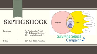


Septic shock is a life-threatening condition that arises when sepsis leads to dangerously low blood pressure and problems in organ function. It results from an infection that causes changes throughout the body. Early recognition and treatment are important, including administering antibiotics within an hour, aggressive fluid resuscitation, and monitoring for organ dysfunction. Goals of management are restoring blood pressure, reversing signs of low perfusion, and treating the underlying infection while avoiding additional organ injury.


![sepsis and septic shock guidelines[12585].pptx](https://cdn.slidesharecdn.com/ss_thumbnails/sepsisandsepticshockguidelines12585-230514040014-1b3555b4-thumbnail.jpg?width=640&height=640&fit=bounds)


![sepsis and septic shock guidelines[12585].pptx](https://cdn.slidesharecdn.com/ss_thumbnails/sepsisandsepticshockguidelines12585-230514035330-c3213eca-thumbnail.jpg?width=640&height=640&fit=bounds)


