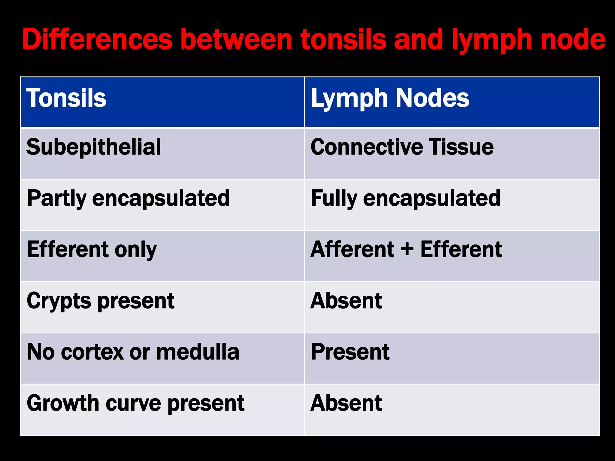
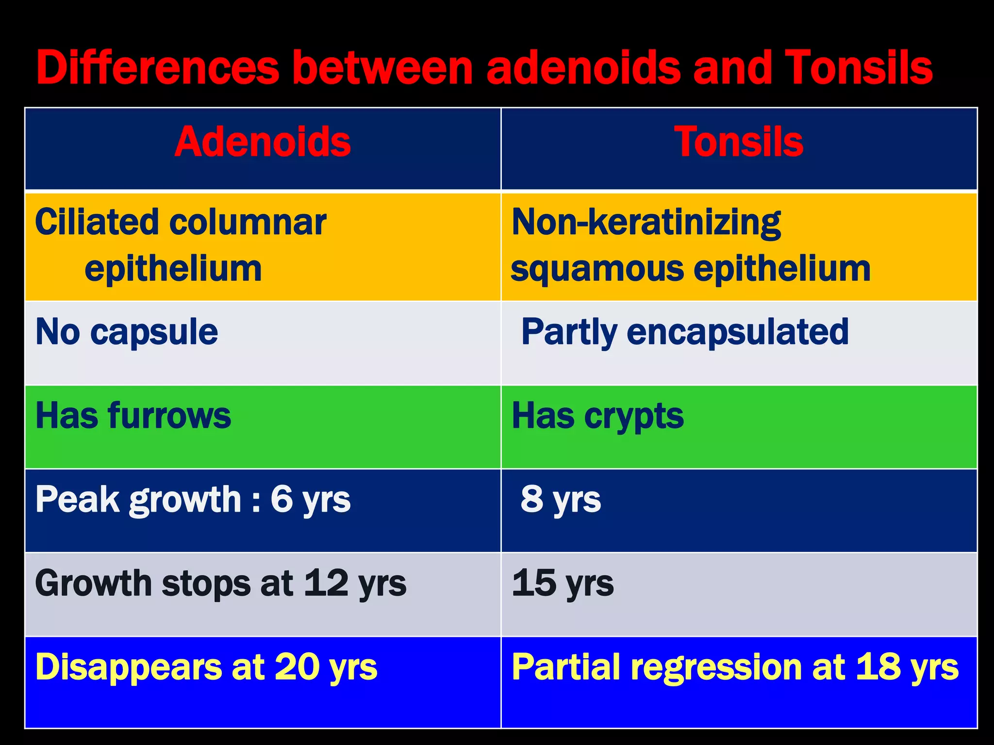
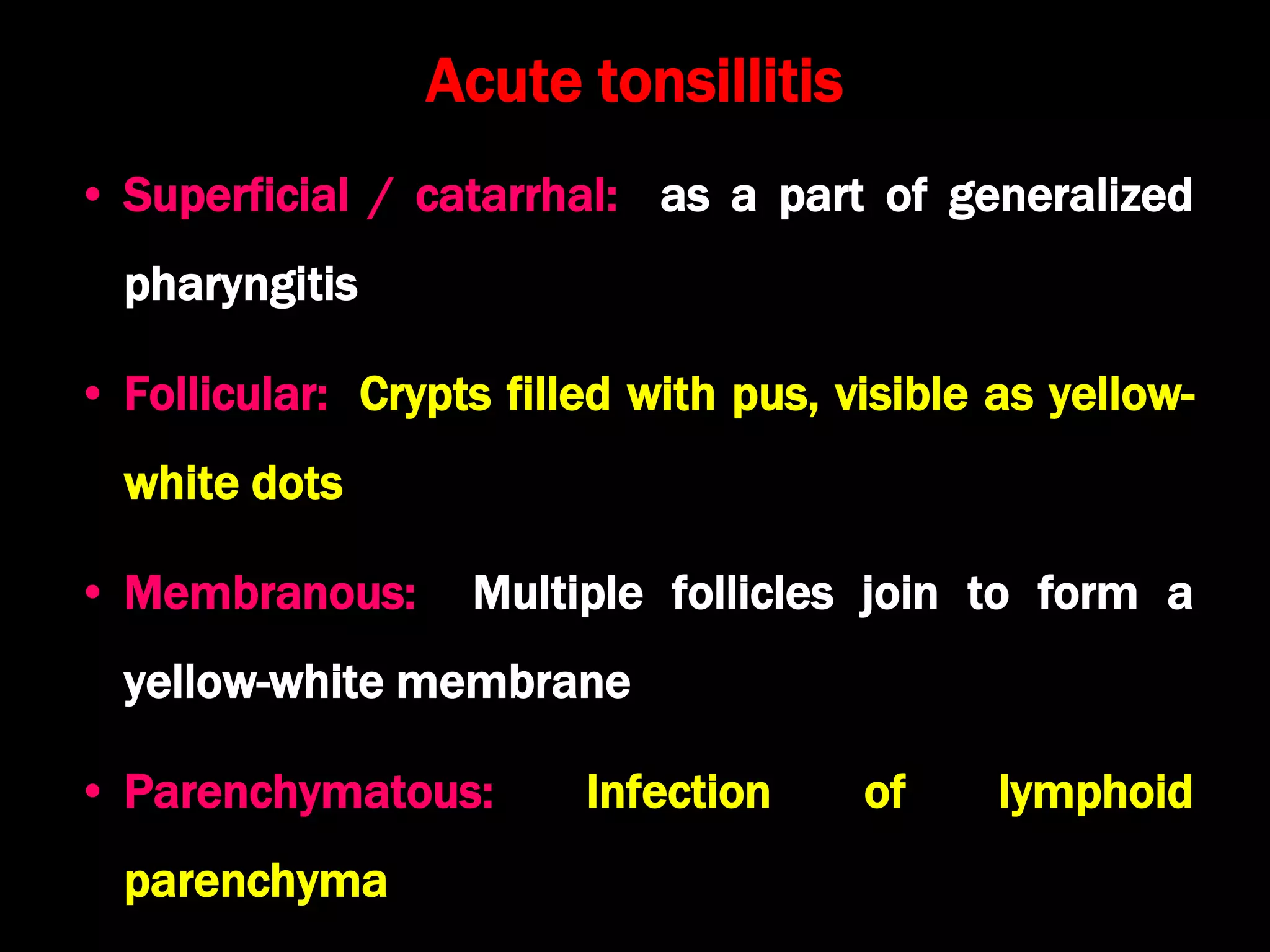
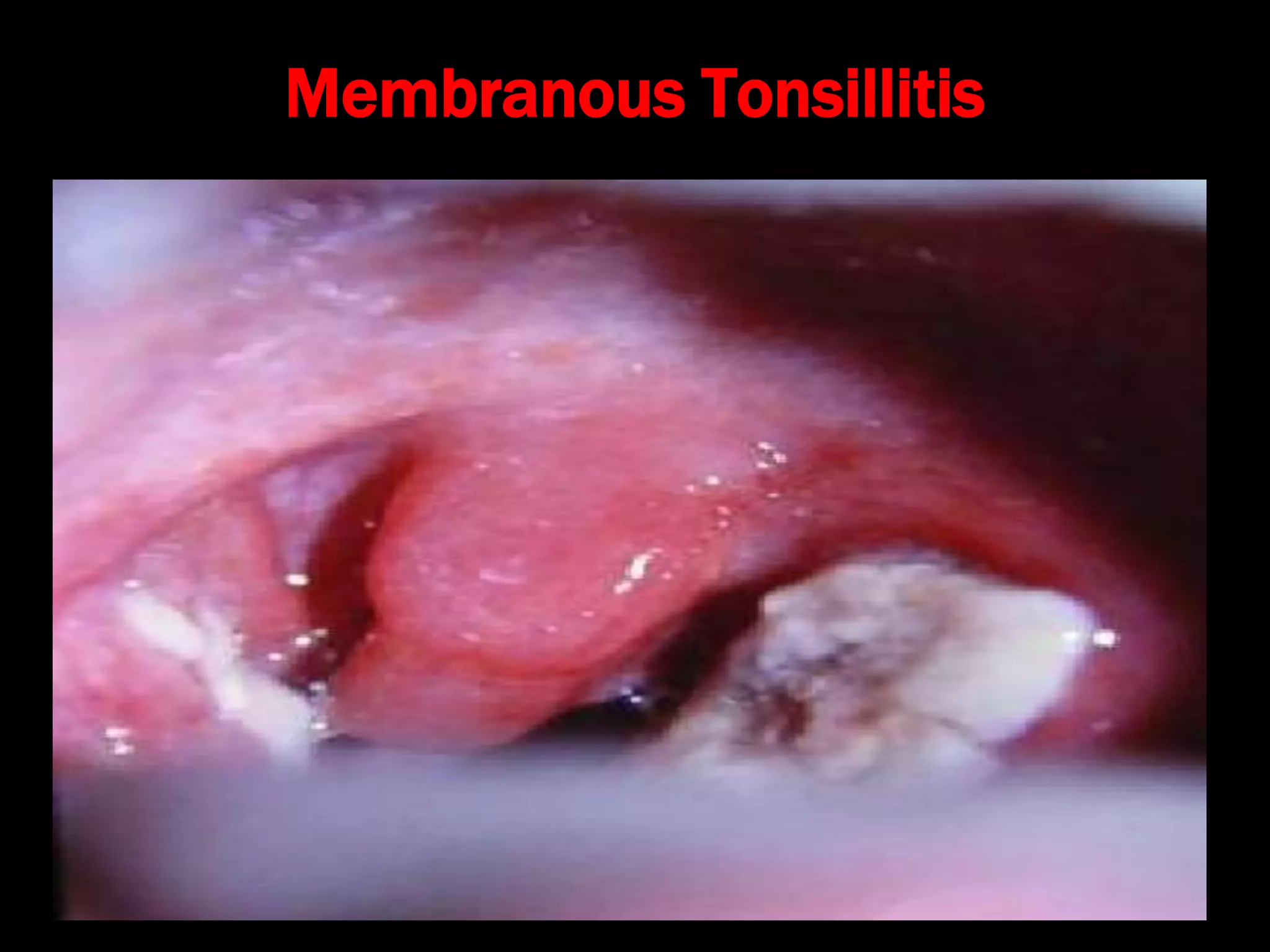
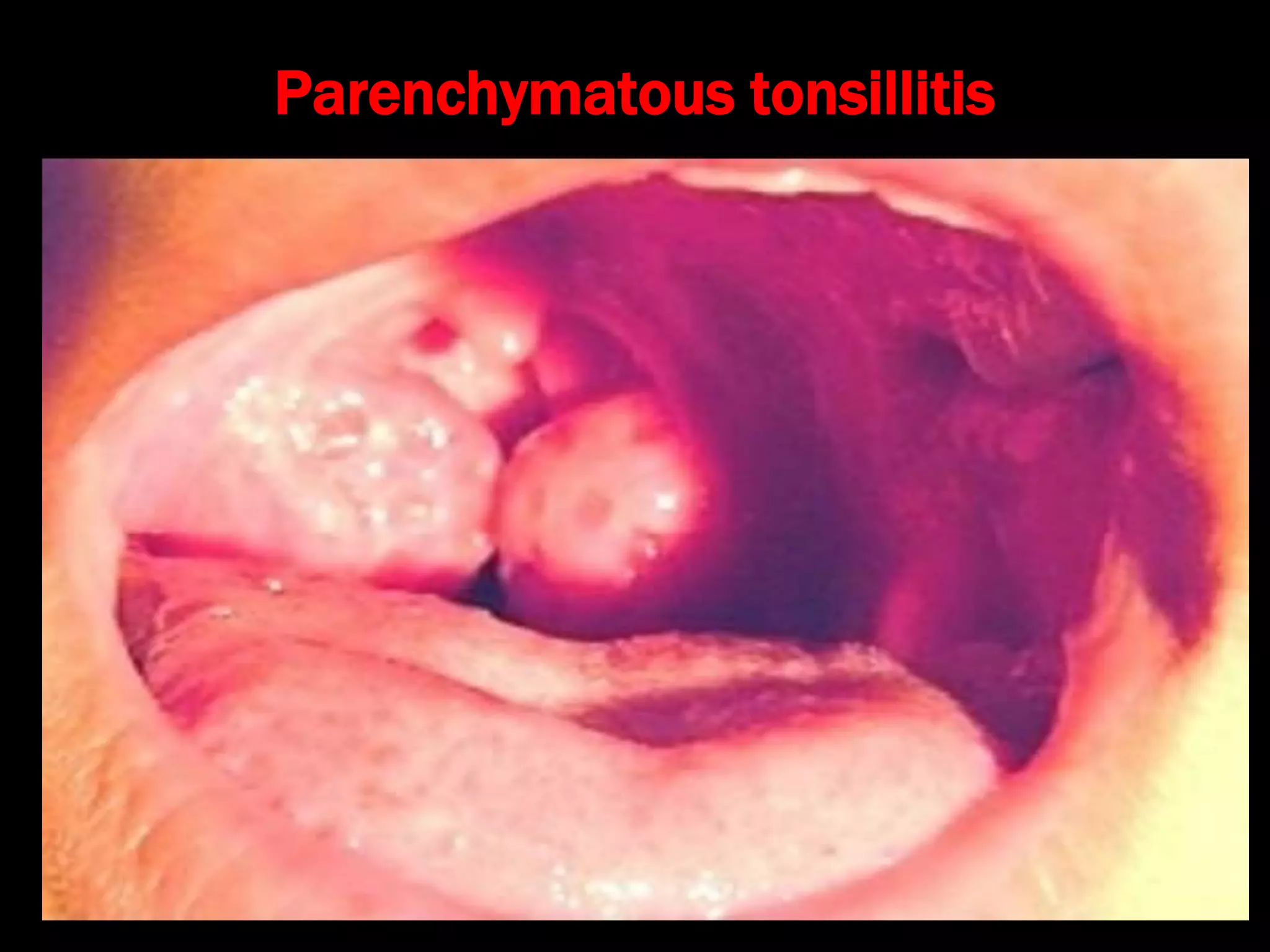
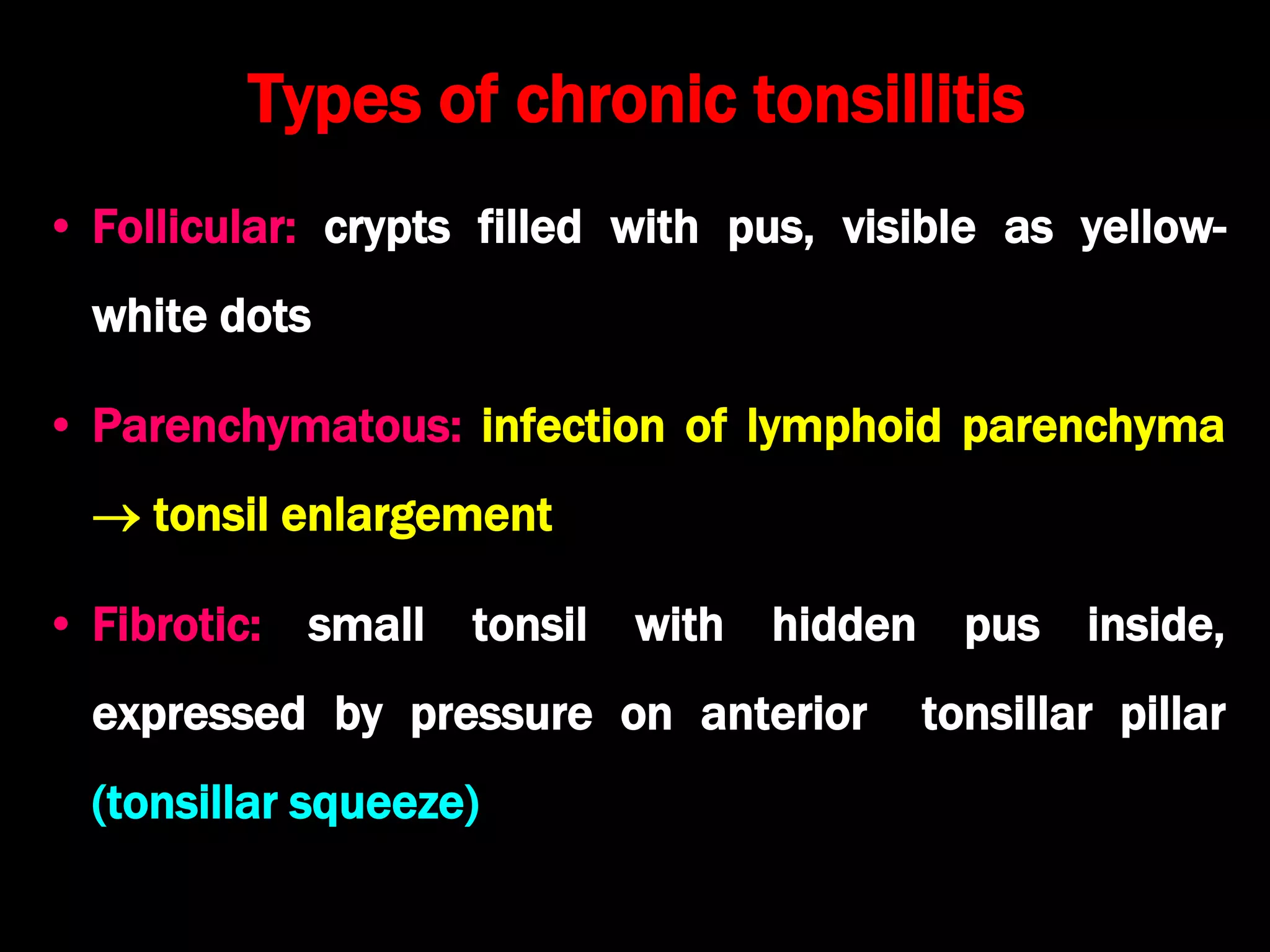
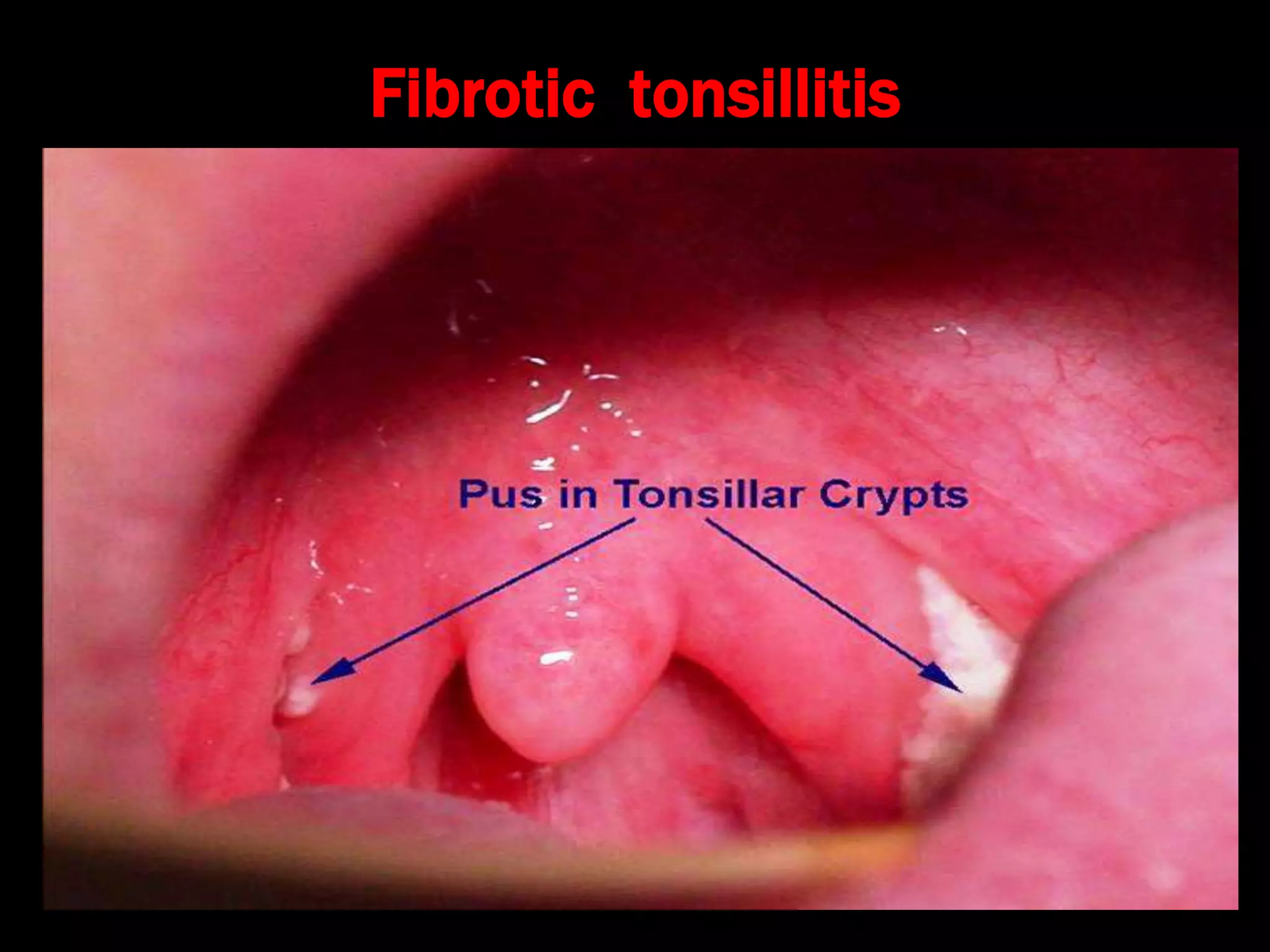
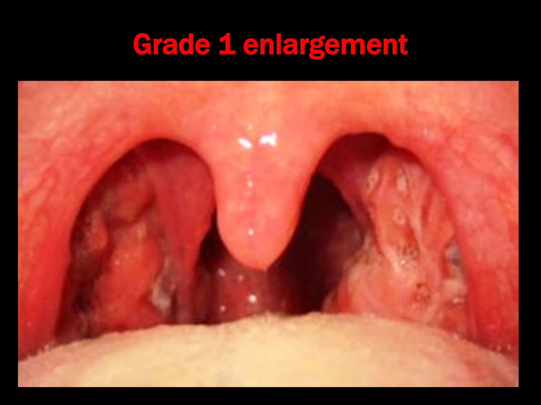
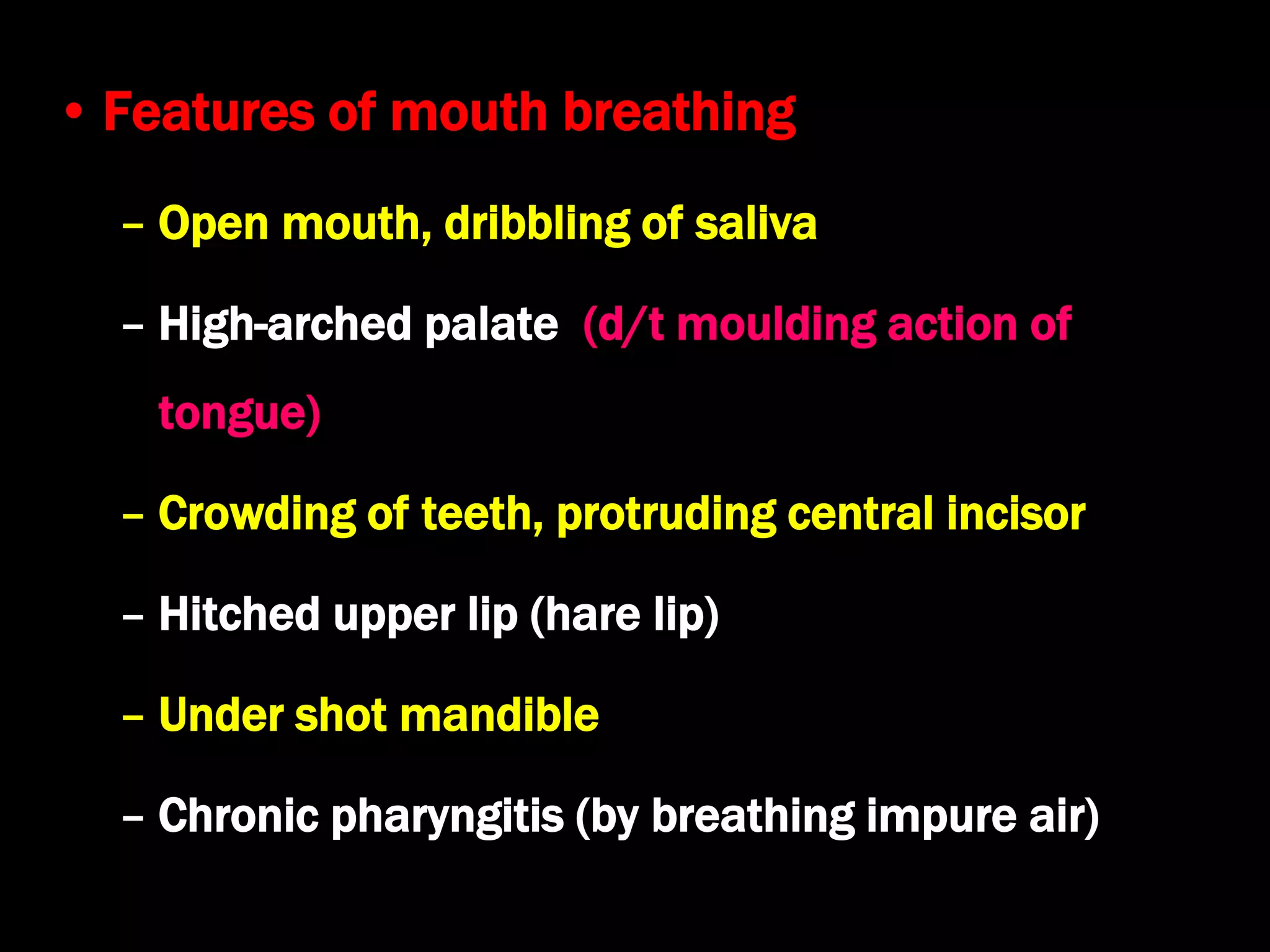
Tonsils and Adenoids
Dr. Krishna Koirala
2020-01-27
• Definition
– Palatine tonsils are dense compact bodies of
lymphoid tissue located in the lateral wall of the
oropharynx, bounded by the palatoglossus muscle
anteriorly and the palatopharyngeus and superior
constrictor muscles posteriorly and laterally
Arterial supply of tonsils
• Lingual artery: Dorsal
linguae branch
• Facial artery
– Tonsillar branch
– Ascending palatine
• Ascending Pharyngeal
artery
• Descending palatine
artery
• Venous drainage
– Para tonsillar vein  common facial vein and
pharyngeal venous plexus  internal jugular vein
• Lymphatic drainage
– Jugulo- digastric lymph node of Woods
• Nerve supply
– Glossopharyngeal nerve and lesser palatine nerve
Relations of tonsillar bed
Relations of tonsillar bed (Inside out)
1. Tonsillar capsule
2. Peritonsillar space with paratonsillar vein
3. Pharyngobasilar fascia , Superior constrictor muscle,
Bucco-pharyngeal fascia
4. Styloid process, muscles, glossopharyngeal nerve
5. Internal carotid artery, tonsillar artery
6. Medial pterygoid, submandibular salivary gland
7. Mandible
Differences between tonsils and lymph node
Tonsils Lymph Nodes
Subepithelial Connective Tissue
Partly encapsulated Fully encapsulated
Efferent only Afferent + Efferent
Crypts present Absent
No cortex or medulla Present
Growth curve present Absent
Differences between adenoids and Tonsils
Adenoids Tonsils
Ciliated columnar
epithelium
Non-keratinizing
squamous epithelium
No capsule Partly encapsulated
Has furrows Has crypts
Peak growth : 6 yrs 8 yrs
Growth stops at 12 yrs 15 yrs
Disappears at 20 yrs Partial regression at 18 yrs
Acute tonsillitis
• Superficial / catarrhal: as a part of generalized
pharyngitis
• Follicular: Crypts filled with pus, visible as yellow-
white dots
• Membranous: Multiple follicles join to form a
yellow-white membrane
• Parenchymatous: Infection of lymphoid
parenchyma
Catarrhal(Superficial) Tonsillitis
Follicular Tonsillitis
Membranous Tonsillitis
Parenchymatous tonsillitis
Types of chronic tonsillitis
• Follicular: crypts filled with pus, visible as yellow-
white dots
• Parenchymatous: infection of lymphoid parenchyma
 tonsil enlargement
• Fibrotic: small tonsil with hidden pus inside,
expressed by pressure on anterior tonsillar pillar
(tonsillar squeeze)
Fibrotic tonsillitis
Signs of tonsillitis
• Congested tonsil and tonsillar pillars
• Enlarged tonsil (except chronic fibrotic type)
• Tonsil squeezed by tongue depressor pressing on
anterior tonsillar pillar  pus comes out in chronic
fibrotic tonsillitis (Irwin Moore sign)
• Jugulo-digastric lymph node enlarged ( tender in
acute tonsillitis)
Grades of tonsillar enlargement
Grade 1 enlargement
Grade 2 enlargement
Grade 3 enlargement
Grade 4 enlargement
Complications of acute tonsillitis
• Local / locoregional
– Recurrent tonsillitis
– Intra-tonsillar abscess
– Peritonsillar abscess (Quinsy)
– Parapharyngeal abscess
– Retropharyngeal abscess
– Otitis media
– Suppurative cervical
lymphadenitis
• Systemic
– Rheumatic fever
– Subacute bacterial
endocarditis (SABE)
– Glomerulonephritis
– Septicemia
Differential diagnosis of white patch on the tonsil
• Membranous tonsillitis
• Faucial diphtheria
• Infectious mononucleosis (Mono spot test)
• Candidiasis (throat swab  Candida albicans)
• Vincent's angina (fusiform bacilli, spirochete)
• Tonsillar neoplasm / leukemia (excision biopsy)
• Agranulocytosis (Peripheral smear)
• Traumatic ulcer (history of trauma)
• Keratosis Pharyngis
Treatment of tonsillitis
• Bed rest
• Adequate hydration
• Systemic antibiotics: ampicillin, erythromycin ,
ceftriaxone, cefuroxime, amoxyclav
• Antihistamines and decongestants
• Analgesics
• Antiseptic gargle
• Treatment of focus of infection
Differences
between
Membranous
Tonsillitis
Diphtheria
Age > 5 yr 2- 5 yr
Onset Acute Insidious
General
Symptoms
More Less
Odynophagia More Less
Temperature High Low
Tachycardia Proportionate Disproportionate
Tonsils Enlarged, congested Normal
Membranous
tonsillitis
Diphtheria
Membrane Bilateral
Whitish yellow
Thin
Limited to tonsil
Easily removed
May be unilateral
Gray
Thick
May go beyond
Bleeds on removal
Culture  Hemolytic
streptococci
Corynebacterium
diphtheriae
Lymph node Jugulo-digastric Generalized (Bull neck)
Treatment of faucial diphtheria
• Isolation and bed rest
• I.V. benzyl penicillin 600 mg q6h
• Diphtheritic anti - toxin infusion in saline
– 20,000 – 40,000 U :  48 hrs duration, tonsillar
– 40,000 – 80,000 U : nasopharynx / larynx
– 80,000 – 120,000 U :  48 hrs, neck edema
• Emergency tracheostomy required for stridor
Tonsillolith and Tonsillar cyst
Recurrent tonsillitis / retention of debris
Blockage of tonsillar crypts
pus and debris
calcify
yellow colored
inclusion cyst
Tonsillolith tonsillar cyst
Tonsillolith and Tonsillar cyst contd…...
• Clinical features
– Halitosis, bitter taste in mouth
– White outgrowths from tonsillar crypts or yellow
cyst in supra-tonsillar cleft
• Treatment
– Asymptomatic  drainage of cyst or manual
expression of tonsillolith
– Severe symptoms  tonsillectomy
Keratosis pharyngis
• Benign , self limiting condition
• Etiology : Smoking, alcohol, vitamin
A deficiency
• O/E:
– Yellowish, horn-like outgrowths
from mucosa of tonsil that cannot
be wiped off
• Histopathology :
– Hypertrophy and
hyperkeratinization of epithelium
– Absence of inflammation
• Treatment:
– Reassurance
– Tonsillectomy in severe cases
D/D of Unilateral tonsillar enlargement
• Tonsillar causes
– Tonsillar malignancy
– Peritonsillar abscess
– Intra-tonsillar abscess
– Tonsillolith
– Tonsillar cyst
– Tonsillar artery aneurysm
– Vincent's angina
• Extra-tonsillar causes
– Parapharyngeal abscess
– Parapharyngeal tumors
– Tumors of deep parotid
lobe
– Internal carotid art.
aneurysm
– Cervical
lymphadenopathy
Adenoids
• Symptomatic, hypertrophic nasopharyngeal
(Luschka's) tonsils
• Adenoids lead to
– Nasal obstruction  Mouth breathing
– Eustachian tube block  OME
• Features like adenoids are also seen in
– Dental mal-occlusion
– B/L nasal block ( Nasal polyps, choanal atresia)
Adenoid facies
• Features of nasal obstruction
– B/L nose block & nasal discharge
– Rhinolalia clausa (flat toneless voice)
– Difficulty in feeding
– Snoring
– Pulmonary hypertension
– Pinched nostrils (due to disuse atrophy)
• Features of mouth breathing
– Open mouth, dribbling of saliva
– High-arched palate (d/t moulding action of
tongue)
– Crowding of teeth, protruding central incisor
– Hitched upper lip (hare lip)
– Under shot mandible
– Chronic pharyngitis (by breathing impure air)
• Features of Eustachian tube block
– Earache
– Conductive deafness (due to O.M.E.)
– Dull, expressionless look
– Inattentive child
• Other Features
– Pectus excavatum
– Nocturnal enuresis
Nasopharyngoscopy
Plain X-ray soft tissue nasopharynx lateral view
Management
• Diagnosis
– Nasopharyngoscopy  rigid / flexible
– Plain X–ray soft tissue nasopharynx lateral view
with head extended  adenoid mass
• Treatment
– Mild symptoms  antihistamine + decongestant
– Severe symptoms  adenoidectomy

Tonsils and adenoids

  • 1.
    Tonsils and Adenoids Dr.Krishna Koirala 2020-01-27
  • 2.
    • Definition – Palatinetonsils are dense compact bodies of lymphoid tissue located in the lateral wall of the oropharynx, bounded by the palatoglossus muscle anteriorly and the palatopharyngeus and superior constrictor muscles posteriorly and laterally
  • 3.
    Arterial supply oftonsils • Lingual artery: Dorsal linguae branch • Facial artery – Tonsillar branch – Ascending palatine • Ascending Pharyngeal artery • Descending palatine artery
  • 4.
    • Venous drainage –Para tonsillar vein  common facial vein and pharyngeal venous plexus  internal jugular vein • Lymphatic drainage – Jugulo- digastric lymph node of Woods • Nerve supply – Glossopharyngeal nerve and lesser palatine nerve
  • 5.
  • 6.
    Relations of tonsillarbed (Inside out) 1. Tonsillar capsule 2. Peritonsillar space with paratonsillar vein 3. Pharyngobasilar fascia , Superior constrictor muscle, Bucco-pharyngeal fascia 4. Styloid process, muscles, glossopharyngeal nerve 5. Internal carotid artery, tonsillar artery 6. Medial pterygoid, submandibular salivary gland 7. Mandible
  • 7.
    Differences between tonsilsand lymph node Tonsils Lymph Nodes Subepithelial Connective Tissue Partly encapsulated Fully encapsulated Efferent only Afferent + Efferent Crypts present Absent No cortex or medulla Present Growth curve present Absent
  • 8.
    Differences between adenoidsand Tonsils Adenoids Tonsils Ciliated columnar epithelium Non-keratinizing squamous epithelium No capsule Partly encapsulated Has furrows Has crypts Peak growth : 6 yrs 8 yrs Growth stops at 12 yrs 15 yrs Disappears at 20 yrs Partial regression at 18 yrs
  • 9.
    Acute tonsillitis • Superficial/ catarrhal: as a part of generalized pharyngitis • Follicular: Crypts filled with pus, visible as yellow- white dots • Membranous: Multiple follicles join to form a yellow-white membrane • Parenchymatous: Infection of lymphoid parenchyma
  • 10.
  • 11.
  • 12.
  • 13.
  • 14.
    Types of chronictonsillitis • Follicular: crypts filled with pus, visible as yellow- white dots • Parenchymatous: infection of lymphoid parenchyma  tonsil enlargement • Fibrotic: small tonsil with hidden pus inside, expressed by pressure on anterior tonsillar pillar (tonsillar squeeze)
  • 15.
  • 16.
    Signs of tonsillitis •Congested tonsil and tonsillar pillars • Enlarged tonsil (except chronic fibrotic type) • Tonsil squeezed by tongue depressor pressing on anterior tonsillar pillar  pus comes out in chronic fibrotic tonsillitis (Irwin Moore sign) • Jugulo-digastric lymph node enlarged ( tender in acute tonsillitis)
  • 18.
  • 19.
  • 20.
  • 21.
  • 22.
  • 23.
    Complications of acutetonsillitis • Local / locoregional – Recurrent tonsillitis – Intra-tonsillar abscess – Peritonsillar abscess (Quinsy) – Parapharyngeal abscess – Retropharyngeal abscess – Otitis media – Suppurative cervical lymphadenitis • Systemic – Rheumatic fever – Subacute bacterial endocarditis (SABE) – Glomerulonephritis – Septicemia
  • 24.
    Differential diagnosis ofwhite patch on the tonsil • Membranous tonsillitis • Faucial diphtheria • Infectious mononucleosis (Mono spot test) • Candidiasis (throat swab  Candida albicans) • Vincent's angina (fusiform bacilli, spirochete) • Tonsillar neoplasm / leukemia (excision biopsy) • Agranulocytosis (Peripheral smear) • Traumatic ulcer (history of trauma) • Keratosis Pharyngis
  • 25.
    Treatment of tonsillitis •Bed rest • Adequate hydration • Systemic antibiotics: ampicillin, erythromycin , ceftriaxone, cefuroxime, amoxyclav • Antihistamines and decongestants • Analgesics • Antiseptic gargle • Treatment of focus of infection
  • 26.
    Differences between Membranous Tonsillitis Diphtheria Age > 5yr 2- 5 yr Onset Acute Insidious General Symptoms More Less Odynophagia More Less Temperature High Low Tachycardia Proportionate Disproportionate Tonsils Enlarged, congested Normal
  • 27.
    Membranous tonsillitis Diphtheria Membrane Bilateral Whitish yellow Thin Limitedto tonsil Easily removed May be unilateral Gray Thick May go beyond Bleeds on removal Culture  Hemolytic streptococci Corynebacterium diphtheriae Lymph node Jugulo-digastric Generalized (Bull neck)
  • 28.
    Treatment of faucialdiphtheria • Isolation and bed rest • I.V. benzyl penicillin 600 mg q6h • Diphtheritic anti - toxin infusion in saline – 20,000 – 40,000 U :  48 hrs duration, tonsillar – 40,000 – 80,000 U : nasopharynx / larynx – 80,000 – 120,000 U :  48 hrs, neck edema • Emergency tracheostomy required for stridor
  • 29.
    Tonsillolith and Tonsillarcyst Recurrent tonsillitis / retention of debris Blockage of tonsillar crypts pus and debris calcify yellow colored inclusion cyst Tonsillolith tonsillar cyst
  • 30.
    Tonsillolith and Tonsillarcyst contd…... • Clinical features – Halitosis, bitter taste in mouth – White outgrowths from tonsillar crypts or yellow cyst in supra-tonsillar cleft • Treatment – Asymptomatic  drainage of cyst or manual expression of tonsillolith – Severe symptoms  tonsillectomy
  • 31.
    Keratosis pharyngis • Benign, self limiting condition • Etiology : Smoking, alcohol, vitamin A deficiency • O/E: – Yellowish, horn-like outgrowths from mucosa of tonsil that cannot be wiped off • Histopathology : – Hypertrophy and hyperkeratinization of epithelium – Absence of inflammation • Treatment: – Reassurance – Tonsillectomy in severe cases
  • 32.
    D/D of Unilateraltonsillar enlargement • Tonsillar causes – Tonsillar malignancy – Peritonsillar abscess – Intra-tonsillar abscess – Tonsillolith – Tonsillar cyst – Tonsillar artery aneurysm – Vincent's angina • Extra-tonsillar causes – Parapharyngeal abscess – Parapharyngeal tumors – Tumors of deep parotid lobe – Internal carotid art. aneurysm – Cervical lymphadenopathy
  • 33.
    Adenoids • Symptomatic, hypertrophicnasopharyngeal (Luschka's) tonsils • Adenoids lead to – Nasal obstruction  Mouth breathing – Eustachian tube block  OME • Features like adenoids are also seen in – Dental mal-occlusion – B/L nasal block ( Nasal polyps, choanal atresia)
  • 34.
  • 35.
    • Features ofnasal obstruction – B/L nose block & nasal discharge – Rhinolalia clausa (flat toneless voice) – Difficulty in feeding – Snoring – Pulmonary hypertension – Pinched nostrils (due to disuse atrophy)
  • 36.
    • Features ofmouth breathing – Open mouth, dribbling of saliva – High-arched palate (d/t moulding action of tongue) – Crowding of teeth, protruding central incisor – Hitched upper lip (hare lip) – Under shot mandible – Chronic pharyngitis (by breathing impure air)
  • 37.
    • Features ofEustachian tube block – Earache – Conductive deafness (due to O.M.E.) – Dull, expressionless look – Inattentive child • Other Features – Pectus excavatum – Nocturnal enuresis
  • 38.
  • 39.
    Plain X-ray softtissue nasopharynx lateral view
  • 40.
    Management • Diagnosis – Nasopharyngoscopy rigid / flexible – Plain X–ray soft tissue nasopharynx lateral view with head extended  adenoid mass • Treatment – Mild symptoms  antihistamine + decongestant – Severe symptoms  adenoidectomy