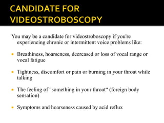
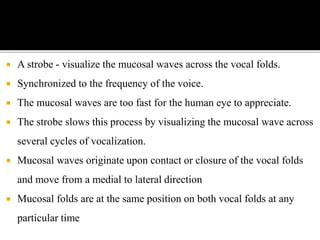
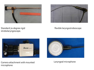
PRESENTER: DR. ARJUN SURESH
 Videostroboscopy - practical and useful technique - clinical
evaluation - visco-elastic properties of phonatory mucosa.
 Painless, OPD-based procedure
 Essential evaluation of laryngeal mucosa, vocal fold motion
biomechanics, and mucosal vibration.
 Helps detect pathology - determine impact on voice and airway
function.
 Method used to visualize vocal fold vibration.
 Uses synchronized, flashing light passed via flexible or rigid
telescope
 Flashes of light are synchronized to vocal fold vibration at
slightly slower speed, allowing examiner to observe it during
sound production in slow motion
 Essential for planning effective phonomicrosurgery.
 Real-time information - nature of vibration, image to detect vocal
pathology, and permanent video record of examination.
 Improves sensitivity of subtle laryngeal diagnoses.
 Helps evaluate:
1. Vocal fold biomechanics
2. Laryngeal mucosa
3. Mucosal Vibration
• Cause of voice dysfunction or hoarseness - vocal cord swelling,
irritations, misuse, growths, polyps or acid reflux.
• To visualize your vocal cords
• To evaluate or detect vocal cord lesions and other irregularities
like inflammation, scar tissue or muscle tension conditions
• To assess swallowing issues, which could be caused by muscle
abnormalities
You may be a candidate for videostroboscopy if you're
experiencing chronic or intermittent voice problems like:
 Breathiness, hoarseness, decreased or loss of vocal range or
vocal fatigue
 Tightness, discomfort or pain or burning in your throat while
talking
 The feeling of "something in your throat“ (foreign body
sensation)
 Symptoms and hoarseness caused by acid reflux
 A strobe - visualize the mucosal waves across the vocal folds.
 Synchronized to the frequency of the voice.
 The mucosal waves are too fast for the human eye to appreciate.
 The strobe slows this process by visualizing the mucosal wave across
several cycles of vocalization.
 Mucosal waves originate upon contact or closure of the vocal folds
and move from a medial to lateral direction
 Mucosal folds are at the same position on both vocal folds at any
particular time
 No absolute CI – patient should have adequate mouth opening
and patent nasal airway.
A videostroboscopic unit:
 A stroboscopic light source and microphone,
 Video camera
 Endoscope
 Video recorder.
Stroboscopy can be performed by using either rigid or flexible
Endoscopes.
Standard 70-degree rigid
strobolaryngoscope.
Camera attachment with mounted
microphone
Laryngeal microphone
flexible laryngostroboscope
 Videostrobolaryngoscopy begins by seating the patient in the
examination chair at a height comfortable for the examiner.
 The patient leans forward with the neck flexed and the head
extended at the atlo-occipital joint (Kirstein position).
 Once the patient is in appropriate position,
 Spray topical anesthesia – posterior part of tongue and
oropharynx.
 Calibrate microphone and hold against thyroid lamina.
 To avoid condensation – dip scope in hot water.
 Open mouth, protrude tongue- scope is inserted.
 Proper focus – visualise subepithelial vasculature of vocal
fold.
 With Vocal folds in focus – ask patient to produce ‘ee’ sound.
 Should be done at low, mid and high frequency pitches and
different volumes.
 Examiner - comment on arytenoid and vocal fold mobility,
glottic closure pattern, mucosal wave, and pliability.
 Ulcerative lesions or masses can also be observed.
1. VOCAL CYST
- Encapsulated, spheroid lesions - mucus or keratin
- Located - lamina propria of the vocal fold.
- Keratin cysts - likely congenital & mucous cysts - likely acquired.
- Generally unilateral, though several may be present at the time of
diagnosis.
- On stroboscopy, region of the cyst - diminished pliability - mucosal wave
does not propagate normally through the region of the cyst.
- Mucosal-wave deficit - size and location of the cyst.
- Illustrated by the fact - small superior-surface cysts minimally affect vocal
function.
 Unilateral or bilateral.
 Represent phonotraumatic pathology - collision forces, shearing
stresses - lamina propria.
 Consistency - gelatinous to fibrotic.
 Glottic closure – compromised - gaps anterior and posterior to the
lesion in maximal closure.
 The vibratory patterns of VC – asymmetric - diminution of vibration
near the lesion.
 Medial surface polyp - disturbs the vibratory pattern - contralateral
vocal fold during closure.
 Bilateral fibrovascular lesions - symmetric sessile masses.
 Occur in the center of the musculomembranous region -
basement-membrane - between the overlying epithelium and
the underlying superficial lamina propria.
 Glottic closure is compromised - high pitch frequencies.
 Mucosal wave - usually preserved bilaterally - pliability and
amplitude - decreased in the region of the nodule.
Stroboscopy
Stroboscopy

Stroboscopy

  • 1.
  • 2.
     Videostroboscopy -practical and useful technique - clinical evaluation - visco-elastic properties of phonatory mucosa.  Painless, OPD-based procedure  Essential evaluation of laryngeal mucosa, vocal fold motion biomechanics, and mucosal vibration.  Helps detect pathology - determine impact on voice and airway function.
  • 3.
     Method usedto visualize vocal fold vibration.  Uses synchronized, flashing light passed via flexible or rigid telescope  Flashes of light are synchronized to vocal fold vibration at slightly slower speed, allowing examiner to observe it during sound production in slow motion
  • 4.
     Essential forplanning effective phonomicrosurgery.  Real-time information - nature of vibration, image to detect vocal pathology, and permanent video record of examination.  Improves sensitivity of subtle laryngeal diagnoses.  Helps evaluate: 1. Vocal fold biomechanics 2. Laryngeal mucosa 3. Mucosal Vibration
  • 5.
    • Cause ofvoice dysfunction or hoarseness - vocal cord swelling, irritations, misuse, growths, polyps or acid reflux. • To visualize your vocal cords • To evaluate or detect vocal cord lesions and other irregularities like inflammation, scar tissue or muscle tension conditions • To assess swallowing issues, which could be caused by muscle abnormalities
  • 6.
    You may bea candidate for videostroboscopy if you're experiencing chronic or intermittent voice problems like:  Breathiness, hoarseness, decreased or loss of vocal range or vocal fatigue  Tightness, discomfort or pain or burning in your throat while talking  The feeling of "something in your throat“ (foreign body sensation)  Symptoms and hoarseness caused by acid reflux
  • 8.
     A strobe- visualize the mucosal waves across the vocal folds.  Synchronized to the frequency of the voice.  The mucosal waves are too fast for the human eye to appreciate.  The strobe slows this process by visualizing the mucosal wave across several cycles of vocalization.  Mucosal waves originate upon contact or closure of the vocal folds and move from a medial to lateral direction  Mucosal folds are at the same position on both vocal folds at any particular time
  • 9.
     No absoluteCI – patient should have adequate mouth opening and patent nasal airway.
  • 10.
    A videostroboscopic unit: A stroboscopic light source and microphone,  Video camera  Endoscope  Video recorder. Stroboscopy can be performed by using either rigid or flexible Endoscopes.
  • 11.
    Standard 70-degree rigid strobolaryngoscope. Cameraattachment with mounted microphone Laryngeal microphone flexible laryngostroboscope
  • 12.
     Videostrobolaryngoscopy beginsby seating the patient in the examination chair at a height comfortable for the examiner.  The patient leans forward with the neck flexed and the head extended at the atlo-occipital joint (Kirstein position).
  • 13.
     Once thepatient is in appropriate position,  Spray topical anesthesia – posterior part of tongue and oropharynx.  Calibrate microphone and hold against thyroid lamina.  To avoid condensation – dip scope in hot water.  Open mouth, protrude tongue- scope is inserted.  Proper focus – visualise subepithelial vasculature of vocal fold.
  • 14.
     With Vocalfolds in focus – ask patient to produce ‘ee’ sound.  Should be done at low, mid and high frequency pitches and different volumes.  Examiner - comment on arytenoid and vocal fold mobility, glottic closure pattern, mucosal wave, and pliability.  Ulcerative lesions or masses can also be observed.
  • 15.
    1. VOCAL CYST -Encapsulated, spheroid lesions - mucus or keratin - Located - lamina propria of the vocal fold. - Keratin cysts - likely congenital & mucous cysts - likely acquired. - Generally unilateral, though several may be present at the time of diagnosis. - On stroboscopy, region of the cyst - diminished pliability - mucosal wave does not propagate normally through the region of the cyst. - Mucosal-wave deficit - size and location of the cyst. - Illustrated by the fact - small superior-surface cysts minimally affect vocal function.
  • 17.
     Unilateral orbilateral.  Represent phonotraumatic pathology - collision forces, shearing stresses - lamina propria.  Consistency - gelatinous to fibrotic.  Glottic closure – compromised - gaps anterior and posterior to the lesion in maximal closure.  The vibratory patterns of VC – asymmetric - diminution of vibration near the lesion.  Medial surface polyp - disturbs the vibratory pattern - contralateral vocal fold during closure.
  • 19.
     Bilateral fibrovascularlesions - symmetric sessile masses.  Occur in the center of the musculomembranous region - basement-membrane - between the overlying epithelium and the underlying superficial lamina propria.  Glottic closure is compromised - high pitch frequencies.  Mucosal wave - usually preserved bilaterally - pliability and amplitude - decreased in the region of the nodule.