Download as PDF, PPTX
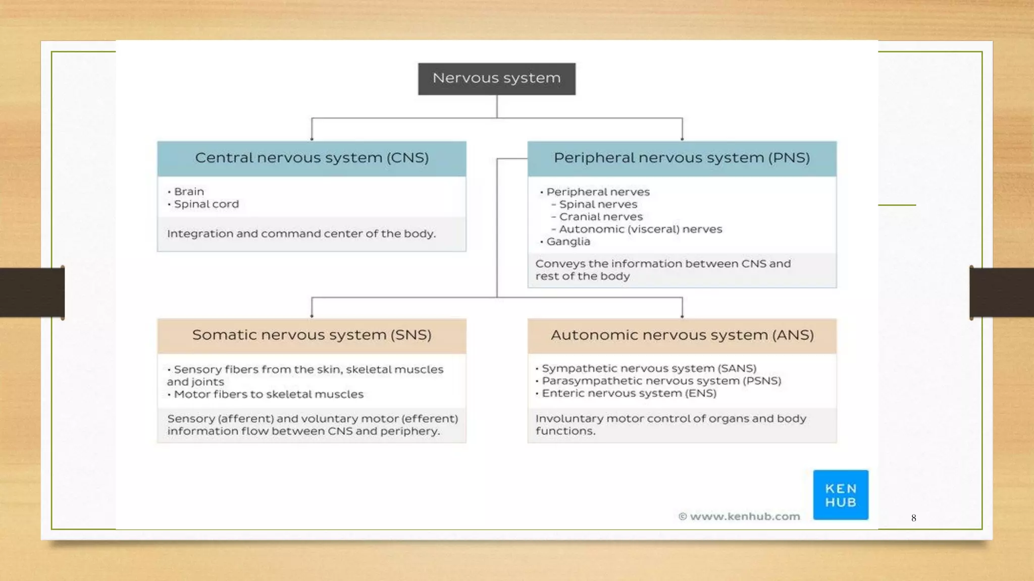
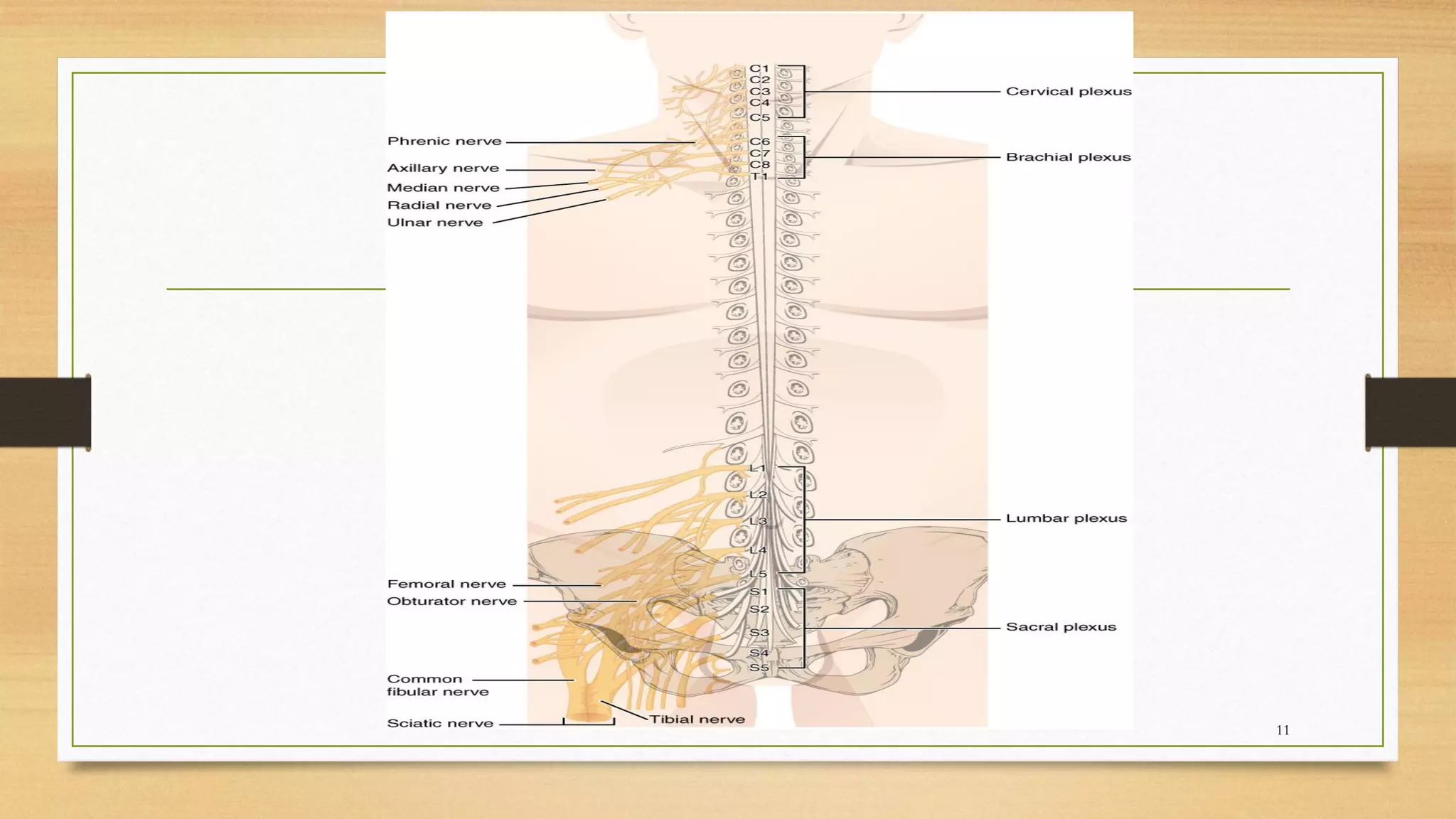

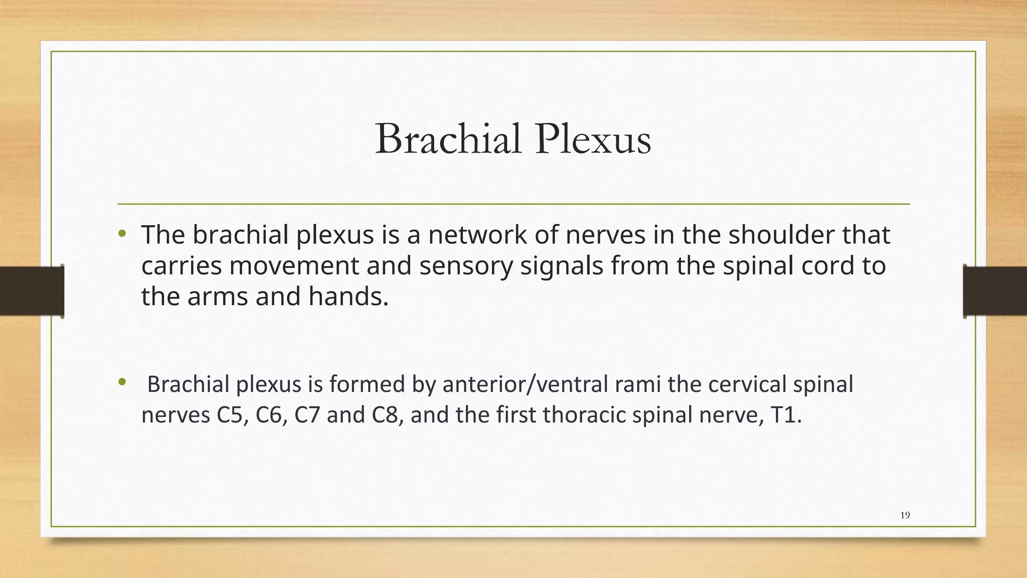
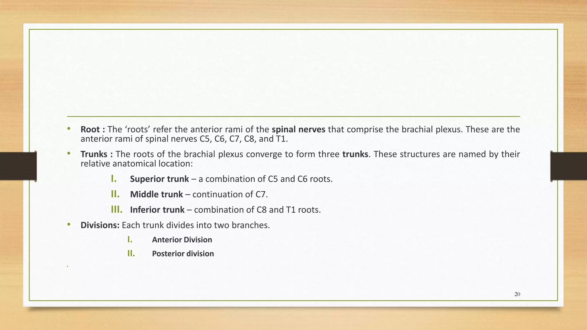
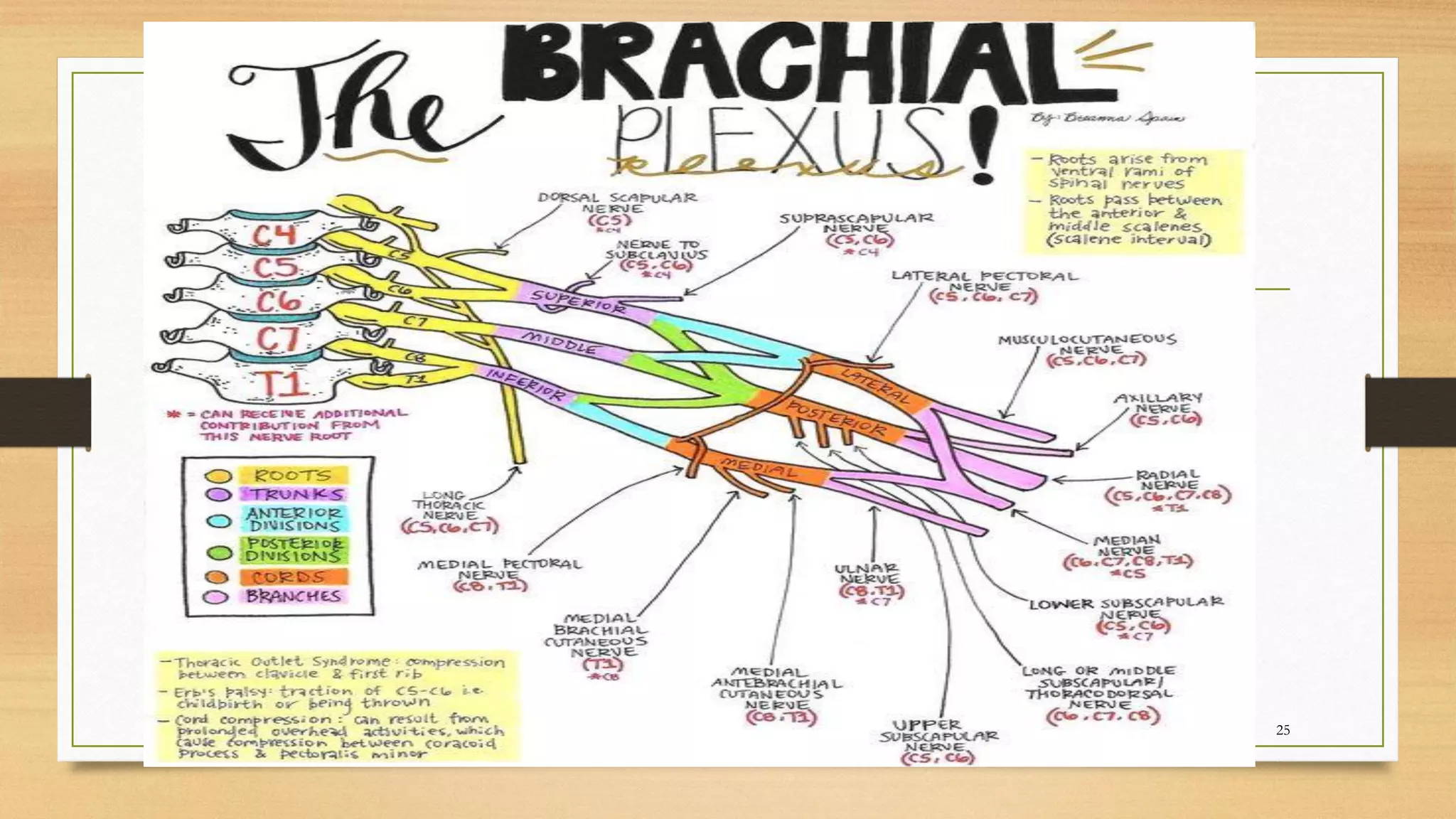
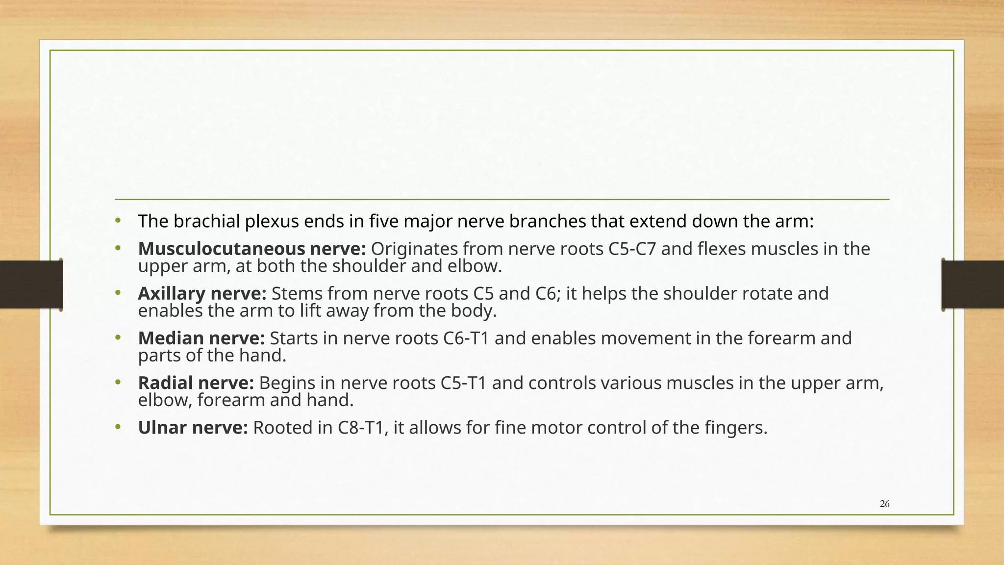
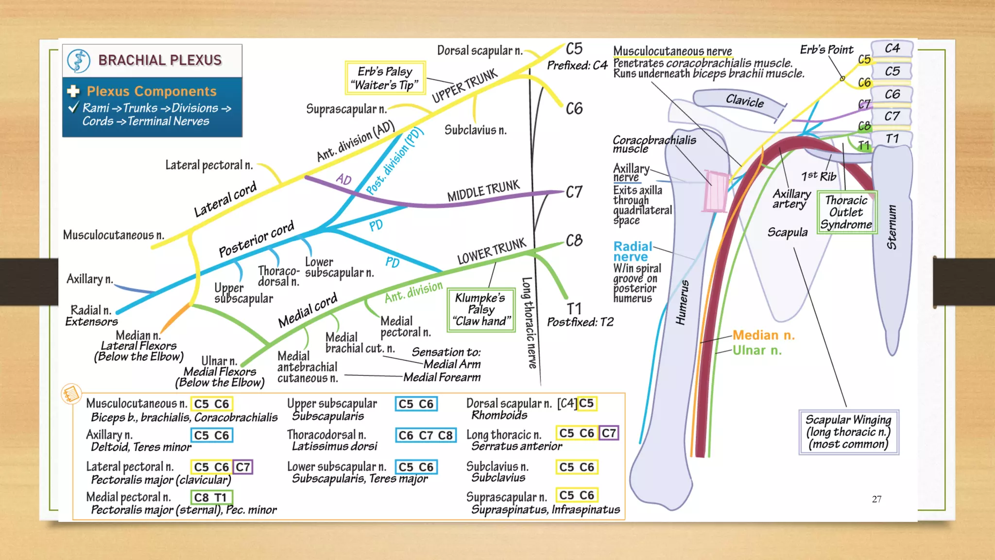

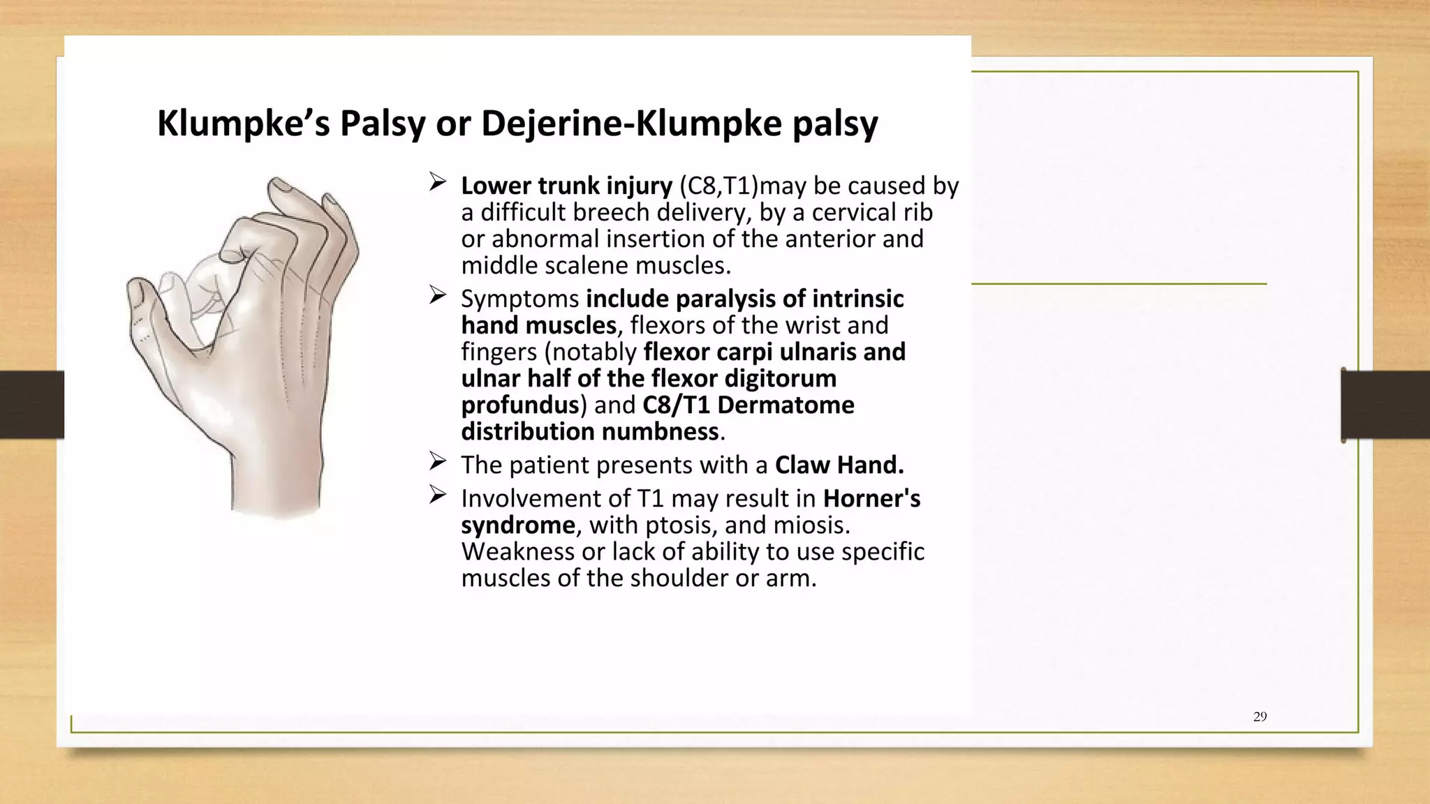

This document provides an overview of the nervous system, including its basic components and classifications. It discusses the central nervous system, which consists of the brain and spinal cord, and the peripheral nervous system. It describes how spinal nerves form from the dorsal and ventral roots of the spinal cord. It also provides details on the brachial plexus, a network of nerves in the shoulder that carries signals from the spinal cord to the arms. It outlines how the brachial plexus is formed from cervical and thoracic spinal nerves and discusses its main branches and functions.


![2-Anatomy of the Spinal Cord [Autosaved].ppt](https://cdn.slidesharecdn.com/ss_thumbnails/2-anatomyofthespinalcordautosaved-240314080102-4f8095d0-thumbnail.jpg?width=640&height=640&fit=bounds)