Human Anatomy is fundamental to every medical and healthcare professional. However, the science of anatomy and effects of stroke are also extremely useful to anyone interested in understanding more about the human body. In this course, you’ll gain an understanding of the basic concepts of anatomy and learn to ‘dissect’ the human body with a logical approach through
Human Anatomy is fundamental to every medical and healthcare professional. However, the science of anatomy and effects of stroke are also extremely useful to anyone interested in understanding more about the human body. In this course, you’ll gain an understanding of the basic concepts of anatomy and learn to ‘dissect’ the human body with a logical approach through
Anterior compartment of leg and Dorsum of foot CIMS
introduction about leg and four how we can differentiate , cutaneous innervation and in the contents like muscles with its blood supply nerve supply and finally will be appplied regarding topic
Anterior compartment of leg and Dorsum of foot CIMS
introduction about leg and four how we can differentiate , cutaneous innervation and in the contents like muscles with its blood supply nerve supply and finally will be appplied regarding topic
The French Revolution, which began in 1789, was a period of radical social and political upheaval in France. It marked the decline of absolute monarchies, the rise of secular and democratic republics, and the eventual rise of Napoleon Bonaparte. This revolutionary period is crucial in understanding the transition from feudalism to modernity in Europe.
For more information, visit-www.vavaclasses.com
Introduction to AI for Nonprofits with Tapp NetworkTechSoup
Dive into the world of AI! Experts Jon Hill and Tareq Monaur will guide you through AI's role in enhancing nonprofit websites and basic marketing strategies, making it easy to understand and apply.
Francesca Gottschalk - How can education support child empowerment.pptxEduSkills OECD
Francesca Gottschalk from the OECD’s Centre for Educational Research and Innovation presents at the Ask an Expert Webinar: How can education support child empowerment?
Embracing GenAI - A Strategic ImperativePeter Windle
Artificial Intelligence (AI) technologies such as Generative AI, Image Generators and Large Language Models have had a dramatic impact on teaching, learning and assessment over the past 18 months. The most immediate threat AI posed was to Academic Integrity with Higher Education Institutes (HEIs) focusing their efforts on combating the use of GenAI in assessment. Guidelines were developed for staff and students, policies put in place too. Innovative educators have forged paths in the use of Generative AI for teaching, learning and assessments leading to pockets of transformation springing up across HEIs, often with little or no top-down guidance, support or direction.
This Gasta posits a strategic approach to integrating AI into HEIs to prepare staff, students and the curriculum for an evolving world and workplace. We will highlight the advantages of working with these technologies beyond the realm of teaching, learning and assessment by considering prompt engineering skills, industry impact, curriculum changes, and the need for staff upskilling. In contrast, not engaging strategically with Generative AI poses risks, including falling behind peers, missed opportunities and failing to ensure our graduates remain employable. The rapid evolution of AI technologies necessitates a proactive and strategic approach if we are to remain relevant.
Biological screening of herbal drugs: Introduction and Need for
Phyto-Pharmacological Screening, New Strategies for evaluating
Natural Products, In vitro evaluation techniques for Antioxidants, Antimicrobial and Anticancer drugs. In vivo evaluation techniques
for Anti-inflammatory, Antiulcer, Anticancer, Wound healing, Antidiabetic, Hepatoprotective, Cardio protective, Diuretics and
Antifertility, Toxicity studies as per OECD guidelines
1. 1
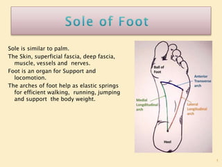
Sole is similar to palm.
The Skin, superficial fascia, deep fascia,
muscle, vessels and nerves.
Foot is an organ for Support and
locomotion.
The arches of foot help as elastic springs
for efficient walking, running, jumping
and support the body weight.
2. 2
The skin of the sole, like that of
the palm is:
1. Thick for protection
2. Firmly adherent to the underlying plantar
aponeurosis and
3. Creased.
These features increase the efficiency of the
grip of the sole on the ground.
3. 3
The skin of the sole, like that of the palm
is:
The skin is mainly supplied by three cutaneous nerves
The nerves are:
a) Medial calcanean branches of the tibial nerve, to the
posterior and medial portions.
b) Branches from the medial plantar nerve to the
larger, anteromedial portion including the medial 3½
digits.
c) Branches from the lateral plantar nerve to the
smaller anterolateral portion including the lateral 1½
digits.d. Small areas on medial and lateral sides are
innervated by saphenous and sural nerves
4. 4
The superficial fascia of the sole is fibrous and
dense.
Fibrous bands bind the skin to the deep fascia or
plantar aponeurosis, and divide the
subcutaneous fat into small tight compartments
which serve as water-cushions and reinforce the
spring-effect of the arches of the foot during
walking, running and jumping.
The fascia is very thick and dense over the
weight-bearing points. It contains cutaneous
nerves and vessels
5.
6. 1. Plantar Aponeurosis in the sole.
2. Deep transverse metatarsal ligament between
Metatarsophalangeal joints.
3. The fibrous flexor sheaths in the toe.
6
7. THICKENED CENTRAL BAND OF THE DEEP FASCIA
IN THE SOLE OF THE FOOT.
7
The aponeurosis is triangular in shape. The
apex is proximal.
It is attached to the medial tubercle of the
calcaneum, proximal to the attachment of the
flexor digitorum brevis.
The base is distal. It divides into five processes
near the heads of the metatarsal bones. The
digital nerves and vessels pass through the
intervals between the processes
PLANTAR APONEUROSIS
8.
9. THICKENED CENTRAL BAND OF THE DEEP FASCIA
IN THE SOLE OF THE FOOT.
9
Each process splits, opposite the metatarso-
phalangeal joints, into a superficial and a deep
slip. The superficial slip is attached to dermis of
skin. The deep slip divides into two parts which
embrace the flexor tendons, and blend with the
fibrous flexor sheaths and with the deep
transverse metatarsal ligaments.
PLANTAR APONEUROSIS
10. THICKENED CENTRAL BAND OF THE DEEP FASCIA
IN THE SOLE OF THE FOOT.
10
Function
1. It fixes the skin of sole.
2. It protects deeper structure.
3. It help to maintaining longitudinal arches of foot.
4. It gives origin to muscles of the first layer of sole.
PLANTAR APONEUROSIS
11. THE MUSCLE OF THE SOLE IS
ARRANGED IN FOUR (4) LAYERS.
11
1. FIRST LAYER
(SUPERFICIAL)
a) FLEXOR DIGITORUM BRVIS
b) ABDUCTOR HALLUCIS
c) ABDUCTOR DIGITI MINIMI
12.
13. 13
Origin :
Medial Tubercle of calcaneum and plantar
aponeurosis.
Insertion :
Middle phalanges of digits 2-5
Nerve supply :
Medial planter nerve
Action :
Flexion on proximal interpharangeal and
Metatarsopharlyngeal joint.
14. 14
Origin :
Medial tubercle of calcaneum, flexor
retinaculum, medial intermuscular septum.
Insertion :
Medial Plantar portion of proximal phalanx
of great toe.
Nerve supply :
Medial Plantar Nerve
Actions :
Abducts great toe at Metatarsopharlyngeal
joint and flexes great toe at
Metatarsopharlyngeal joint.
15. 15
Origin :
Medial and lateral processes of posterior
calcaneal tuberosity
Insertion :
Lateral side of base of proximal phalanx of
5th toe and 5th metatarsal
Nerve supply : Lateral plantar (S2,3)
Action : Flexes and abducts 5th toe. Support
lateral longitudinal arch
17. 17
Origin: From upper two thirds of the medial
part of the posterior surface below the soleal
line.
Insertion: The muscle divides into four
tendons. Each is inserted into the plantar
surface of distal phalanx of second to fifth
digit.
Nerve Supply: Tibial nerve (S2, S3)
Action : Plantar flexion of lateral four toes
and of ankle. Maintains medial longitudinal
arch.
18.
19. Origin :
It arises by two heads:
a. Medial head is large and fleshy; it arises from
the medial concave surface of the calcaneum
b. Lateral head is smaller and tendinous; it
arises from the calcaneum in front of the
lateral tubercle.The two heads unite at an
acute angle.
Insertion :
The muscle fibres are inserted into the lateral
side of the flexor digitorum longus
19
20.
21. 21
Nerve supply :
Main trunk of lateral plantar nerve.
Action :
Straightens the pull of the long flexor
muscles.
Flexes the toes through the long
tendons.
22. Origin:
Lower three-fourths of the posterior
surface of fibula except lowest 2.5 cm
and adjoining interosseous membrane.
Insertion: plantar surface of the base of
distal phalanx of the great toe.
Nerve Supply: Tibial nerve
Function: Plantar flexor of the big toe,
plantar flexor of ankle joint, maintains
medial longitudinal arch.
22
23.
24. Origin: They arise from the tendons of theflexor digitorum
longus.
The first lumbrical is unipennate, and theothers are
bipennate First lumbrical arises from medial side of 1st tendon
of flexor digitorum longus.
Second lumbrical arises from adjacent sides of 1st and
2nd tendons of flexor digitorum longus.
Third lumbrical arises from adjacent sides of 2nd and
3rd tendons of flexor digitorum longus.
Fourth lumbrical arises from adjacent sides of 3rd and
4th tendons of flexor digitorum longus.
24
26. Insertion : Their tendons pass forwards on the medial sides of
the metatarsophalangeal joints of the lateral four toes,
and then dorsally for insertion into the extensor
expansion.
Nerve supply : The first muscle by the medial plantar nerve;
and the other three by the deep branch of lateral
plantar nerve
Action : They maintain extension of the digits at the inter-
phalangeal joints so that in walking and running the
toes do not buckle under
26
29. Origin:
It arises by a Y-shaped tendon:a.
The lateral limb, from the medial
part of the plantar surface of the
cuboid bone, behind the groove
for the peroneus longus and from
the adjacent side of the lateral
cuneiform boneb. The medial
limb is a direct continuation of
the tendon of tibialis posterior
into the foot.
29
30. 30
Insertion : The muscle splits into
medial and lateral parts, each of
which ends in a tendon. Each tendon
is inserted into the corresponding
side of the base of the proximal
phalanx of the great toe.
Nerve supply : Medial Plantar Nerve
Actions : Flexes the proximal phalanx
at the metatarsophalangeal joint of
the great toe.
31. Origin : It arises by two heads:
a. The oblique head is large, and arises
from the bases of the second, third, and
fourth metatarsals, from the sheath of the
tendon of the peroneus longus
b. The transverse head is small, and arises
from the deep metatarsal ligament, and the
plantar ligaments of the
metatarsophalangeal joints of the third,
fourth and fifth toes (transverse head has no
bony origin)
31
32.
33. 33
Insertion : On the lateral side of the
base of the proximal phalanx of the
big toe, in common with the lateral
tendon of the flexor hallucis brevis.
Nerve supply : Deep Branch of lateral
plantar nerve, which terminates in this
muscle.
Actions : Adductor of great toe
towards second toe. Maintains
transverse arches of foot.
34. Origin :
a. Base of the fifth metatarsal
bone
b. Sheath of the tendon of the
peroneus longus
Insertion : Into the lateral side of
the base of the proximal phalanx
of the little toe.
34
35.
36. 36
Nerve supply : Superficial branch of
lateral plantar nerve
Action : Flexes the proximal phalanx at
the metatarsophalangeal joint of the
little toe
39. 39
Origin : Bases and medial sides of third,
fourth and fifth metatarsals.
Insertion : Medial sides of bases of
proximal phalanges and dorsal
digital/extensor expansions of 3rd, 4th
and5th toes
Nerve supply : First and second by lateral
plantar (deep branch). Third by lateral
plantar (superficial branch)
Function : Adductors of third, fourth and
fifth toes toward the axis. Flexor of
metatarsophalangeal and extensor of
interphalangeal joints of third, fourth and
fifth toes
40. 40
Origin :
Adjacent sides of metatarsal bones
Insertion :
Bases of proximal phalanges and
dorsal digital expansion of toes;
first on medial side of 2nd toe;
second on lateral side of 2nd toe;
third on lateral side of 3rd toe and
fourth on lateral side of 4th toe.
41. 41
Nerve supply :
First, second, third by lateral plantar
(deep branch), fourth dorsal
interosseous by superficial branch of
lateral plantar.
Function : Abductors of toes from axis
of second toe. First and second cause
medial and lateral abduction of second
toe. Third and fourth for abduction of
3rd and 4th toes
42. Origin : Posterior surfaces of leg bones
Insertion : Tuberosity of navicular
Nerve supply : Tibial Nerve
Function : Plantar Flexion of ankle
42
Tibialis
Posterior
43. Origin:
Upper part of lateral surface of fibula
Insertion :
Base of 1st metatarsal
Nerve Supply :
Superficial peroneal nerve
Function : Evertor of foot
43
44. origin and course :.
Largest terminal branch of Tibial nerve. It passes
forwards between Abductor hallucis and flexor
digitorum brevis and divides into its branches.
Root Value : L4, L5, S1
Branches:
The Muscular branches supply the Four muscle.
1. Abductor hallucis
2. Flexor digitorum brevis
3. Flexor hallucis brevis
4. First lumbrical muscle
44
45. Branches
45
Its muscular branches supply four muscles as follows.
1. The abductor hallucis.
2. The flexor digitorum brevis.
3. The flexor hallucis brevis receives a branch from the first
digital nerve.
4. The first lumbrical muscle receives a branch from the
second digital nerve.
47. Branches
47
Cutaneous branches supply the skin of the medial part of the sole, and
of the medial 3½ toes through four digital branches.
The first digital nerve supplies the medial side of the great toe.
The second nerve supplies the adjacent sides of the first and second
toes.
The third nerve supplies the adjacent sides of the second and third
toes.
The fourth nerve supplies the adjacent sides of the third and fourth
toes.
Each digital nerve gives off a dorsal branch which supplies structures
around the nail of the digit concerned.
Articular branches supply joints of the tarsus and metatarsus.
48. 48
Origin:
Small terminal branch of Tibial nerve.
Passes laterally and forwards till base of fifth Metatarsal,
where it divides Superficial and Deep branches.
Its Root value is Ventral Primary Rami of S2,S3, its
supply 14
muscles of the sole.
49. Branches :
The main trunk supplies two muscles-the flexor
digitorum accessorius and the abductor digiti minimi, and
the skin of the sole.
The main trunk ends by dividing into superficial and
deep branches.
The superficial branch divides into two branches-
lateral and medial.
The lateral branch supplies three muscles-flexor digiti
minimi brevis, the third plantar and fourth dorsal
interossei, and the skin on the lateral side of the little toe.
50. Branches :
The medial branch communicates with the medial
plantar nerve, and supplies the skin lining the fourth
interdigital cleft.
The deep branch supplies nine muscles, including the
second, third and fourth lumbricals; first, second and
third dorsal interossei; first and second plantar interossei
and adductor hallucis.
51. 51
Beginning, Course and Termination :
Medial plantar artery is a smaller terminal branch of the posterior tibial
artery. It lies along the medial border of foot and divides into branches.
52. 52
Branches
It gives off cutaneous, muscular branches to the overlying skin and to
the adjoining muscles, and three small superficial digital branches that
end by joining the first, second and third plantar metatarsal arteries
which are branches of the plantar arch
53.
54. 54
Beginning, Course and Termination :
Lateral plantar artery is the larger terminal
branch of the posterior tibial artery. At the
base of the fifth metatarsal bone, it gives a
superficial branch and then continues as
the plantar arch
55.
56. 56
Branches :
Muscular branches supply the adjoining muscles. Cutaneous branches
supply the skin and fasciae of the lateral part of the sole. Anastomotic
branches reach the lateral border of the foot and anastomose with
arteries on the dorsum of the foot. A calcanean branch is occasionally
given off to the skin of the heel.
57. 57
Beginning, Course and Termination :
Plantar arch is formed by the direct continuation of the lateral plantar
artery after it has given off the superficial branch and is completed
medially by the dorsalis pedis artery. It extends from the base of the fifth
metatarsal bone to the proximal part of the first intermetatarsal space,
and lies between the third and fourth layers of the sole. It is
accompanied by venae comitantes. The deep branch of the lateral
plantar nerve lies in the concavity of the plantar arch
58. 58
Branches of the Plantar Arch :
1. Four plantar metatarsal arteries run distally, one in each
intermetatarsal space. Each artery ends by dividing into two plantar
digital branches for adjacent sides of two digits.
The first artery also gives off a branch to the medial side of the great
toe. The lateral side of the little toe gets a direct branch from the
lateral plantar artery.
59.
60. 60
Branches of the plantar arch :
2. The plantar arch gives off three proximal perforating arteries that pass
through the second, third and fourth intermetatarsal spaces and
communicate with the dorsal metatarsal arteries which are the
branches of the arcuate artery.
The distal end of each plantar metatarsal artery gives off a distal
perforating artery which joins the distal part of the corresponding
dorsal metatarsal artery
61. Plantar fasciitis occurs in policemen due to stretching
of the plantar aponeurosis. This results in pain in the
heel region, especially during standing.
62. A neuroma may be formed on the branch of medial plantar nerve
between 3rd and 4th metatarsal bones. It is called Morton's neuroma
This causes pain between third and
fourth metatarsals. It may be also due
to pressure on digital nerve between
3rd and 4th metatarsals. Any of the
digital nerves, especially the one in the
third interdigital cleft may develop
neuroma. This is a painful condition
63. Fracture of shaft of 2nd/3rd/4th/metatarsal bones is called 'march
fracture. It is seen in army personnel, policemen as they have to
march a lot. It occurs due to decalcification and vascular necrosis.
Toes may be spread out
or splayed.
Longitudinal arches are
exaggerated leading to
pes cavus
64. Normal architecture of foot is subjected to insults due to 'high heels'.
Females apparently look taller, smarter but may suffer from sprains
and dislocations of the ankle joint
65. If medial border of foot is raised, person
walks on lateral border of foot. The
condition is called 'talipes varus’
If lateral border of foot is raised, person
walks on medial border of foot. The
condition is called 'talipes valgus' .
66. If foot is dorsiflexed, person walks
on the hee lcondition is called
'talipes calcaneus’ .
If foot is plantar flexed, person
walks on toes. The condition is
called 'talipes equinus' .
67. Most common is talipes equinovarus in
which theheel is medial, the foot is plantar
flexed and invertedwith high medial
longitudinal arch