Download to read offline


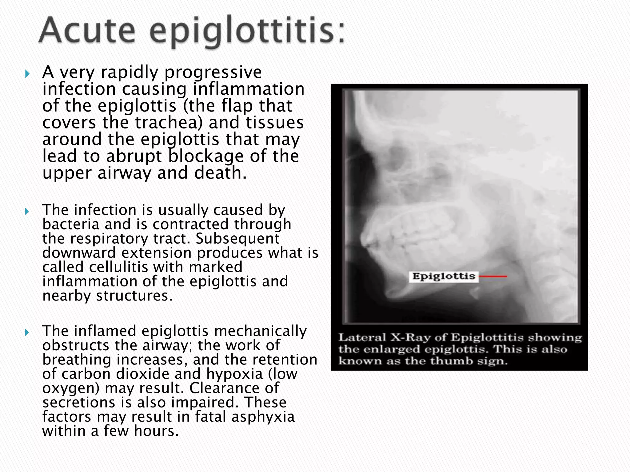


The document discusses the structure and function of the human respiratory tract, outlining its segments, common infections, and related diseases. It covers conditions such as pharyngitis, diphtheria, and various types of pneumonia, detailing symptoms, transmission methods, and treatment options. Prevention measures, particularly vaccination recommendations, are emphasized for managing respiratory infections and diseases.


