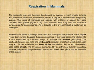
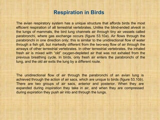
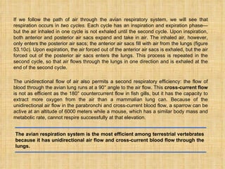
Specialities in Birds respiratory system: Air sacs, specialized parabronchi , Unidirectional flow
Benifits of air sacs, Benefit of 2 respiratory cycles
Bird-like respiratory systems in dinosaurs
Rate of breathings in birds
Air sacs are thin walled non-muscular,non-vascular,non-elastic and bladder like membrane connected to the lungs
Air sacs are only the inflated extensions of the mucous membrane of some blindly ending bronchioles
Some of them extended to bones
In pigeon there are nine major air sacs
One of them is unpaired and others are paired
Unpaired air sac is interclavicular
Paired sac include cervical,anterior thoracic,posterior thoracic and abdominal air sacs
Abdominal and posterior thoracic sacs get filled with fresh air during inspiration.so they are called inspiratory air sacs
Others get filled with fresh air during expiration
The respiratory system (also respiratory apparatus, ventilatory system) is a biological system consisting of specific organs and structures used for gas exchange in animals and plants. The anatomy and physiology that make this happen varies greatly, depending on the size of the organism, the environment in which it lives and its evolutionary history. In land animals the respiratory surface is internalized as linings of the lungs.[1] Gas exchange in the lungs occurs in millions of small air sacs called alveoli in mammals and reptiles, but atria in birds. These microscopic air sacs have a very rich blood supply, thus bringing the air into close contact with the blood.[2] These air sacs communicate with the external environment via a system of airways, or hollow tubes, of which the largest is the trachea, which branches in the middle of the chest into the two main bronchi. These enter the lungs where they branch into progressively narrower secondary and tertiary bronchi that branch into numerous smaller tubes, the bronchioles. In birds the bronchioles are termed parabronchi. It is the bronchioles, or parabronchi that generally open into the microscopic alveoli in mammals and atria in birds. Air has to be pumped from the environment into the alveoli or atria by the process of breathing which involves the muscles of respiration.
The main function of gills is respiration...In gills, there are many hair like projections called gill filaments..in gill filaments, there are number of lamella, from transfer of gases and water occur..
Why do animals need to breathe?
Breathing is important to organisms because cells require energy (oxygen) to move, reproduce and function. Breath also expels carbon dioxide, which is a by-product of cellular processes within the bodies of animals.
Respiration is the process of releasing energy from food and this takes place inside the cells of the body.
The process of respiration involves taking in oxygen (of air) into cells, using it for releasing energy by burning food, and then eliminating the waste products (carbon dioxide and water) from the body.
Respiration is essential for life because it provides energy for carrying out all the life processes which are necessary to keep the organisms alive.
The energy produced during respiration is stored in the form of ATP (Adenosine Tri- Phosphate) molecules in the cells of the body and used by the organism as when required.
KEY POINTS
Life started in an anaerobic environment in the so called ‘primodial broth’ (a mixture of organic molecules.
Subsequently, oxygen strangely enough became an crucial factor for aerobic metabolism especially in the higher life forms.
The rise of an oxygenic environment was an important event in the diversification of life.
It evoked a dramatic shift from inefficient to sophisticated oxygen dependent oxidizing ecosystems.
Anaerobic fermentation, the metabolic process that prevailed for the first about 2 billion years of the evolution of life, was a very inefficient way of extracting energy from organic molecules. Ex: A molecule of glucose, e.g., produces only two molecules of ATP (≈ 15 kCal) compared with 36 ATP molecules (≈ 263 kCal) in oxygenic respiration.
Aerobic metabolism must have developed at a critical point when the partial pressure of oxygen rose from an initial level to one adequately high to drive it passively across the cell membrane.
Respiration is a complex and highly integrated biomechanical, physiological, and behavioral processes.
The transfer of O2 occurs through a flow of tissue barriers and compartments by diffusion down a partial pressure gradient, which drops to about zero at the mitochondrial level.
Acquisition of molecular oxygen (O2) from the external fluid media (water and air) and the discharge of carbon dioxide (CO2) into the same milieu is the primary role of respiration.
The respiratory system is a biological system consisting of specific organs and structures.
In aquatic animals such as fish respiration takes place through special respiratory organs called gills, however lung fish respiration takes place through lungs. Gills are present on both the sides of the head of fish. The gills are covered by gill covers also called operculum. When the fish open its mouth, water is drawn into the buccal cavity and passed through the gills. The gills contain special type of cells that absorb the oxygen present in water. The absorbed oxygen is then supplied to all the cells of body through blood. In the cells, oxygen is converted into carbon dioxide and returned back to gills through blood. Ultimately, the gills release the carbon dioxide in water passing through them.
Respiration in Fish
The gills of fish are very efficient; it is estimated gills can extract about 80% oxygen dissolved in water. In addition to the respiratory organs, the gills have an important role in maintaining the right balance of salts in the body.
Automate machine for rescue operation for childeSAT Journals
Abstract
The aim of the project is to rescue of trapped child in to the boar well by continues monitoring & supply of necessary items to survive using technical method. These bore wells in turn have started to take many innocent lives. Bores which yielded water and subsequently got depleted are left uncovered. Small children without noticing the hole dug for the bore well slip in and get trapped. To aid in such rescue we have proposed a system. The Methods to keep a child alive in a bore should take in to consideration the lack of oxygen, increased temperatures and humidity, which produces hyperthermia. These problems are addressed with fresh air delivery with or without oxygen. A hand-powered equipment to deliver fresh air inside bore is being designed. This method brings down temperature and delivers fresh air. Visualizing the child is made possible with infrared waterproof cameras and a portable high resolution TV Monitor. This is light weight machine that will go down into the bore well pipe and save the child life systematically. This machine will go down the bore well and perform the action. This robot type machine can stay alive trapped body from bore well until parallel hall will be dug up which will work as minimum amount of time and save the child life systematically.
Keywords: human control machine, hand power equipment, infrared camera, oxygen controlling system, labview software
Respiratory System
The Respiratory System Essay
Respiratory System Analysis
Respiratory System
The Cardiovascular And Respiratory Systems Essay
The Respiratory System
Respiratory System Research Paper
Respiratory System Essay
Respiratory System
Respiratory System
Cardiovascular And Respiratory System Essay
Anatomy And Physiology Of The Respiratory System
The Respiratory System Essay
The Five Key Functions Of The Respiratory System
Respiratory Diseases: Emphysema Essay
Human Respiratory System Essay
Essay on The Respiratory System
Respiratory Essay
Tom Selleck Health: A Comprehensive Look at the Iconic Actor’s Wellness Journeygreendigital
Tom Selleck, an enduring figure in Hollywood. has captivated audiences for decades with his rugged charm, iconic moustache. and memorable roles in television and film. From his breakout role as Thomas Magnum in Magnum P.I. to his current portrayal of Frank Reagan in Blue Bloods. Selleck's career has spanned over 50 years. But beyond his professional achievements. fans have often been curious about Tom Selleck Health. especially as he has aged in the public eye.
Follow us on: Pinterest
Introduction
Many have been interested in Tom Selleck health. not only because of his enduring presence on screen but also because of the challenges. and lifestyle choices he has faced and made over the years. This article delves into the various aspects of Tom Selleck health. exploring his fitness regimen, diet, mental health. and the challenges he has encountered as he ages. We'll look at how he maintains his well-being. the health issues he has faced, and his approach to ageing .
Early Life and Career
Childhood and Athletic Beginnings
Tom Selleck was born on January 29, 1945, in Detroit, Michigan, and grew up in Sherman Oaks, California. From an early age, he was involved in sports, particularly basketball. which played a significant role in his physical development. His athletic pursuits continued into college. where he attended the University of Southern California (USC) on a basketball scholarship. This early involvement in sports laid a strong foundation for his physical health and disciplined lifestyle.
Transition to Acting
Selleck's transition from an athlete to an actor came with its physical demands. His first significant role in "Magnum P.I." required him to perform various stunts and maintain a fit appearance. This role, which he played from 1980 to 1988. necessitated a rigorous fitness routine to meet the show's demands. setting the stage for his long-term commitment to health and wellness.
Fitness Regimen
Workout Routine
Tom Selleck health and fitness regimen has evolved. adapting to his changing roles and age. During his "Magnum, P.I." days. Selleck's workouts were intense and focused on building and maintaining muscle mass. His routine included weightlifting, cardiovascular exercises. and specific training for the stunts he performed on the show.
Selleck adjusted his fitness routine as he aged to suit his body's needs. Today, his workouts focus on maintaining flexibility, strength, and cardiovascular health. He incorporates low-impact exercises such as swimming, walking, and light weightlifting. This balanced approach helps him stay fit without putting undue strain on his joints and muscles.
Importance of Flexibility and Mobility
In recent years, Selleck has emphasized the importance of flexibility and mobility in his fitness regimen. Understanding the natural decline in muscle mass and joint flexibility with age. he includes stretching and yoga in his routine. These practices help prevent injuries, improve posture, and maintain mobilit
263778731218 Abortion Clinic /Pills In Harare ,sisternakatoto
263778731218 Abortion Clinic /Pills In Harare ,ABORTION WOMEN’S CLINIC +27730423979 IN women clinic we believe that every woman should be able to make choices in her pregnancy. Our job is to provide compassionate care, safety,affordable and confidential services. That’s why we have won the trust from all generations of women all over the world. we use non surgical method(Abortion pills) to terminate…Dr.LISA +27730423979women Clinic is committed to providing the highest quality of obstetrical and gynecological care to women of all ages. Our dedicated staff aim to treat each patient and her health concerns with compassion and respect.Our dedicated group ABORTION WOMEN’S CLINIC +27730423979 IN women clinic we believe that every woman should be able to make choices in her pregnancy. Our job is to provide compassionate care, safety,affordable and confidential services. That’s why we have won the trust from all generations of women all over the world. we use non surgical method(Abortion pills) to terminate…Dr.LISA +27730423979women Clinic is committed to providing the highest quality of obstetrical and gynecological care to women of all ages. Our dedicated staff aim to treat each patient and her health concerns with compassion and respect.Our dedicated group of receptionists, nurses, and physicians have worked together as a teamof receptionists, nurses, and physicians have worked together as a team wwww.lisywomensclinic.co.za/
TEST BANK for Operations Management, 14th Edition by William J. Stevenson, Ve...kevinkariuki227
TEST BANK for Operations Management, 14th Edition by William J. Stevenson, Verified Chapters 1 - 19, Complete Newest Version.pdf
Flu Vaccine Alert in Bangalore Karnatakaaddon Scans
As flu season approaches, health officials in Bangalore, Karnataka, are urging residents to get their flu vaccinations. The seasonal flu, while common, can lead to severe health complications, particularly for vulnerable populations such as young children, the elderly, and those with underlying health conditions.
Dr. Vidisha Kumari, a leading epidemiologist in Bangalore, emphasizes the importance of getting vaccinated. "The flu vaccine is our best defense against the influenza virus. It not only protects individuals but also helps prevent the spread of the virus in our communities," he says.
This year, the flu season is expected to coincide with a potential increase in other respiratory illnesses. The Karnataka Health Department has launched an awareness campaign highlighting the significance of flu vaccinations. They have set up multiple vaccination centers across Bangalore, making it convenient for residents to receive their shots.
To encourage widespread vaccination, the government is also collaborating with local schools, workplaces, and community centers to facilitate vaccination drives. Special attention is being given to ensuring that the vaccine is accessible to all, including marginalized communities who may have limited access to healthcare.
Residents are reminded that the flu vaccine is safe and effective. Common side effects are mild and may include soreness at the injection site, mild fever, or muscle aches. These side effects are generally short-lived and far less severe than the flu itself.
Healthcare providers are also stressing the importance of continuing COVID-19 precautions. Wearing masks, practicing good hand hygiene, and maintaining social distancing are still crucial, especially in crowded places.
Protect yourself and your loved ones by getting vaccinated. Together, we can help keep Bangalore healthy and safe this flu season. For more information on vaccination centers and schedules, residents can visit the Karnataka Health Department’s official website or follow their social media pages.
Stay informed, stay safe, and get your flu shot today!
Lung Cancer: Artificial Intelligence, Synergetics, Complex System Analysis, S...Oleg Kshivets
RESULTS: Overall life span (LS) was 2252.1±1742.5 days and cumulative 5-year survival (5YS) reached 73.2%, 10 years – 64.8%, 20 years – 42.5%. 513 LCP lived more than 5 years (LS=3124.6±1525.6 days), 148 LCP – more than 10 years (LS=5054.4±1504.1 days).199 LCP died because of LC (LS=562.7±374.5 days). 5YS of LCP after bi/lobectomies was significantly superior in comparison with LCP after pneumonectomies (78.1% vs.63.7%, P=0.00001 by log-rank test). AT significantly improved 5YS (66.3% vs. 34.8%) (P=0.00000 by log-rank test) only for LCP with N1-2. Cox modeling displayed that 5YS of LCP significantly depended on: phase transition (PT) early-invasive LC in terms of synergetics, PT N0—N12, cell ratio factors (ratio between cancer cells- CC and blood cells subpopulations), G1-3, histology, glucose, AT, blood cell circuit, prothrombin index, heparin tolerance, recalcification time (P=0.000-0.038). Neural networks, genetic algorithm selection and bootstrap simulation revealed relationships between 5YS and PT early-invasive LC (rank=1), PT N0—N12 (rank=2), thrombocytes/CC (3), erythrocytes/CC (4), eosinophils/CC (5), healthy cells/CC (6), lymphocytes/CC (7), segmented neutrophils/CC (8), stick neutrophils/CC (9), monocytes/CC (10); leucocytes/CC (11). Correct prediction of 5YS was 100% by neural networks computing (area under ROC curve=1.0; error=0.0).
CONCLUSIONS: 5YS of LCP after radical procedures significantly depended on: 1) PT early-invasive cancer; 2) PT N0--N12; 3) cell ratio factors; 4) blood cell circuit; 5) biochemical factors; 6) hemostasis system; 7) AT; 8) LC characteristics; 9) LC cell dynamics; 10) surgery type: lobectomy/pneumonectomy; 11) anthropometric data. Optimal diagnosis and treatment strategies for LC are: 1) screening and early detection of LC; 2) availability of experienced thoracic surgeons because of complexity of radical procedures; 3) aggressive en block surgery and adequate lymph node dissection for completeness; 4) precise prediction; 5) adjuvant chemoimmunoradiotherapy for LCP with unfavorable prognosis.
Report Back from SGO 2024: What’s the Latest in Cervical Cancer?bkling
Are you curious about what’s new in cervical cancer research or unsure what the findings mean? Join Dr. Emily Ko, a gynecologic oncologist at Penn Medicine, to learn about the latest updates from the Society of Gynecologic Oncology (SGO) 2024 Annual Meeting on Women’s Cancer. Dr. Ko will discuss what the research presented at the conference means for you and answer your questions about the new developments.
HOT NEW PRODUCT! BIG SALES FAST SHIPPING NOW FROM CHINA!! EU KU DB BK substit...GL Anaacs
Contact us if you are interested:
Email / Skype : kefaya1771@gmail.com
Threema: PXHY5PDH
New BATCH Ku !!! MUCH IN DEMAND FAST SALE EVERY BATCH HAPPY GOOD EFFECT BIG BATCH !
Contact me on Threema or skype to start big business!!
Hot-sale products:
NEW HOT EUTYLONE WHITE CRYSTAL!!
5cl-adba precursor (semi finished )
5cl-adba raw materials
ADBB precursor (semi finished )
ADBB raw materials
APVP powder
5fadb/4f-adb
Jwh018 / Jwh210
Eutylone crystal
Protonitazene (hydrochloride) CAS: 119276-01-6
Flubrotizolam CAS: 57801-95-3
Metonitazene CAS: 14680-51-4
Payment terms: Western Union,MoneyGram,Bitcoin or USDT.
Deliver Time: Usually 7-15days
Shipping method: FedEx, TNT, DHL,UPS etc.Our deliveries are 100% safe, fast, reliable and discreet.
Samples will be sent for your evaluation!If you are interested in, please contact me, let's talk details.
We specializes in exporting high quality Research chemical, medical intermediate, Pharmaceutical chemicals and so on. Products are exported to USA, Canada, France, Korea, Japan,Russia, Southeast Asia and other countries.
Recomendações da OMS sobre cuidados maternos e neonatais para uma experiência pós-natal positiva.
Em consonância com os ODS – Objetivos do Desenvolvimento Sustentável e a Estratégia Global para a Saúde das Mulheres, Crianças e Adolescentes, e aplicando uma abordagem baseada nos direitos humanos, os esforços de cuidados pós-natais devem expandir-se para além da cobertura e da simples sobrevivência, de modo a incluir cuidados de qualidade.
Estas diretrizes visam melhorar a qualidade dos cuidados pós-natais essenciais e de rotina prestados às mulheres e aos recém-nascidos, com o objetivo final de melhorar a saúde e o bem-estar materno e neonatal.
Uma “experiência pós-natal positiva” é um resultado importante para todas as mulheres que dão à luz e para os seus recém-nascidos, estabelecendo as bases para a melhoria da saúde e do bem-estar a curto e longo prazo. Uma experiência pós-natal positiva é definida como aquela em que as mulheres, pessoas que gestam, os recém-nascidos, os casais, os pais, os cuidadores e as famílias recebem informação consistente, garantia e apoio de profissionais de saúde motivados; e onde um sistema de saúde flexível e com recursos reconheça as necessidades das mulheres e dos bebês e respeite o seu contexto cultural.
Estas diretrizes consolidadas apresentam algumas recomendações novas e já bem fundamentadas sobre cuidados pós-natais de rotina para mulheres e neonatos que recebem cuidados no pós-parto em unidades de saúde ou na comunidade, independentemente dos recursos disponíveis.
É fornecido um conjunto abrangente de recomendações para cuidados durante o período puerperal, com ênfase nos cuidados essenciais que todas as mulheres e recém-nascidos devem receber, e com a devida atenção à qualidade dos cuidados; isto é, a entrega e a experiência do cuidado recebido. Estas diretrizes atualizam e ampliam as recomendações da OMS de 2014 sobre cuidados pós-natais da mãe e do recém-nascido e complementam as atuais diretrizes da OMS sobre a gestão de complicações pós-natais.
O estabelecimento da amamentação e o manejo das principais intercorrências é contemplada.
Recomendamos muito.
Vamos discutir essas recomendações no nosso curso de pós-graduação em Aleitamento no Instituto Ciclos.
Esta publicação só está disponível em inglês até o momento.
Prof. Marcus Renato de Carvalho
www.agostodourado.com
Ethanol (CH3CH2OH), or beverage alcohol, is a two-carbon alcohol
that is rapidly distributed in the body and brain. Ethanol alters many
neurochemical systems and has rewarding and addictive properties. It
is the oldest recreational drug and likely contributes to more morbidity,
mortality, and public health costs than all illicit drugs combined. The
5th edition of the Diagnostic and Statistical Manual of Mental Disorders
(DSM-5) integrates alcohol abuse and alcohol dependence into a single
disorder called alcohol use disorder (AUD), with mild, moderate,
and severe subclassifications (American Psychiatric Association, 2013).
In the DSM-5, all types of substance abuse and dependence have been
combined into a single substance use disorder (SUD) on a continuum
from mild to severe. A diagnosis of AUD requires that at least two of
the 11 DSM-5 behaviors be present within a 12-month period (mild
AUD: 2–3 criteria; moderate AUD: 4–5 criteria; severe AUD: 6–11 criteria).
The four main behavioral effects of AUD are impaired control over
drinking, negative social consequences, risky use, and altered physiological
effects (tolerance, withdrawal). This chapter presents an overview
of the prevalence and harmful consequences of AUD in the U.S.,
the systemic nature of the disease, neurocircuitry and stages of AUD,
comorbidities, fetal alcohol spectrum disorders, genetic risk factors, and
pharmacotherapies for AUD.
The prostate is an exocrine gland of the male mammalian reproductive system
It is a walnut-sized gland that forms part of the male reproductive system and is located in front of the rectum and just below the urinary bladder
Function is to store and secrete a clear, slightly alkaline fluid that constitutes 10-30% of the volume of the seminal fluid that along with the spermatozoa, constitutes semen
A healthy human prostate measures (4cm-vertical, by 3cm-horizontal, 2cm ant-post ).
It surrounds the urethra just below the urinary bladder. It has anterior, median, posterior and two lateral lobes
It’s work is regulated by androgens which are responsible for male sex characteristics
Generalised disease of the prostate due to hormonal derangement which leads to non malignant enlargement of the gland (increase in the number of epithelial cells and stromal tissue)to cause compression of the urethra leading to symptoms (LUTS
micro teaching on communication m.sc nursing.pdfAnurag Sharma
Microteaching is a unique model of practice teaching. It is a viable instrument for the. desired change in the teaching behavior or the behavior potential which, in specified types of real. classroom situations, tends to facilitate the achievement of specified types of objectives.
Title: Sense of Taste
Presenter: Dr. Faiza, Assistant Professor of Physiology
Qualifications:
MBBS (Best Graduate, AIMC Lahore)
FCPS Physiology
ICMT, CHPE, DHPE (STMU)
MPH (GC University, Faisalabad)
MBA (Virtual University of Pakistan)
Learning Objectives:
Describe the structure and function of taste buds.
Describe the relationship between the taste threshold and taste index of common substances.
Explain the chemical basis and signal transduction of taste perception for each type of primary taste sensation.
Recognize different abnormalities of taste perception and their causes.
Key Topics:
Significance of Taste Sensation:
Differentiation between pleasant and harmful food
Influence on behavior
Selection of food based on metabolic needs
Receptors of Taste:
Taste buds on the tongue
Influence of sense of smell, texture of food, and pain stimulation (e.g., by pepper)
Primary and Secondary Taste Sensations:
Primary taste sensations: Sweet, Sour, Salty, Bitter, Umami
Chemical basis and signal transduction mechanisms for each taste
Taste Threshold and Index:
Taste threshold values for Sweet (sucrose), Salty (NaCl), Sour (HCl), and Bitter (Quinine)
Taste index relationship: Inversely proportional to taste threshold
Taste Blindness:
Inability to taste certain substances, particularly thiourea compounds
Example: Phenylthiocarbamide
Structure and Function of Taste Buds:
Composition: Epithelial cells, Sustentacular/Supporting cells, Taste cells, Basal cells
Features: Taste pores, Taste hairs/microvilli, and Taste nerve fibers
Location of Taste Buds:
Found in papillae of the tongue (Fungiform, Circumvallate, Foliate)
Also present on the palate, tonsillar pillars, epiglottis, and proximal esophagus
Mechanism of Taste Stimulation:
Interaction of taste substances with receptors on microvilli
Signal transduction pathways for Umami, Sweet, Bitter, Sour, and Salty tastes
Taste Sensitivity and Adaptation:
Decrease in sensitivity with age
Rapid adaptation of taste sensation
Role of Saliva in Taste:
Dissolution of tastants to reach receptors
Washing away the stimulus
Taste Preferences and Aversions:
Mechanisms behind taste preference and aversion
Influence of receptors and neural pathways
Impact of Sensory Nerve Damage:
Degeneration of taste buds if the sensory nerve fiber is cut
Abnormalities of Taste Detection:
Conditions: Ageusia, Hypogeusia, Dysgeusia (parageusia)
Causes: Nerve damage, neurological disorders, infections, poor oral hygiene, adverse drug effects, deficiencies, aging, tobacco use, altered neurotransmitter levels
Neurotransmitters and Taste Threshold:
Effects of serotonin (5-HT) and norepinephrine (NE) on taste sensitivity
Supertasters:
25% of the population with heightened sensitivity to taste, especially bitterness
Increased number of fungiform papillae
These simplified slides by Dr. Sidra Arshad present an overview of the non-respiratory functions of the respiratory tract.
Learning objectives:
1. Enlist the non-respiratory functions of the respiratory tract
2. Briefly explain how these functions are carried out
3. Discuss the significance of dead space
4. Differentiate between minute ventilation and alveolar ventilation
5. Describe the cough and sneeze reflexes
Study Resources:
1. Chapter 39, Guyton and Hall Textbook of Medical Physiology, 14th edition
2. Chapter 34, Ganong’s Review of Medical Physiology, 26th edition
3. Chapter 17, Human Physiology by Lauralee Sherwood, 9th edition
4. Non-respiratory functions of the lungs https://academic.oup.com/bjaed/article/13/3/98/278874
1. RESPIRATORY SYSTEM The word respiration describes two processes. Internal or cellular respiration is the process by which glucose or other small molecules are oxidized to produce energy: this requires oxygen and generates carbon dioxide. External respiration (breathing) involves simply the stage of taking oxygen from the air and returning carbon dioxide to it.
2. Respiration in Mammals The metabolic rate, and therefore the demand for oxygen, is much greater in birds and mammals, which are endothermic and thus require a more efficient respiratory system. The lungs of mammals are packed with millions of alveoli, tiny sacs clustered like grapes (figure 53.9). This provides each lung with an enormous surface area for gas exchange. Air is brought to the alveoli through a system of air passages. Inhaled air is taken in through the mouth and nose past the pharynx to the larynx (voice box), where it passes through an opening in the vocal cords, the glottis, into a tube supported by C-shaped rings of cartilage, the trachea (windpipe). The trachea bifurcates into right and left bronchi (singular, bronchus ), which enter each lung and further subdivide into bronchioles that deliver the air into blind-ended sacs called alveoli. The alveoli are surrounded by an extremely extensive capillary network. All gas exchange between the air and blood takes place across the walls of the alveoli.
3.
4. The branching of bronchioles and the vast number of alveoli combine to increase the respiratory surface area ( A in Fick’s Law) far above that of amphibians or reptiles. In humans, there are about 300 million alveoli in each of the two lungs, and the total surface area available for diffusion can be as much as 80 square meters, or about 42 times the surface area of the body. Respiration in mammals will be considered in more detail in a separate section later. Mammalian lungs are composed of millions of alveoli that provide a huge surface area for gas exchange. Air enters and leaves these alveoli through the same system of airways.
5. Respiration in Birds The avian respiratory system has a unique structure that affords birds the most efficient respiration of all terrestrial vertebrates. Unlike the blind-ended alveoli in the lungs of mammals, the bird lung channels air through tiny air vessels called parabronchi, where gas exchange occurs (figure 53.10 a ). Air flows through the parabronchi in one direction only; this is similar to the unidirectional flow of water through a fish gill, but markedly different from the two-way flow of air through the airways of other terrestrial vertebrates. In other terrestrial vertebrates, the inhaled fresh air is mixed with “old” oxygen-depleted air that was not exhaled from the previous breathing cycle. In birds, only fresh air enters the parabronchi of the lung, and the old air exits the lung by a different route. The unidirectional flow of air through the parabronchi of an avian lung is achieved through the action of air sacs, which are unique to birds (figure 53.10 b ). There are two groups of air sacs, anterior and posterior. When they are expanded during inspiration they take in air, and when they are compressed during expiration they push air into and through the lungs.
6.
7. If we follow the path of air through the avian respiratory system, we will see that respiration occurs in two cycles. Each cycle has an inspiration and expiration phase—but the air inhaled in one cycle is not exhaled until the second cycle. Upon inspiration, both anterior and posterior air sacs expand and take in air. The inhaled air, however, only enters the posterior air sacs; the anterior air sacs fill with air from the lungs (figure 53.10 c ). Upon expiration, the air forced out of the anterior air sacs is exhaled, but the air forced out of the posterior air sacs enters the lungs. This process is repeated in the second cycle, so that air flows through the lungs in one direction and is exhaled at the end of the second cycle. The unidirectional flow of air also permits a second respiratory efficiency: the flow of blood through the avian lung runs at a 90° angle to the air flow. This cross-current flow is not as efficient as the 180° countercurrent flow in fish gills, but it has the capacity to extract more oxygen from the air than a mammalian lung can. Because of the unidirectional air flow in the parabronchi and cross-current blood flow, a sparrow can be active at an altitude of 6000 meters while a mouse, which has a similar body mass and metabolic rate, cannot respire successfully at that elevation. The avian respiration system is the most efficient among terrestrial vertebrates because it has unidirectional air flow and cross-current blood flow through the lungs.
8. Structures and Mechanisms of Breathing In mammals, inspired air travels through the trachea, bronchi, and bronchioles to reach the alveoli, where gas exchange occurs. Each alveolus is composed of an epithelium only one cell thick, and is surrounded by blood capillaries with walls that are also only one cell layer thick. There are about 30 billion capillaries in both lungs, or about 100 capillaries per alveolus. Thus, an alveolus can be visualized as a microscopic air bubble whose entire surface is bathed by blood. Because the alveolar air and the capillary blood are separated by only two cell layers, the distance between the air and blood is only 0.5 to 1.5 micrometers, allowing for the rapid exchange of gases by diffusion by decreasing d in Fick’s Law. The blood leaving the lungs, as a result of this gas exchange, normally contains a partial oxygen pressure (PO2) of about 100 millimeters of mercury. As previously discussed, the PO2 is a measure of the concentration of dissolved oxygen—you can think of it as indicating the plasma oxygen. Because the PO2 of the blood leaving the lungs is close to the PO2 of the air in the alveoli (about 105 mm Hg), the lungs do a very effective, but not perfect, job of oxygenating the blood. After gas exchange in the systemic capillaries, the blood that returns to the right side of the heart is depleted in oxygen, with a PO2 of about 40 millimeters of mercury. These changes in the PO2 of the blood, as well as the changes in plasma carbon dioxide (indicated as the PCO2), are shown in figure 53.11.
9.
10. The outside of each lung is covered by a thin membrane called the visceral pleural membrane. A second, parietal pleural membrane lines the inner wall of the thoracic cavity. The space between these two membranes, the pleural cavity, is normally very small and filled with fluid. This fluid links the two membranes in the same way a thin film of water can hold two plates of glass together, effectively coupling the lungs to the thoracic cavity. The pleural membranes package each lung separately—if one collapses due to a perforation of the membranes, the other lung can still function. Mechanics of Breathing As in all other terrestrial vertebrates except amphibians, air is drawn into the lungs by the creation of a negative, or sub atmospheric, pressure. In accordance with Boyle’s Law, when the volume of a given quantity of gas increases its pressure decreases. This occurs because the volume of the thorax is increased during inspiration (inhalation), and the lungs likewise expand because of the adherence of the visceral and parietal pleural membranes. When the pressure within the lungs is lower than the atmospheric pressure, air enters the lungs.
11. The thoracic volume is increased through contraction of two sets of muscles: the external intercostals and the diaphragm. During inspiration, contraction of the external intercostal muscles between the ribs raises the ribs and expands the rib cage. Contraction of the diaphragm, a convex sheet of striated muscle separating the thoracic cavity from the abdominal cavity, causes the diaphragm to lower and assume a more flattened shape. This expands the volume of the thorax and lungs while it increases the pressure on the abdomen (causing the belly to protrude). You can force a deeper inspiration by contracting other muscles that insert on the sternum or rib cage and expand the thoracic cavity and lungs to a greater extent (figure 53.12 a ). The thorax and lungs have a degree of elasticity —they tend to resist distension and they recoil when the distending force subsides. Expansion of the thorax and lungs during inspiration places these structures under elastic tension. It is the relaxation of the external intercostal muscles and diaphragm that produces unforced expiration, because it relieves that elastic tension and allows the thorax and lungs to recoil. You can force a greater expiration by contracting your abdominal muscles and thereby pressing the abdominal organs up against the diaphragm (figure 53.12 b ).
12.
13. Breathing Measurements A variety of terms are used to describe the volume changes of the lung during breathing. At rest, each breath moves a tidal volume of about 500 milliliters of air into and out of the lungs. About 150 milliliters of the tidal volume is contained in the tubular passages (trachea, bronchi, and bronchioles), where no gas exchange occurs. The air in this anatomical dead space mixes with fresh air during inspiration. This is one of the reasons why respiration in mammals is not as efficient as in birds, where air flow through the lungs is one-way. The maximum amount of air that can be expired after a forceful, maximum inspiration is called the vital capacity. This measurement, which averages 4.6 liters in young men and 3.1 liters in young women, can be clinically important, because an abnormally low vital capacity may indicate damage to the alveoli in various pulmonary disorders. For example, in emphysema, a potentially fatal condition usually caused by cigarette smoking, vital capacity is reduced as the alveoli are progressively destroyed.
14. If breathing is excessive for a particular metabolic rate, so that the blood PCO2 is abnormally lowered, the person is said to be hyperventilating. Perhaps surprisingly, the increased breathing that occurs during moderate exercise is not necessarily hyperventilation, because the faster breathing is matched to the faster metabolic rate, and blood gas measurements remain normal. The next section describes how breathing is regulated to keep pace with metabolism. A person normally breathes at a rate and depth that properly oxygenate the blood and remove carbon dioxide, keeping the blood PO2 and PCO2 within a normal range. If breathing is insufficient to maintain normal blood gas measurements (a rise in the blood PCO2 is the best indicator), the person is hypoventilating. Humans inspire by contracting muscles that insert on the rib cage and by contracting the diaphragm. Expiration is produced primarily by muscle relaxation and elastic recoil. As a result, the blood oxygen and carbon dioxide levels are maintained in a normal range through adjustments in the depth and rate of breathing.
15. Mechanisms That Regulate Breathing Each breath is initiated by neurons in a respiratory control center located in the medulla oblongata, a part of the brain stem (see chapter 54). These neurons send impulses to the diaphragm and external intercostal muscles, stimulating them to contract, and contractions of these muscles expand the chest cavity, causing inspiration. When these neurons stop producing impulses, the inspiratory muscles relax and expiration occurs. Although the muscles of breathing are skeletal muscles, they are usually controlled automatically. This control can be voluntarily overridden, however, as in hypoventilation (breath holding) or hyperventilation. A proper rate and depth of breathing is required to maintain the blood oxygen and carbon dioxide levels in the normal range. Thus, although the automatic breathing cycle is driven by neurons in the brain stem, these neurons must be responsive to changes in blood PO2 and PCO2 in order to maintain homeostasis. You can demonstrate this mechanism by simply holding your breath. Your blood carbon dioxide immediately rises and your blood oxygen falls. After a short time, the urge to breathe induced by the changes in blood gases becomes overpowering. This is due primarily to the rise in blood carbon dioxide, as indicated by a rise in PCO2, rather than to the fall in oxygen levels.
16. A rise in PCO2 causes an increased production of carbonic acid (H2CO3), which is formed from carbon dioxide and water and acts to lower the blood pH (carbonic acid dissociates into HCO3 - and H+, thereby increasing blood H+ concentration). A fall in blood pH stimulates neurons in the aortic and carotid bodies, which are sensory structures known as peripheral chemoreceptors in the aorta and the carotid artery. These receptors send impulses to the respiratory control center in the medulla oblongata, which then stimulates increased breathing. The brain also contains chemoreceptors, but they cannot be stimulated by blood H+ because the blood is unable to enter the brain. After a brief delay, however, the increased blood PCO2 also causes a decrease in the pH of the cerebrospinal fluid (CSF) bathing the brain. This stimulates the central chemoreceptors in the brain (figure 53.13). The peripheral chemoreceptors are responsible for the immediate stimulation of breathing when the blood PCO2 rises, but this immediate stimulation only accounts for about 30% of increased ventilation. The central chemoreceptors are responsible for the sustained increase in ventilation if PCO2 remains elevated. The increased respiratory rate then acts to eliminate the extra CO2, bringing the blood pH back to normal (figure 53.14).
17.
18. A person cannot voluntarily hyperventilate for too long. The decrease in plasma PCO2 and increase in pH of plasma and CSF caused by hyperventilation extinguish the reflex drive to breathe. They also lead to constriction of cerebral blood vessels, causing dizziness. People can hold their breath longer if they hyperventilate first, because it takes longer for the CO2 levels to build back up, not because hyperventilation increases the PO2 of the blood. Actually, in people with normal lungs, PO2 becomes a significant stimulus for breathing only at high altitudes, where the PO2 is low. Low PO2 can also stimulate breathing in patients with emphysema, where the lungs are so damaged that blood CO2 can never be adequately eliminated. Breathing serves to keep the blood gases and pH in the normal range and is under the reflex control of peripheral and central chemoreceptors. These chemoreceptors sense the pH of the blood and cerebrospinal fluid, and they regulate the respiratory control center in the medulla oblongata of the brain.