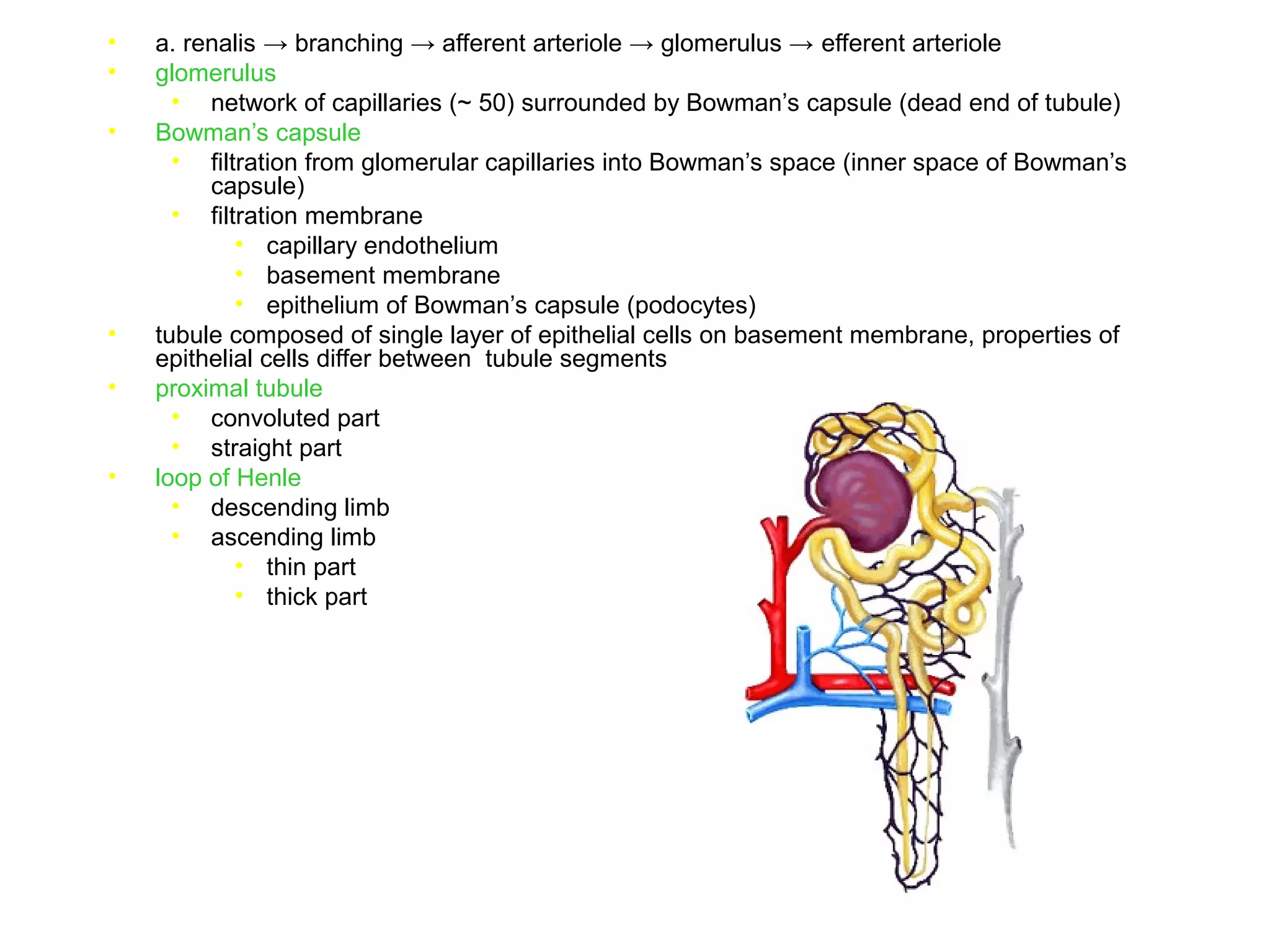
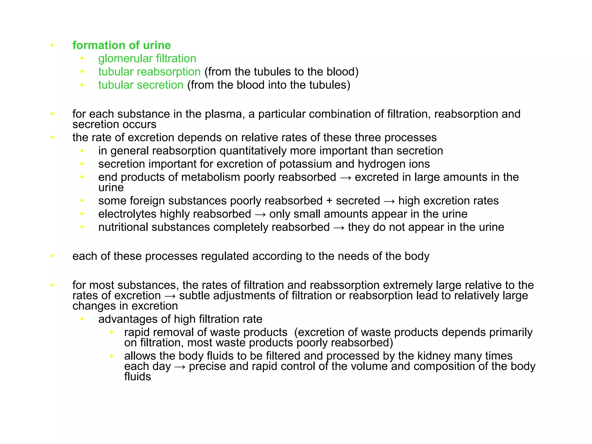
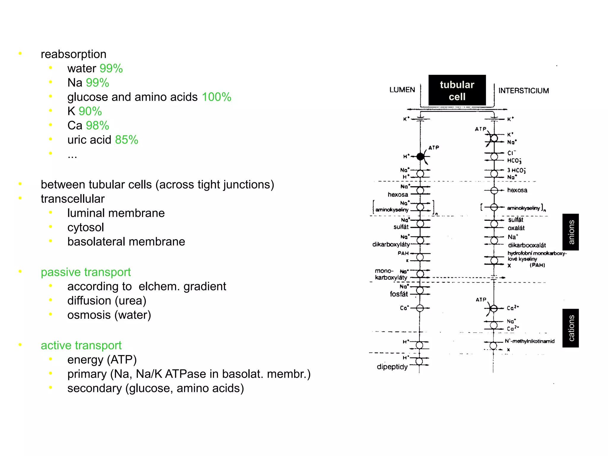
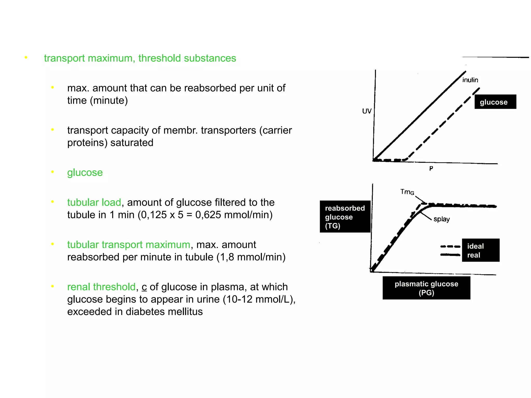
The document discusses the physiology of the kidney. It provides an overview of renal functions such as regulating water, electrolyte and acid-base balance, and excreting waste products. It describes the structure of the kidney including nephrons, glomeruli and tubules. It discusses glomerular filtration, the processes of filtration, reabsorption and secretion, and how clearance is used to measure glomerular filtration rate and renal blood flow.
Renal blood flow (The Guyton and Hall physiology)Maryam Fida
In an average 70-kilogram man, the combined blood flow through both kidneys is about 1100 ml/min, or about 22 per cent of the cardiac output. Two kidneys makes about 0.4 % of total body weight but receive very high blood flow as compared with other body organ. The purpose of additional blood flow is to supply sufficient plasma for high rates of GF which is essential for regulating body fluid volumes & solute concentrations.
Characteristics of the renal blood flow:
1, High blood flow. 1100 ml/min, or 22 percent of the cardiac output. 94% to the cortex.
2, Two capillary beds
High hydrostatic pressure in glomerular capillary (about 60 mmHg) and low hydrostatic pressure in peritubular capillaries (about 13 mmHg)
Blood flow to renal medulla is supplied by vasa recta.
Blood flow in vasa recta of medulla is very low as compared to blood flow in cortex.
Blood flow in renal medulla is 1-2 % of total renal blood flow.
Vasa recta are important to form concentrated urine.
A comprehensive presentation on glomerular filtration rate (GFR) & renal blood flow and how these entities are impacted by intrinsic and extrinsic regulation.
This was presented by the author in the finals of the physiology seminar presentation in medical school.
Tubular reabsorption (The Guyton and Hall physiology)Maryam Fida
It is the second step of urine formation.
It is defined as;
“ The process by which water and other substances are transported by renal tubules back to blood is called Tubular Reabsorption”.
Tubular reabsorption is highly selective.
Some substances like glucose and amino acids are completely absorbed from tubules. So, the urinary excretion is zero.
Ions such as Na+, Cl-, HCO3- are highly absorbed but rate of absorption and excretion varies, according to body needs.
Materials Not Reabsorbed
Nitrogenous waste products
Urea
Uric acid
Creatinine
Excess water
I am a medical student. I have one friend who is persuing his MBBS degree in Taishan Medical UNiversity. I got these notes from him.
These notes are by Dr. Bikesh, He is a famous lecturer of TMU.
These notes have helped me a lot and i also watch his lecture videos , which are great; highly simple and huge content.
I am uploading with Renal physiology. If you want some other topics i would upload for you.
"Let the Knowledge be spread" Dr. Bikesh
Reabsorption In Renal Tubule (The Guyton and Hall physiology)Maryam Fida
Features of PCTPCT have high capacity of active & passive re-absorption.
This is due to special cellular features of epithelial cells.
They have increased no. of mitochondria due to high metabolic activity.
brush border on luminal (apical) side.
Brush border contains protein carrier molecules to transport Na+ by co-transport mechanism with other substances (a.acids, glucose etc).
Additional sodium is transported by COUNTER-TRANSPORT that reabsorb sodium while secreting hydrogen.
About 65 % of filtered load of Na+ & water is reabsorbed in PCT.
A lower % age of Cl- is also absorbed.
In 1st half of PC tubules, Na+ is re-absorbed by co-transport along with glucose, a.acids and other solutes.
In 2nd half of PC tubules, mainly Na+ is reabsorbed with Cl- and some of glucose + a.acids remain un-absorbed.
2nd half of PCT has high conc of Cl- (140 mEq/L) as compared to 1st half (105 mEq/L).
The basics of autoregulation of Gloemrular filtration rate. This ppt deals with basic renal physiology, tubuloglomerular feedback, myogenic reflex, juxtaglomerular apparatus and renin angiotensin aldosterone system in brief. P.S.- The ppt has animations so kindly view in slide/presentation mode
Loop of Henle with its complex anatomy and even more complicated physiology has long remained an enigma to researchers all around the world. Here we discuss about the functional anatomy and the transport characteristics of Loop of Henle.
renal system
a general and a quick learning tip for the renal physiology
useful for undergraduates and can be a quick revising ppt for post graduate as well
these slides are prepared to understand Urinary system IN EASY WAY Important links- NOTES- https://mynursingstudents.blogspot.com/ youtube channel https://www.youtube.com/c/MYSTUDENTSU... CHANEL PLAYLIST- ANATOMY AND PHYSIOLOGY-https://www.youtube.com/playlist?list=PL93S13oM2gAPM3VTGVUXIeswKJ3XGaD2p COMMUNITY HEALTH NURSING- https://www.youtube.com/playlist?list=PL93S13oM2gAPyslPNdIJoVjiXEDTVEDzs CHILD HEALTH NURSING- https://www.youtube.com/playlist?list=PL93S13oM2gANcslmv0DXg6BWmWN359Gvg FIRST AID- https://www.youtube.com/playlist?list=PL93S13oM2gAMvGqeqH2ZTklzFAZhOrvgP HCM- https://www.youtube.com/playlist?list=PL93S13oM2gAM7mZ1vZhQBHWbdLnLb-cH9 FUNDAMENTALS OF NURSING- https://www.youtube.com/playlist?list=PL93S13oM2gAPFxu78NDLpGPaxEmK1fTao COMMUNICABLE DISEASES- https://www.youtube.com/playlist?list=PL93S13oM2gAOWo4IwNjLU_LCuhRN0ZLeb ENVIRONMENTAL HEALTH- https://www.youtube.com/playlist?list=PL93S13oM2gAPkI6LvfS8Zu1nm6mZi9FK6 MSN- https://www.youtube.com/playlist?list=PL93S13oM2gAOdyoHnDLAoR_o8M6ccqYBm HINDI ONLY- https://www.youtube.com/playlist?list=PL93S13oM2gAN4L-FJ3s_IEXgZCijGUA1A ENGLISH ONLY- https://www.youtube.com/playlist?list=PL93S13oM2gAMYv2a1hFcq4W1nBjTnRkHP facebook profile- https://www.facebook.com/suresh.kr.lrhs/ FACEBOOK PAGE- https://www.facebook.com/My-Student-S... facebook group NURSING NOTES- https://www.facebook.com/groups/24139... FOR MAKING EASY NOTES YOU CAN ALSO VISIT MY BLOG – BLOGGER- https://mynursingstudents.blogspot.com/ Instagram- https://www.instagram.com/mystudentsu... Twitter- https://twitter.com/student_system?s=08
#Nephrons, #kidney, #urine, #BORN,#ASSESSMENT, #APPEARENCE,#PULSE,#GRIMACE,#REFLEX,#RESPIRATION,#RESUSCITATION,#NEWBORN,#BABY,#VIRGINIA, #APGAR, #OXYGEN,#CYANOSIS,#OPTICNERVE, #SARACHNA,#MYSTUDENTSUPPORTSYSTEM, #rashes,#nursingclasses, #communityhealthnursing,#ANM, #GNM, #BSCNURING,#NURSINGSTUDENTS, #WHO,#NURSINGINSTITUTION,#COLLEGEOFNURSING,#nursingofficer,#COMMUNITYHEALTHOFFICER
2. Overview of renal functions
• regulation of water and electrolyte balances
• ability to match the excretion to the intake
• enormous capacity (for sodium from 1/10 to 10 times normal)
• regulation of acid-base balance
• excreting acids and regulation of body fluid buffer stores
• the only means for elimination of certain acids (sulfuric, phosphoric acid)
• excretion of metabolic waste products
• urea (metabolism of amino acids)
• creatinine (muscle creatine)
• uric acid (nucleic acids)
• end products of hemoglobin breakdown (bilirubin)
• metabolites of hormones
• removal of foreign chemicals
• toxins
• pesticides
• drugs
• food additives
3. Overview of renal functions
• regulation of arterial pressure
• a dominant role in long-term regulation
• excretion of variable amounts of water and electrolytes
• contribution to short-term regulation
• secretion of vasoactive factors (renin)
• secretion of hormones
• erythropoietin
• production of red blood cells
• hypoxia
• renin
• regulation of arterial pressure
• 1,25-dihydroxyvitamin D3
• hydroxylation on position 1
• calcium and phosphate regulation
• gluconeogenesis
• in prolonged fasting
• in fasting comparable to liver
5. Structure of kidney and of nephron
• paired organs
• retroperitoneum
• 150 g each
• hilum
• renal artery and vein
• lymphatics
• nerve supply
• ureter
• outer cortex
• inner medulla
• multiple renal pyramids
• from border between cortex and
medulla to papilla
• papilla projects into the renal pelvis
• ~1 million functional units = nephrons in
each kidney
• each nephron capable of forming urine
• kidney cannot regenerate nephrons
• after age of 40 reduction of the number
of nephrons by 10% every 10 years
• loss compensated by adaptive
changes in remaining nephrons
proximal tubule
distal tubule
cortex
outer medulla
inner medulla
collecting duct
loop of Henle, thin
descendent limb
loop of Henle, thick
ascendent limb
6. Structure of kidney and of nephron
• nephron
• glomerulus
• filtration of primary filtrate from
the blood
• tubule
• filtered fluid converted to urine by
reabsorption and secretion
• glomerulus (cortex)
• Bowman’s capsule (cortex)
• proximal tubule (cortex)
• Henle’s loop (medulla)
• distal tubule (cortex)
• collecting duct (cortex, medulla)
• cortical nephrons
• glomerulus close to kidney surface
• short loop of Henle
• entire tubular system surrounded by an
extensive network of peritubular
capillaries
• juxtamedullary nephrons
• glomerulus near the medulla
• long loop of Henle
• specialized peritubular capillaries (vasa
recta) extend downward into the
medulla, side by side with the loops of
Henle
proximal tubule
distal tubule
cortex
outer medulla
inner medulla
collecting duct
loop of Henle, thin
descendent limb
loop of Henle, thick
ascendent limb
7. • a. renalis → branching → afferent arteriole → glomerulus → efferent arteriole
• glomerulus
• network of capillaries (~ 50) surrounded by Bowman’s capsule (dead end of tubule)
• Bowman’s capsule
• filtration from glomerular capillaries into Bowman’s space (inner space of Bowman’s
capsule)
• filtration membrane
• capillary endothelium
• basement membrane
• epithelium of Bowman’s capsule (podocytes)
• tubule composed of single layer of epithelial cells on basement membrane, properties of
epithelial cells differ between tubule segments
• proximal tubule
• convoluted part
• straight part
• loop of Henle
• descending limb
• ascending limb
• thin part
• thick part
8. • juxtaglomerular apparatus
• thick ascending limb of the loop of Henle passes between and contacts afferent and
efferent arterioles
• epithelial tubular cells at the point of contact are more dense and are called macula
densa
• wall of afferent arteriole in region of contact contains secretory cells – juxtaglomerular
cells – secrete renin
• complex of juxtaglom. cells and macula densa = juxtaglomerular apparatus
• distal convoluted tubule
• collecting ducts
prox. tub
glom.
aff. art.
juxt. app.
dist. tub.
Bow.
caps. eff. art.
9. • a. renalis → branching → afferent arteriole → glomerulus → efferent arteriole
• efferent arteriole → peritubular capillary network (a second capillary network)
• network surrounding cortical portions of the tubule
• eventually rejoin to form veins
• in cortical nephrons the entire tubular system surrounded by peritubular capillaries
• in juxtamedullary nephrons long efferent arterioles extend down to the outer medulla
and then divide into specialized peritubular capillaries = long and straight capillary
loops that extend downward into the medulla along the loop of Henle = vasa recta
• essential for the formation of concentrated urine
10. • formation of urine
• glomerular filtration
• tubular reabsorption (from the tubules to the blood)
• tubular secretion (from the blood into the tubules)
• for each substance in the plasma, a particular combination of filtration, reabsorption and
secretion occurs
• the rate of excretion depends on relative rates of these three processes
• in general reabsorption quantitatively more important than secretion
• secretion important for excretion of potassium and hydrogen ions
• end products of metabolism poorly reabsorbed → excreted in large amounts in the
urine
• some foreign substances poorly reabsorbed + secreted → high excretion rates
• electrolytes highly reabsorbed → only small amounts appear in the urine
• nutritional substances completely reabsorbed → they do not appear in the urine
• each of these processes regulated according to the needs of the body
• for most substances, the rates of filtration and reabssorption extremely large relative to the
rates of excretion → subtle adjustments of filtration or reabsorption lead to relatively large
changes in excretion
• advantages of high filtration rate
• rapid removal of waste products (excretion of waste products depends primarily
on filtration, most waste products poorly reabsorbed)
• allows the body fluids to be filtered and processed by the kidney many times
each day → precise and rapid control of the volume and composition of the body
fluids
12. • glomerular filtration membrane
• capillary endothelium – perforated by fenestrae, diameter ~100 nm
• basement membrane – a meshwork of collagen and proteoglycan fibrillae
• prevents filtration of proteins
• in part due to negative electrical charge associated with proteoglycans
• epithelium of Bowman’s capsule – podocytes, many finger-like projections,
projections form slits (slit-pores)
• free filtration for neutral molecules with diameters up to 4 nm, for neutral molecules
with diameters >8 nm filtration approches zero
• low molecular weight (<10 000) molecules
• albumin (mol. weight 69 000, 6 nm) does not pass (negative el. charge)
• net filtration pressure
• (+) hydrostatic pressure in glom. capillaries (60 mm Hg)
• (-) hydrostatic pressure in Bowman’s capsule (18 mm Hg)
• (-) osmotic pressure of plasma proteins (32 mm Hg – average,
• beginning 28 mm Hg, end 36 mm Hg)
• (+) osmotic pressure of proteins in Bowman’s space (0 mm Hg)
• FP = 60 - 18 - 32 = 10 mm Hg
• composition of glomerular filtrate
• similar to plasma, EXCEPT that it has no significant amount of proteins (about 1/240
the protein in the plasma)
• Donnan equilibrium effect – in GF anions (other than proteins) about 5% higher
and cations about 5% lower than in plasma
• glomerular filtration rate
• 180 l/day, 125 ml/ min
• (-) decrease of filtration area, decrease of plasma volume, obstruction in urine flow
13. • clearance
• volume of plasma, from which the substance is completely cleared by the kidney per unit of
time
• ≥ 0
• ≤ plasma flow through the kidneys
1
22
1
C
CV
V
×
=
2211 CVCV ×=×
X
X
X
P
VU
C
×
=
14. • determination of glomerular filtration rate
• substance freely filterable, in the tubule not reabsorbed, not secreted, not metabolized
• clearance = glomerular filtration rate
• inulin
• fructose polymer
• not endogenous → administration necessary
• GF = V x Uin / Pin
• 125 ml/min
• clearance of creatinine
• endogenous
• secreted and reabsorbed in tubule, secretion prevails (+10%)
• determination of plasma levels falsely higher (other substances included by the method)
• clearance corresponds to GFR
• both V x Ucr and Pcr falsely higher
• the errors cancel each other
15. • determination of the plasma and blood flow through the kidneys
• substance freely filterable, in the tubule not reabsorbed, not metabolized, but secreted so much that
the plasma is completely cleared from that substance by the time the plasma leaves the kidneys
• clearance = plasma flow through the kidneys
• PAH (para-aminohippuric acid)
• PAH clearance (650 ml/min)
• extraction ratio of PAH = 0,9
• plasma flow through the kidneys: 650 / 0,9 = 720 ml/min
• hematocrit 0,45
• blood flow through the kidneys: 720 / 0,55 = 1310 ml/min
• filtration fraction
• fraction of plasma that filters through the glomerular membrane
• 125 / 720 = 0,17
• 0,16 - 0,20
16. • physiological control of glomerular filtration and renal blood flow
• nervous regulation
• sympathetic
• blood vessels including afferent and efferent arterioles richly innervated by sympathetic
nerve fibers
• strong activation of renal sympathetic nerves → constriction of arterioles and reduction of
renal blood flow and GFR
• mild sympathetic activation → little effect
• probably most important during severe acute disturbances lasting min – hours (severe
hemorrhage, brain ischemia)
• humoral regulation
• norepinephrine, epinephrine
• constriction of afferent and efferent arterioles → reduction of blood flow and GFR
• angiotensin II
• preferentially constricts efferent arteriole
• increases the glomerular hydrostatic pressure while reducing the renal blood flow
• production of angiotensin II increases in conditions of decreased arterial pressure or
volume depletion → in these circumstances angiotensinn II prevents decreases in
glomerular hydrostatic pressure and GFR
• NO
• released by vascular endothelium
• decreases renal vascular resistance
• probably prevents excessive vasoconstriction of the kidney, thus allowing normal
excretion of sodium and water
17. • autoregulation of glomerular filtration and renal blood flow
• keeps the renal blood flow and GFR relatively constant, despite marked changes in arterial
blood pressure
• autoregulation in other organs ensures normal delivery of oxygen and nutrients and removal of
the waste products
• in the kidney the normal blood flow much higher than required for these functions
• the main function of autoregulation in the kidney is to maintain constant GFR and to allow a
precise control of excretion of water and solutes
• tubuloglomerular feedback
• feedback mechanism that links changes in sodium chloride concentration at the macula
densa with the control of renal arteriolar resistance
• keeps a relatively constant delivery of sodium chloride to the distal tubule
• function of juxtaglomerular complex
• decreased GFR → slow flow rate in the loop of Henle → increased reabsorption of
sodium and chloride in the ascending limb → reduction in concentration of sodium
chloride at the macula densa → signal from the macula densa
→ decrease in resistance of afferent arteriole
→ renin release from the juxtaglomerular cells → angitensin II constricts efferent
arteriole
• feedback mechanism to both afferent and efferent arteriole increases glomerular
hydrostatic pressure and normalizes GFR
• myogenic autoregulation
• general ability of blood vessels to resist stretching during increased arterial pressure by
contraction of vascular smooth muscle
• importance in kidney uncertain
• additional adaptive mechanism in the tubule allows to increase reabsorption when GFR rises
(glomerulotubular balance – see later in Tubular function)
19. • reabsorption
• water 99%
• Na 99%
• glucose and amino acids 100%
• K 90%
• Ca 98%
• uric acid 85%
• ...
• between tubular cells (across tight junctions)
• transcellular
• luminal membrane
• cytosol
• basolateral membrane
• passive transport
• according to elchem. gradient
• diffusion (urea)
• osmosis (water)
• active transport
• energy (ATP)
• primary (Na, Na/K ATPase in basolat. membr.)
• secondary (glucose, amino acids)
tubular
cell
cationsanions
20. • transport maximum, threshold substances
• max. amount that can be reabsorbed per unit of
time (minute)
• transport capacity of membr. transporters (carrier
proteins) saturated
• glucose
• tubular load, amount of glucose filtered to the
tubule in 1 min (0,125 x 5 = 0,625 mmol/min)
• tubular transport maximum, max. amount
reabsorbed per minute in tubule (1,8 mmol/min)
• renal threshold, c of glucose in plasma, at which
glucose begins to appear in urine (10-12 mmol/L),
exceeded in diabetes mellitus
reabsorbed
glucose
(TG)
ideal
real
plasmatic glucose
(PG)
glucose
21. • active transport
• primary
• against electrochemical gradient
• hydrolysis of ATP
• sodium-potassium ATPase
• hydrogen ATPase
• hydrogen-potassium ATPase
• calcium ATPase
• sodium
• extensive sodium-potassium ATPase system on the basolateral membrane
• trasports sodium out of the cell into the interstitium
• potassium trasported from the interstitium to the inside of the cell
• maintains low intracellular sodium and high intracellular potassium and negative
potential of -70 mV
• pumping of sodium out of the cell across the basolateral membrane favors passive
diffusion of sodium across the luminal membrane
• concentration gradient (12 mM/L vs. 140 mM/L)
• electrical gradient (negative intracellular potential)
• in most parts of the tubule
• in proximal tubule additional mechanisms for moving large amounts of sodium
• extensive brush border on the luminal side ( surface area)
• carrier proteins for facilitated diffusion through the luminal membrane
22. • active transport
• secondary
• two or more substances interact with a membrane protein and are co-transported across the
membrane
• one substance moves down its electrochemical gradient and the energy released is used to
drive another substance
• glucose, amino acids in the proximal tubule
• carrier protein in the brush border combines with sodium ion and glucose or amino
acid molecule and transports them inside
• exit of glucose or amino acid across the basolateral membrane by facilitated diffusion
• depends on the primary active transport (sodium-potassium ATPase in basolateral
membrane) to maintain an electrochemical gradient for sodium
• secondary active secretion
• often countertransport of the substance with sodium ions
• energy liberated from the downhill movement of of one substance (sodium) enables
uphill movement of a second substance in the opposite direction
• secretion of hydrogen ions coupled to sodium reabsorption in the luminal membrane
• pinocytosis
• reabsorption of large molecules (protein)
• proximal tubule
• protein attaches to the mebrane → this portion of membrane invaginates to the interior →
vesicle formed containing protein → protein digested into amino acids
• requires energy – considered a form of active transport
23. • passive transport
• passive water reabsorption by osmosis
• transport of solutes creates a concentration gradient that causes osmosis of water
• in the same direction as transport of solutes
• both paracellular (across tight junctions) and transcellular pathway (especially in proximal
tubule)
• passive reabsorption of chloride, urea and other solutes
• passive chloride reabsorption coupled to reabsorption of sodium by way of electrical and
chloride concentration gradient
• reabsorption of sodium leaves the inside of lumen negative compared to interstitial
fluid → paracellular diffusion of chloride ions
• osmosis of water → concentration gradient for chloride
• urea also passively reabsorbed but to a much lesser extent than chloride
• reabsorption of water → concentration gradient for urea
• urea does nor permeate the tubule that well → about one half of urea that is filtered is
passively reabsorbed
• the remainder passes into the urine
• creatinine
• almost no reabsorption (molecule is larger than that of urea)
• virtually all creatinine filtered is excreted in the urine
24. • proximal tubule
• high capacity for active and passive reabsorption
• proximal tubule epithelial cells
• high metabolic activity
• large numbers of mitochondria
• extensive brush border on the luminal side and labyrinth of intercellular and basal channels
→ large membrane surface area
• large numbers of transport proteins for both co-transport (glucose, amino acids) and
counter-transport (hydrogen)
• sodium-potassium pump provides the force for reabsorption of sodium, chloride and water
throughout the proximal tubule, but there are diferences in the mechanisms of transport through
the luminal membrane
• the first half of proximal tubule
• sodium reabsorbed by co-transport with glucose, amino acids
• the second half of proximal tubule
• little glucose and amino acids left, increased concentration of chloride ions
• sodium mainly reabsorbed with chloride
• concentrations of solutes
• amount of sodium decreases markedly but concentration of sodium (an total osmolarity)
remains relatively constant
• water reabsorption keeps pace with sodium reabsorption (high permeability for water)
• concentrations of glucose, amino acids, bicarbonate decrease
• reabsorption faster than that of water
• concentration of urea increases
25. • proximal tubule
• secretion of organic acids and bases (the waste products of metabolism)
• site for secretion of bile salts, oxalate, urate, catecholamines…
• secretion + filtration + lack of reabsorption (in any portion of the tubule) → rapid excretion
• secretion of potentially harmful drugs or toxins
• rapid removal from the blood
• for certain drugs the rapid clearance makes difficult to maintain a therapeutically effective
concentration (penicillin, salicylates)
26. • loop of Henle
• 3 functionally different segments
• descending thin segment
• ascending thin segment
• ascending thick segment
• descending thin segment
• no brush border, few mitochondria, low metabolic activity
• high permeability for water
• low permeability for most solutes
• mainly simple diffusion of substances through the wall
• ascending thin segment
• no brush border, few mitochondria, low metabolic activity
• low reabsorption capacity
• virtually impermeable for water
• ascending thick segment
• thick epithelial cells with high metabolic activity and active reabsorption of sodium, chloride
and potassium
• considerable reabsorption of other ions (calcium, magnesium, bicarbonate)
• high expression of of sodium potassium pump in the basolateral membrane
• in the luminal membrane the main transport mechanism for sodium is
1-sodium 2-chloride 1-potassium cotransporter
• the luminal membrane also contains the sodium-hydrogen countertransport mechanism
• virtually impermeable for water → tubular fluid becomes very dilute
27. • distal tubule
• juxtaglomerular complex at the beginning of the distal tubule
• the first half of the distal dubule
• highly convoluted
• similar characteristics as the thick ascending segment of the loop of Henle
• high reabsorption of the ions (sodium, chloride, potassium)
• virtually impermeable to water and urea
• often referred as the diluting segment
• the second half of the distal tubule and the subsequent cortical collecting duct have similar
functional characteristics
• late distal tubule and cortical collecting duct
• principal cells
• reabsorb sodium and water and secrete potassium
• transport dependent od the sodium-potassium pump
in the basolateral membrane
• intercalated cells
• reabsorb potassium and secrete hydrogen
• hydrogen-ATPase
• hydrogen-potassium ATPase (less important)
• carbonic anhydrase
HH22O + COO + CO22
HH22COCO33
carbonic anhydrasecarbonic anhydrase
HH++
+ HCO+ HCO33
--
28. • late distal tubule and cortical collecting duct
• impermeable to urea
• reabsorption of sodium (and secretion of potassium) controlled by hormones (aldosterone)
• a key role of intercalated cells in acid-base regulation
• secretion of hydrogen ions against a large concentration gradient (1:1000)
• permeability to water controlled by ADH
• in the absence of ADH virtually impermeable to water
• medullary collecting duct
• cuboidal cells with smooth surface and relatively few mitochondria
• permeability to water controlled by ADH
• permeable to urea
• capable of secreting hydrogen ions against a large concentration gradient
29. • urea
• passive reabsorption (fac. diffusion) in prox. tubule and medullary part of collecting
duct
• other parts of tubule are impermeable for urea
• due to reabsorption of water in loop of Henle and in distal tubule the concentration of
urea in tubular fluid increases
• in the medullary part of collecting duct reabsorption of urea according to the
concentration gradient → urea contributes to the hyperosmolarity of medulla (up to
400-500 mM in the presence of ADH)
• reabsorption of urea in the medullary part of collecting duct increased by ADH
• directly: regulation of urea transporter
• indirectly: in the presence of ADH the inner part of collecting duct becomes
highly permeable for water → reabsorption of water → increase of urea
concentration in tubular fluid → reabsorption of urea
• tubular secretion
• transport from peritubular capillaries into tubular lumen
• beside glomerular filtration the second pathway into the tubule
• the most important secreted ions: K, H
• organic compounds (creatinine)
• foreign chemicals (penicillin)
30. • regulation of tubular reabsorption
• glomerulotubular balance
• intrinsic ability of tubules to increase their reabsorption rate in response to increased tubular
load (inflow)
• it helps to prevent overloading of the distal tubular segments when GFR increases
• peritubular capillary and renal interstitial fluid physical forces
• fluid and electrolytes reabsorbed from the tubules to the renal interstitium and further into
the peritubular capillaries
• net reabsorptive force across the peritubular capillaries (sum of physical forces)
• hydrostatic pressure inside the peritubular capillaries (-)
• hydrostatic pressure in the renal interstitium outside the capillaries (+)
• colloid osmotic pressure of the peritubular capillary plasma proteins (+)
• colloid osmotic pressure of the proteins in the renal interstitium (-)
• -13+6+32-15=+10 mmHg
• regulation of peritubular capillary physical forces
• peritubular capillary hydrostatic pressure
• arterial pressure (↑)
• resistance of the afferent and the efferent arteriole (↓)
• colloid osmotic pressure of the plasma
• systemic plasma colloid osmotic pressure (↑)
• filtration fraction (↑)
31. • regulation of tubular reabsorption
• peritubular capillary and renal interstitial fluid physical forces
• renal interstitial hydrostatic and colloid osmotic pressures
• changes in peritubular capillary physical forces influence tubular reabsorption by
changing the physical forces in the renal interstitium
• decrease in peritubular reabsorptive forces → reduction of the uptake of fluid and
solutes from the interstitium to the capillaries → ↑ renal interstitial fluid hydrostatic
pressure and ↓interstitial fluid colloid osmotic pressure → decreased reabsorption of
fluid from the renal tubules into the interstitium
• through changes in the hydrostatic and colloid osmotic pressures of the renal
interstitium, the uptake of water and solutes by the peritubular capillaries is closely
matched to the net reabsorption of water and solutes from the tubular lumen to the
interstitium
32. • regulation of tubular reabsorption
• effect of arterial pressure on urine output - pressure natriuresis and pressure diuresis
• increases in arterial pressure often cause increases in urinary excretion of sodium and water
• due to autoregulation (see in GFR chapter) changes in arterial pressure usually have only a
small effect on renal blood flow and GFR
• slight increase in GFR may contribute to pressure natriuresis and diuresis
• greater effect, when GFR autoregulation impaired (kidney disease)
• decreased percentage of the filtered load of sodium and water that is reabsorbed by the
tubules
• results from a slight increase in peritubular capillary hydrostatic pressure and
subsequent increase in renal interstitial fluid hydrostatic pressure
• reduced formation of angiotensin II
33. • regulation of tubular reabsorption
• hormonal control of reabsorption
• aldosterone
• increases sodium reabsorption and potassium secretion
• the primary site of aldosterone action – the principal cells of the cortical collecting duct
• increased expression of sodium-potassium pump in the basolateral membrane and
channels in the luminal membrane
• angiotensin II
• increases sodium and water reabsorption
• stimulates aldosterone secretion
• constricts efferent arteriole
• reduction of peritubular capillary hydrostatic pressure
• by reducing the renal blood flow it raises filtration fraction in the glomerulus and
the colloid osmotic pressure in the peritubular capillaries
• stimulates sodium reabsorption in proximal tubule (stimulation of sodium-
potassium pump in the basolateral membrane and sodium-hydrogen exchange
in the luminal membrane)
• ADH
• increases water reabsorption
• increases water permeability of the distal tubule and collecting duct
• atrial natriuretic peptide
• secreted by atrial cardiac cells, when distended
• inhibits reabsorption of sodium and water, especially in the collecting duct
34. • regulation of tubular reabsorption
• hormonal control of reabsorption
• PTH
• increases tubular reabsorption of calcium, especially in the thick ascending limb of the
loop of Henle and in the distal tubule
• inhibits phosphate reabsorption in the proximal tubule
• stimulates magnesium reabsorption by the loop of Henle
• sympathetic nervous system activation
• decreases sodium and water excretion
• constriction of both afferent and efferent arterioles → reduction of GFR
• increases sodium reabsorption in the proximal tubule and the thick ascending limb of
the loop of Henle
• increases renin release
36. • to a large extent, the osmolarity (concentration of sodium) regulated by the amount of extracellular
water
• body water controlled by:
• fluid intake
• thirst and salt appetite determine intakes of water and salt
• renal excretion of water
• kidney eliminates excess water by excreting dilute urine
• kidney conserves water by excreting concentrated urine
• renal feedback mechanisms to control extracellular fluid sodium concentration and
osmolarity
37. • reabsorption of water in tubular segments
• proximal tubule: 60-70%
• loop of Henle
• descendent limb permeable
• ascendent limb relatively impermeable
• 20%
• distal tubule
• relatively impermeable
• 5%
• collecting ducts
• controlled by antidiuretic hormone (ADH,
vasopresin) – increases the number of water
channels in luminal membrane of tubular
cells
• 10% in cortical part of collect. ducts
• 4,7% in medullar part of collect. ducts
• osmolality of urine in human up to 1200-1400 mM
– 99,7% of filtered water reabsorbed, 0,3% of
filtered water excreted in urine (0,5 l /day)
• in the absence of ADH the urine osmolality can
decrease to 50 mM – 13% of filtered water
excreted in urine (23 l /day)
fraction in tubular fluid
glucose
water
creatinine
prox. tub. dist. tub.Henle loop coll. duct
38. • excreting excess water by forming a dilute urine
• to excrete excess water
• it is necessary to dilute the filtrate
• solutes reabsorbed to a greater extent than water
• proximal tubule
• fluid remains isoosmotic
• descending limb of loop of Henle
• reabsorption of water by osmosis
• tubular fluid reaches equilibrium with surrounding interstitial fluid, which is hypertonic
• ascending limb of loop of Henle (especially thick segment)
• sodium, chloride, potassium reabsorbed
• impermeable to water
• tubular fluid becomes more dilute (about 100 mM/L when entering the distal tubule)
• distal tubule and collecting duct
• additional reabsorption of sodium and chloride
• in the absence of ADH
• impermeable to water
• tubular fluid becomes even more diluted (as low as 50 mM/L)
• fluid leaving the loop of Henle and early distal tubule is always dilute, regardless of the level of
ADH
• in the absence of ADH the urine is further diluted and large volume of dilute urine is excreted
39. • conserving water by excreting a concentrated urine
• water is constantly lost from the body through various routes (lungs, gastrointestinal tract, skin,
kidney)
• the loss matched by the fluid intake
• kidney is capable to form a small volume of concentrated urine
• minimizes the intake of fluid required to maintain homeostasis
• concentrated urine is formed by continuing to excrete solutes while increasing water reabsorption
and decreasing the volume of the urine formed
• obligatory urine volume
• about 600 mM of solutes must be excreted each day
• for a maximal urine concentrating ability of 1200 mM/L the minimal volume of urine that must
be excreted (=obligatory urine volume) equals 0.5 l per day
• requirements for excreting a concentrated urine
• high level of ADH
• increases permeability of the distal tubules and collecting ducts to water
• high osmolarity of the renal medullary interstitial fluid
• provides osmotic gradient for water reabsorption
• the urine concentrating ability is limited by the level of ADH and by the degree of
hyperosmolarity of the renal medulla
• countercurrent mechanism
• loops of Henle (countercurrent multiplier)
• vasa recta (countercurrent exchanger)
40. • conserving water by excreting a concentrated urine
• countercurrent multiplier (loop of Henle)
• osmolarity of the interstitial fluid in the medulla
progressively increases to 1200-1400 mM/L in the
pelvic tip of the medulla (osmotic stratification of the
renal medulla)
• high solute concentration in the medulla is maintained
by a balanced inflow and outflow of solutes and water
• major factors that contribute to the buildup of solute
concentration
• active transport of sodium ions and cotransport of potassium, chloride and other ions
out of the thick ascending limb
• active transport of ions from the collecting ducts
• passive diffusion of large amounts of urea from the inner medullary collecting ducts to
the medullary interstitium
• limited diffusion of water from the medullary tubules
• active transport from the thick ascending loop of Henle is capable of establishing about a
200-mM concentration gradient between the tubular lumen and the interstitial fluid
• this limit to the gradient is due to paracellular diffusion back into the tubule
• repetitive reabsorption of sodium chloride by the thick ascending loop of Henle and
continued inflow of new sodium chloride from the proximal tubule allows eventually raising
the intersitial osmolarity
41. • conserving water by excreting a concentrated urine
• countercurrent multiplier (loop of Henle)
• repetitive reabsorption of sodium chloride by the thick
ascending loop of Henle and continued inflow of new
sodium chloride from the proximal tubule allows
eventually raising the intersitial osmolarity
• active pump of the thick ascending limb → a 200-mM/L
gradient between tubular and interstitial fluids
• tubular fluid in the descending limb and the interstitial fluid
quickly reach equilibrium
• both 400 mM/L
• osmosis of water from the descending limb
• interstitial osmolarity maintained at 400 mM/L (continuing active transport)
• hyperosmotic fluid from the descending limb flows to the loop of Henle and enters the ascending
limb
• active pump of the thick ascending limb again creates a 200-mM/L gradient
• more and more solutes pumped into interstitium multipliing the concentration gradient
• repeated over and over with the net effect of adding more and more solutes to the medulla
• continuous inflow of isotonic tubular flow from the proximal tubule and osmosis of the water that
is more pronounced in the upper layers responsible for the cortex-medullar gradient (osmotic
stratification of the medulla)
42. • conserving water by excreting a concentrated urine
• role of distal tubule and collecting duct
• hypotonic fluid enters the distal tubule
• early distal tubule further dilutes the fluid
• impermeable to water
• active reabsorption of sodium chloride
• cortical collecting duct
• reabsorption of water dependent on ADH
• high plasma levels of ADH
• collecting duct becomes highly permeable to water
• reabsorption of water into cortex interstitium → removed by rapidly flowing
peritubular capillaries
• medullary collecting duct
• further water reabsorption from the tubular fluid but the total amount of water
reabsorbed relatively small compared to the cortex
• reabsorbed water removed by vasa recta
• high levels of ADH
• fluid at the end of collecting ducts has the same osmolarity as the interstitial fluid
(1200-1400 mM/L)
• by reabsorbing as much water as possible the kidney forms a highly concentrated urine
43. • conserving water by excreting a concentrated urine
• contribution of urea
• urea contributes about 40% of the osmolarity
(500 mM/L) when the kidney is forming a maximally
concentrated urine
• when blood levels of ADH are high → large amounts
of urea passively reabsorbed from the medullary
collecting duct
• due to reabsorption of water in loop of Henle and in distal tubule the concentration of urea in
tubular fluid increases
• in the medullary part of collecting duct reabsorption of urea according to the concentration
gradient → urea contributes to the hyperosmolarity of medulla (up to 400-500 mM in the
presence of ADH)
• reabsorption of urea in the medullary part of collecting duct increased by ADH
• directly: regulation of urea transporter
• indirectly: in the presence of ADH the inner part of collecting duct becomes highly
permeable for water → reabsorption of water → increase of urea concentration in
tubular fluid → reabsorption of urea
44. • conserving water by excreting a concentrated urine
• contribution of urea
• normal person excretes 40-50% of the filtered load
of urea
• urea excretion determined mainly by 2 factors:
• concentration in the plasma
• GFR
• recirculation of urea
• in proximal tubule concentration of urea increases
• a small reabsorption of urea takes place
• urea less permeant than water
• in thin segments of the loop of Henle the concentration of urea continues to rise
• water reabsorption
• diffusion of urea into the thin loop of Henle from the medullary interstitium
• high concentration of urea in the medulary collecting duct → diffusion to the medullary
interstitium → a moderate share of urea eventually diffuses into the thin loop of Henle → urea
can recirculate through the terminal parts of the tubular sytem several times before is excreted
• each time around the circuit contributes to hgher concentration of urea
• additional mechanism for forming hyperosmotic medulla
• economic use of the most abundant waste product excreted by the kidney
45. • conserving water by excreting a concentrated urine
• countercurrent exchange in the vasa recta
• normal circulation would correct the hyperosmolarity, BUT:
• due to the U-shape the vasa recta serve as countercurrent exchanger – solutes go from
interstitial fluid into descendent limb and then go back to the interstitial fluid from ascendent
limb (water always goes in the opposite direction) → wash out of solutes from medulla is
minimized
• medullary blood flow is low (1-2% of the total renal blood flow)
• vasa recta do not create the medullary hyperosmolarity but do prevent it from being dissipated
46. • mechanism of water reabsorption
• passive
• dependent on Na reabsorption
• active reabsorption of Na into interstitial fluid → increased concentration of water in tubular fluid +
decreased concentration of water in interstitial fluid → diffusion of water (osmosis) from tubular
fluid to interstitial fluid (transcellular and between cells)
• transport through water channels (transcellular)
• protein aquaporin I – in proximal tubule, ADH-insensitive
• protein aquaporin II – in collecting duct, ADH-sensitive
• antidiuretic hormone
• secreted by posterior pituitary
• binds to receptors (V2)→ increase of cAMP concentration in tubul. cells → endosomes with protein
water channels connect with the luminal membrane → number of water channels increases →
increased permeability of luminal membrane for water
• diabetes insipidus
• loss of ability to produce ADH (damage of hypothalamus by tumor, trauma...)
• central diabetes insipidus
• defective kidney receptors
• nephrogenic diabetes insipidus
• polyuria (excessive excretion of diluted urine)
• polydipsia (excessive drinking of water)
• polydipsia keeps the patient in otherwise good health
• decreased water intake leads to serious dehydration
47. • water diuresis
• results from drinking big volume of hypotonic fluid
• due to inhibition of ADH secretion
• the main cause is decreased osmolality of plasma after water reabsorption – registered by
osmoreceptors in hypothalamus
• osmotic diuresis
• increased urine volume due to big amount of non-reabsorbed solutes in the tubule
• in diabetes mellitus – glucose staying in tubule (after exceeding the tubular transport maximum)
leads to polyuria
• water diuresis – excretion of solutes is not increased, whereas in osmotic diuresis the increased
excretion of solutes is primary cause of polyuria
• in water diuresis the volume of water reabsorbed in prox. tubule is normal, in osmotic diuresis the
reabsorption of water in prox. tubule and in loop of Henle is decreased
49. • excretion of sodium
• reabsorption of sodium in tubular segments
• proximal tubule: 60-70%
• loop of Henle
• thick part of ascending limb - 27%
• distal tubule (final part) and collecting ducts
• reabsorption of Na variable
• controlled by aldosterone
• mechanism of reabsorption
• Na/K ATPase in basolateral membrane of all tubular
cells
• active transport maintains the intracellular Na
concentration low → passive diffusion of Na through
luminal membrane into the cell
• mechanism of passive movement of Na through
luminal membrane different in different segments:
• prox. tubule – transporters for cotransport of Na
and glucose, Na and amino acids, antiport of Na
and H
• asc. limb of loop of Henle – transporter for Na, K,
2 Cl cotransport
• distal convoluted tubule – transporter for Na, Cl
cotransport
• collecting duct - Na channel
fraction in tubular fluid
glucose
water
creatinine
prox. tub. dist. tub.Henle loop coll. duct
50. • excretion of sodiumexcretion of sodium
• aldosteronealdosterone
• produced by adrenal cortexproduced by adrenal cortex
• acts in distal tubule (final part) and collecting ductacts in distal tubule (final part) and collecting duct
• stimulates reabsorption of Na and secretion of K (through Na/K ATPase the Na reabsorption and Kstimulates reabsorption of Na and secretion of K (through Na/K ATPase the Na reabsorption and K
secretion linked)secretion linked)
• in the absence of aldosterone all Na coming to the final part of distal tubule (35 g/day) excreted, atin the absence of aldosterone all Na coming to the final part of distal tubule (35 g/day) excreted, at
high level of aldosterone all Na reabsorbedhigh level of aldosterone all Na reabsorbed
• it acts via nucleus – increases expression of transport proteins – Na/K ATPase in basolateralit acts via nucleus – increases expression of transport proteins – Na/K ATPase in basolateral
membrane and Na and K channels in luminal membranemembrane and Na and K channels in luminal membrane
fraction in tubular fluid
glucose
water
creatinine
prox. tub. dist. tub.Henle loop coll. duct
51. • excretion of potassium
• reabsorption of potassium
• proximal tubule: 65%
• thick part of ascending limb of loop of Henle: 27%
• in distal tubule and collecting duct the active secretion of K takes place (beside low
reabsorption)
• most of the daily variation in potassium excretion is caused by changes in potassium secretion in
the distal and collecting tubules
• secretion of potassium
• associated with reabsorption of Na
• active transport of Na from tubular cell to interstitial fluid associated with transport of K to
cell and K is then (along concentration gradient) secreted to tubular fluid
• potassium secreted by principal cells in the late distal tubules and cortical collecting ducts
• 90% of epithelial cells in these regions
• 2-step process
• sodium-potassium pump in the basolateral membrane
• passive diffusion through the luminal membrane through potassium channels
• controlled by aldosterone
• increases secretion of K (and reabsorption of Na)
• potassium reabsorbed by intercalated cells in the late distal tubules and cortical collecting ducts
• in circumstances associated with severe potassium depletion
• hydrogen-potassium ATPase in the luminal membrane
• reabsorbs potassium in exchange for hydrogen
• under normal conditions a small role in controlling the excretion of potassium
52. • excretion of potassium
• regulation of potassium secretion
• plasma potassium concentration
• aldosterone
• tubular flow rate
• hydrogen ions
• plasma potassium concentration
• secretion stimulated by an increased plasma concentration
• direct stimulation of sodium-potassium pump
• increased gradient from the interstitial fluid to the cell interior (reduction of backleak)
• stimulation of aldosterone secretion
• aldosterone
• increases expression of transport proteins – Na/K ATPase in basolateral membrane and Na
and K channels in luminal membrane
• the rate of aldosterone secretion by the adrenal cortex controlled by extracellular potassium
• increase in plasma potassium concentration stimulates aldosterone secretion
• distal tubular flow rate
• a rise in distal tubular flow stimulates potassium secretion
• increased flow keeps the tubular fluid potassium concentration low
• acidosis
• increase in hydrogen ion concentration reduces potassium secretion
• increased hydrogen ion concentration reduces the activity of the sodium-potassium
pump
53. • excretion of chloride
• reabsorption of chloride
• reabsorption of chloride associated with reabsorption of Na
• directly
• Na, K, 2 Cl cotransport in loop of Henle
• Na, Cl cotransport in distal tubule
• indirectly
• electrical gradient
• reabsorption of sodium leaves the inside of lumen negative compared to
interstitial fluid
• concentration gradient
• osmosis of water (following sodium reabsorption) → concentration gradient for
chloride
54. • excretion of calcium
• calcium filtered and reabsorbed in the kidney but not secreted
• only ionized plasma calcium filtered at the glomerulus
• normally 99% of the filtered calcium reabsorbed
• 65% reabsorbed in proximal tubule
• 25-30% reabsorbed in the loop of Henle
• 4-9% reabsorbed in the distal tubule and collecting duct
• regulation of calcium reabsorption
• PTH
• increases reabsorption in the thick ascending loop of Henle and distal tubule
• in proximal tubule calcium reabsorption parallels that of sodium and water
• factors that influence sodium an water reabsorption influence also calcium
reabsorption
• extracellular volume expansion reduces proximal sodium and water
reabsorption as well as calcium reabsorption
• acidosis
• stimulates calcium reabsorption in the distal tubule
55. • excretion of phospate
• overflow mechanism
• transport maximum for phosphate reabsorption (0.1 mM/min)
• less than this amount is filtered → all is reabsorbed
• more than this amount is filtered → the excess is excreted
• regulation of phosphate reabsorption
• PTH
• promotes bone resorption → increases phosphate plasma levels
• decreases the renal transport maximum → greater proportion of tubular phosphate
excreted in the urine
• excretion of magnesium
• kidneys normally excrete about 10-15% of the filtered magnesium
• regulation of excretion mainly by changing tubular reabsorption
• proximal tubule
• reabsorption of 25% of the filtered magnesium
• loop of Henle
• the primary site of reabsorption
• reabsorption of 65% of the filtered magnesium
• distal tubule and collecting duct
• small amount (less than 5%) reabsorbed
57. • tubuloglomerular feedback
• maintains constant load for distal tubule
• based on composition of filtrate entering distal tubule the glomerular filtration rate is adjusted
• sensor – macula densa of juxtaglomerular apparatus
• low GF → high reabsorption of Na and Cl in ascending limb of loop of Henle → decreased ionic
concentration at macula densa
• → dilatation of afferent arteriole → increased GF
• → secretion of renin by juxtaglomerular cells → cleaves angiotensin I from plasmatic
angiotensinogen → conversion of angiotensin I to angiotensin II (angiotensin converting
enzyme – especially in lung capillaries, but also in kidneys) → constriction of efferent
arteriole (more sensitive to effects of angiotensin II than afferent arteriole) → increase in
glomerular pressure → increased GF
• glomerulotubular balance
• increased glomerular filtration increases tubular reabsorption of solutes and water → ratio of
reabsorbed solutes remains constant
• high transport capacity of proximal tubule
• one of contributing factors: oncotic pressure in peritubular capillaries
• increased glom. filtration → increased oncotic pressure in plasma flowing to efferent arteriole
and peritubular capillaries → increased tubular reabsorption
Autoregulation mechanismsAutoregulation mechanisms
58. • sodium balance
• Na - crucial factor of ECF volume (the most abundant extracellular cation)
• regulation mechanisms ensure that the amount of Na excreted corresponds to the amount
received – sodium balance
• regulation of glomerular filtration
• decrease of Na (and water) in the body → decrease of GF (= decreased excretion of Na and
water)
• baroreceptors contribute (arterial, venous, atrial)
Regulation of Na excretionRegulation of Na excretion
↓↓ Na and HNa and H22OO
↓↓ plasma volumeplasma volume
↓↓ venous pressurevenous pressure
↓↓ atrial pressureatrial pressure
↓↓ arterial pressurearterial pressure
↑↑ activity of renal symp. nervesactivity of renal symp. nerves
↑↑ constriction of afferent arteriolesconstriction of afferent arterioles
↓↓ glomerular capillary pressureglomerular capillary pressure
↓↓ decrease of GFdecrease of GF
baroreceptorsbaroreceptors
KidneyKidney
59. • regulation of reabsorption
• aldosterone
• stimulates reabsorption of Na and secretion of K in collecting duct
• delay in the effect (45 min) – time necessary for synthesis of transport proteins
• production of aldosterone by adrenal cortex controlled by extracellular K concentration
(increased K conc. → increased secretion of aldosterone)
• renin-angiotensin
• angiotensin II is a potent stimulator of adosterone secretion + increases Na reabsorption in
prox. tubule
• secretion of renin controlled by 3 inputs:
• atrial natriuretic factor
• secreted by cardiac atrial cells at distension of atria (increased Na and ECF volume)
• inhibits reabsorption of Na + increases GF → increases excretion of Na
Regulation of Na excretionRegulation of Na excretion
↓↓ plasma volumeplasma volume
↓↓ GFGF
↓↓ flow to macula densaflow to macula densa
↓↓ blood pressureblood pressure
↑↑ activity of renal symp. nervesactivity of renal symp. nerves
juxtaglomerular cellsjuxtaglomerular cells
(=intrarenal baroreceptors)(=intrarenal baroreceptors)
↑↑ production of reninproduction of renin
60. • extracellular K concentration important for function of excitable tissues
• increased extracell. K
• membrane depolarization
• cardiac arrhythmias
• decreased extracell. K
• membrane hyperpolarization
• weakness of skeletal muscles
• changes in K excretion determined mainly by changes in K secretion in collecting duct
• K secretion controlled by aldosterone
• production of aldosterone by adrenal cortex controlled by extracellular K concentration (increased
K → increased production of aldosterone)
• hyperproduction of aldosterone
• primary aldosteronism (tumor of zona glomerulosa of adrenal cortex)
• hypokalemia → muscle weakness, paralysis
• lack of aldosterone
• Addison’s disease
• chronic hypotension (→ shock)
• hyperkalemia → often cardiac death
Regulation of K excretionRegulation of K excretion
61. • regulation of glomerular filtration
• the same as for Na
• regulation of water reabsorption
• antidiuretic hormone
• ADH synthesized in cell bodies of hypothalamic neurons(ncl. supraopticus, paraventricularis),
transported in axons (axonal transport) to nerve endings in posterior pituitary gland, where it is
secreted to circulation
• ADH increases water reabsorption in distal tubule/collecting duct
• regulation of ADH production
• baroreceptors
• decreased blood pressure → decreased production of impulses by baroreceptors →
hypothalamic neurons - increased ADH secretion
• osmoreceptors
• specialized neuronal cells in anterior hypothalamus (outside of blood-brain barrier in
circumventricular organs – OVLT, subfornical organ)
• react to changes in Na concentration, insensitive to changes in concentration of other ions
(K, glucose, urea)
• increased osmolality → increased production of impulses → increased ADH secretion
• other stimuli
• nausea
• nicotine, morphine (stimulate ADH secretion)
• alcohol (inhibits ADH secretion)
Regulation of water excretionRegulation of water excretion
62. • thirst
• stimuli:
• increased osmolality of plasma (registered by osmoreceptors)
• decreased volume of extracell. fluid (registered by baroreceptors)
• angiotensin II stimulates thirst directly
• thirst center located in hypothalamus near ADH neurons
• anteroventral region of the third ventricle (AV3)
• circumventricular organs
• subfornical organ
• organum vasculosum laminae terminalis (OVLT)
• preoptic nucleus
• between the circumventricular organs
• multiple connections with the two circumventricular organs and the
supraoptic nuclei (ADH production) and blood pressure control centers of
the medulla
• neurons of thirst center
respond to osmolality
changes as osmoreceptors
• other stimuli (dry mouth)
osmolalityosmolality
baroreceptorsbaroreceptors
ECF volumeECF volume
osmoreceptorsosmoreceptors
thirst centerthirst center
drinkingdrinking
↑↑angiotensin IIangiotensin II
64. • plasma pH
• negative decadic logarithm of H concentration
• normal pH of arterial blood 7.4±0.04 (concentration of 0,00004 mM)
• acidosis – high concentration of H (pH below 7.36)
• alkalosis – low concentration of H (pH above 7.44)
• sources of H
• CO2
• CO2 + H2O ↔ H2CO3 ↔ HCO3
-
+ H+
• normally no net gain of H (in pulmonary capillaries the reaction goes in opposite direction)
• net gain in hypoventilation, respiratory disease
• production of acids
• phosphoric, sulfuric acids
• catabolism of proteins
• physical activity (lactate )
• gastrointestinal secretions
• vomitus – loss of H (acidic stomach secretion)
• other secretions alkalic (low H, high HCO3
-
) – loss (diarrhea) represents gain of H (loss of
bicarbonate – same net result as gain of H – see reaction above)
• kidney
• can remove H as well as add H
• 3 lines of defense against changes in hydrogen ion concentration
• buffer systems of body fluids
• respiratory system
• kidney
65. • buffer systems
• can reversibly bind H+
• A-
+ H+
↔ AH
• strong acid - strong tendency to dissociate into ions, i.e. to discharge H+
(HCl)
• strong base – reacts powerfully with H+
(OH-
)
• weak acid – weak tendency to dissociate (to discharge H+
) (H2CO3)
• weak base – reacts weakly with H+
(HCO3
-
)
• buffer system usually consists of weak acid and weak base (H2CO3 a HCO3
-
)
• strong acid (base) is converted to weak acid (base)
• HCl + HCO3
-
→ H2CO3 + Cl-
• major buffer systems of the body
• bicarbonate (extracellular)
• protein (hemoglobin) and phosphate (intracellular)
• bicarbonate buffer system
• CO2 + H2O ↔ H2CO3 ↔ HCO3
-
+ H+
• H2CO3 – very weak acid: 1) weak dissociation into H+
and HCO3
-
2) 99,75% of H2CO3 in solution dissociates into
H2O and CO2
• HCl + HCO3
-
→ H2CO3 + Cl-
• NaOH + H2CO3 → NaHCO3 + H2O
• Henderson-Hasselbalch equation
• pH = 6.1 + log (HCO3
-
/ CO2)
• system regulated by respiratory system (CO2) and by kidney (HCO3
-
)
66. • kidney
• 3 basic mechanisms of the regulation
• secretion of hydrogen ions
• filtration and reabsorption of bicarbonate ions
• production of new bicarbonate ions
• reabsorption of bicarbonate and excretion of hydrogen ions both accomplished through the
hydrogen ion secretion by the tubule
• if more hydrogen ions secreted than bicarbonate ions filtered → a net loss of acid
• if more bicarbonate filtered than hydrogen secreted → a net loss of base
67. • H secretion
• H secreted in prox. tubule, thick segment of ascending limb of loop of Henle, distal tubule and
collecting duct
• prox. tub. - Na/H antiport (exchanger) – Na gradient provides energy for transport of H in opposite
direction
• dist. tub. and collect. duct – H-ATPase (proton pump, Na-independent) in intercalated cells,
stimulated by aldosterone
• H secretion determined by concentration of CO2 in extracellular fluid (CO2↑ → H secretion↑)
• most of secretion (84%) takes place in prox. tubule
• the lowest urine pH – 4.5 (in collecting duct)
• limiting pH (transport system in human
• cannot transport against bigger gradient
• bicarbonate reabsorption
• tubule for bicarbonate relatively impermeable
• reabsorption is indirect – ion entering
interstitial fluid is different from ion
removed from tubular fluid
• reaction between bicarbonate filtered and
hydrogen secreted → formation of H2CO3
and dissociation into H20 and CO2 → CO2
diffuses into the tubular cell → formation of
new H2C03 and dissociation into hydrogen
and bicarbonate → diffusion of bicarbonate
through the basolateral membrane
• titration of bicarbonate ions by H+
ions
• in norm. conditions H secretion
and bicarbonate filtration almost equal
(slight excess of H excreted in urine)
• incomplete titration is the basic
mechanism, by which kidney corrects
acidosis or alkalosis (excess ion excreted
into urine)
HH22O + COO + CO22
HH22COCO33
carboanhydrasecarboanhydrase
HH++
+ HCO+ HCO33
--
HCOHCO33
--
+ H+ H++
HCOHCO33
--
HH22COCO33
HH22O + COO + CO22
HCOHCO33
--
lumenlumen interstit.interstit.
filtrationfiltration
68. • renal compensation of acidosis
• pH = 6.1 + log (HCO3
-
/ CO2)
• acidosis – increase in CO2 in ECF → increased secretion of H by tubul. cells
• secretion of H exceeds filtration of bicarbonate → excess H excreted in urine
• increased H secretion leads to increased influx of bicarbonate from tubul. cells into ECF →
correction of acidosis
• reaction of H with tubular buffers
• increased H secretion would quickly reach the maximal H concentration (pH of 4.5) attainable in
tubular fluid → buffer system necessary
• tubular fluid contains 2 buffer systems:
• phosphate: HPO4
2-
+ H+
↔ H2PO4
-
• ammonia
• quantitatively more important
• in proximal tubule, thick ascending loop of Henle, distal tubule
• glutamine actively transported into tubular cells
• glutamine metabolized into 2 ammonium ions (NH4
+
) and 2 bicarbonate ions
• NH4
+
secreted by a counter-transport in exchange for Na and bicarbonate moves
across the basolateral membrane
• for each molecule of glutamine 2 ammonium ions (NH4
+
) are secreted and 2
bicarbonate ions reabsorbed (generation of new bicarbonate)
• in collecting tubule
• collecting ducts permeable to ammonia (NH3)
• NH3 diffuses into tubular fluid and combines with H+
secreted to form NH4
+
• for each NH4
+
excreted a new bicarbonate is generated and added to the blood
• when hydrogen ions secreted into the tubular fluid combine with buffers other than bicarbonate →
generation of new bicarbonate that enters the blood
• when there are excess hydrogen ions → kidneys reabsorb all the filtered bicarbonate and
generate new bicarbonate
• renal compensation of alkalosis
• increased filtration of bicarbonate + decreased secretion of H → excretion of bicarbonate in urine
69. • respiratory acidosis
• decreased alveolar ventilation → increase in CO2 in ECF → increase in H concentration
• obstruction of airways, pneumonia
• respiratory alkalosis
• increased alveolar ventilation → decreased CO2 in ECF → decreased H concentration
• hyperventilation
• metabolic acidosis and alkalosis
• primary cause is not respiratory
• metabolic acidosis
• kidney does not remove the acidic products of metabolism (renal failure)
• excessive production of acidic products (diabetes mellitus)
• loss of alkalic secretions (diarrhea)
• metabolic alkalosis
• loss of acidic secretions (vomiting of gastric contents)
• excessive production of aldosterone (increased reabsorption of Na associated with increased
secretion of H – Na/H exchanger, stimulation of H-ATPase)
71. • ureter
• the wall contains smooth muscle fibers (longitudinal, circular, spiral)
• increased pressure in renal pelvis → induction of peristaltic contraction (1 – 5x per min), which
shifts urine to the bladder
• ureter courses the bladder wall obliquely and then passes 1 – 2 cm beneath the bladder mucosa
→ increased pressure in the bladder during micturition closes the ureter and prevents the
backflow
• well supplied with pain nerve fibers
• ureter blocked (by a stone) → reflex constriction with severe pain
• pain induces sympathetic reflex back to the kidney → constriction of renal arterioles to
decrease urine output (ureterorenal reflex)
• bladder
• the wall contains smooth muscle fibers (longitudinal, circular, spiral) – detrusor muscle
• contraction of detrusor muscle empties the bladder
• detrusor muscle at transition of bladder and urethra functions as sphincter – internal sphincter –
prevents bladder emptying untill the pressure is raised above the critical level
• beyond the outlet the urethra is surrounded by skeletal muscle – external sphincter – under
voluntary control
• bladder innervation
• pelvic nerves (S2-S4) - contain sensory fibers (register the degree of stretch in bladder wall)
• parasympathetic fibers (innervate detrusor muscle)
• pudendal nerves (S2-S4) - contain skeletal motor fibers for external sphincter
• hypogastric nerves (L1-L3) - contain sympathetic fibers (probably for blood vessels)
72. • micturition reflex
• filling of the bladder → contraction of the detrusor muscle
• activation of stretch receptors → sens. fibers of pelvic nerves → sacral spinal
segments (reflex center) → parasympathetic fibers of pelvic nerves → detrusor muscle
• contraction (and pressure) gradually increases, reaches maximum (several s – 1 min), then the
contraction ceases
• the cycle repeats, with filling more frequent and stronger contraction
• sufficiently strong contraction (volume of ~400 ml) → internal sphincter opens → activation of
stretch receptors of this region → sacral center (inhibition of motor neurons for external sphincter)
→ pudendal nerves → inhibition of external sphincter contraction
• if this inhibition is more potent than the voluntary constrictor signal → urination
200 400 Volume (ml)
10
20
30
Pressure(cmH2O)
73. • supraspinal regulation of micturition
• micturition reflex – spinal reflex, controlled by higher centers:
• facilitory center (pons) and inhibitory center (midbrain)
• cortical centers – mainly inhibitory
• higher centers:
• keep the micturition reflex partially inhibited
• prevent micturition, even if micturition reflex occurs (by controlling the contraction of external
sphincter)
• if micturition desired,
• facilitate sacral centers of micturition reflex
• inhibit contraction of external sphincter
• + contraction of abdominal muscles to increase pressure + relaxation of pelvic
muscles
• voluntary control
• learned during childhood
• filling of bladder → activation of stretch receptors → spinal cord → brain (sense of bladder
fullness + urge to urinate) → prevention of urination
→ initiation of urination