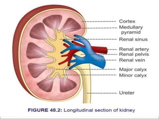
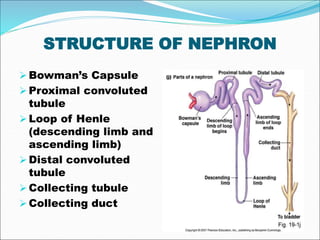
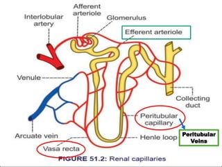
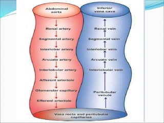
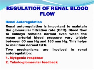
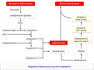
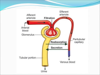
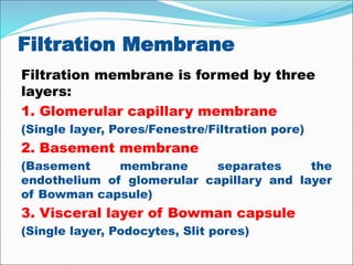
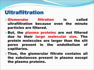
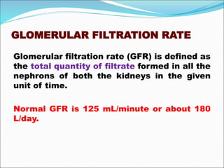
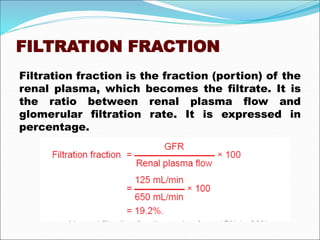
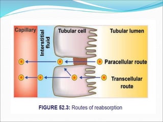
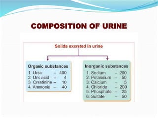
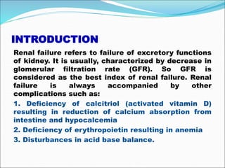
Juxtaglomerular apparatus (The Guyton and Hall physiology)Maryam Fida
JUXTAGLOMERULAR APPARATUS
Juxtaglomerular apparatus is a specialized organ situated near the glomerulus of each nephron (juxta = near).
1..MACULA DENSA
Macula densa is the end portion of thick ascending segment. It is situated between afferent and efferent arterioles of the same nephron. It is very close to afferent arteriole.
Macula densa is formed by tightly packed cuboidal epithelial cells.
2..EXTRAGLOMERULAR MESANGIAL CELLS
Extraglomerular mesangial cells are situated in the triangular region bound by afferent arteriole, efferent arteriole and macula densa. These cells are also called agranular cells, lacis cells or Goormaghtigh cells.
3. Glomerular Mesangial Cell
The basics of autoregulation of Gloemrular filtration rate. This ppt deals with basic renal physiology, tubuloglomerular feedback, myogenic reflex, juxtaglomerular apparatus and renin angiotensin aldosterone system in brief. P.S.- The ppt has animations so kindly view in slide/presentation mode
Cardiac myocytes are short
branched striated muscle cells
Connected with gap junctions
gap junctions transmit
electrical activity between cells
So, cardiac myocytes act as
a single functional unit
(syncitium)1. Rhythmicity
2. Excitability
3. Conductivity
4. Contractility
This presentation is all about Renal System and it's Physiological Processes with complete description.
This is a presentation file for medical students of all disciplines.
This presentation includes mechanism of excretion, ultra filteration, Reabsorption, secretion into kidney. Formation of urine. as well as introduction of osmoregulation and mechanism in aquatic fishes including fresh water fish, marine fish, eusturine fish and migratory fish.
KIDNEY IS A VITAL ORGAN IN HUMAN BEINGS. EVERY HUMAN HAS A PAIR OF KIDNEYS WHICH HELP TO EXCRETE OUT WASTE PRODUCTS FROM THE BODY IN THE FORM OF URINE...
URINE IS FORMED IN KIDNEY BY THREE STEPS WHICH ARE
(1) FILTRATION.
(2) ABSORPTION
(3) SECRETION
Kidney is a vital organ. Each individual has a pair of kidneys .
kidney is bean shaped organ on either side of your spine, below your ribs and behind your belly. Each kidney is about 4 - 5 inches long, roughly the size of a large fist.
The kidney job is to filter the blood.
kidney is reddish brown in color.
kidneys are also called as retro-peritoneal organ.
There are three layers of tissues that surrounds kidney
1. renal capsule
2. adipose capsule
3. renal fascia.
CNS Introduction, Neurons, Type of Neurons and functions, Neuroglia and types, Receptors and their types, Synapse, Neurotransmitters and their functions
These simplified slides by Dr. Sidra Arshad present an overview of the non-respiratory functions of the respiratory tract.
Learning objectives:
1. Enlist the non-respiratory functions of the respiratory tract
2. Briefly explain how these functions are carried out
3. Discuss the significance of dead space
4. Differentiate between minute ventilation and alveolar ventilation
5. Describe the cough and sneeze reflexes
Study Resources:
1. Chapter 39, Guyton and Hall Textbook of Medical Physiology, 14th edition
2. Chapter 34, Ganong’s Review of Medical Physiology, 26th edition
3. Chapter 17, Human Physiology by Lauralee Sherwood, 9th edition
4. Non-respiratory functions of the lungs https://academic.oup.com/bjaed/article/13/3/98/278874
micro teaching on communication m.sc nursing.pdfAnurag Sharma
Microteaching is a unique model of practice teaching. It is a viable instrument for the. desired change in the teaching behavior or the behavior potential which, in specified types of real. classroom situations, tends to facilitate the achievement of specified types of objectives.
Title: Sense of Taste
Presenter: Dr. Faiza, Assistant Professor of Physiology
Qualifications:
MBBS (Best Graduate, AIMC Lahore)
FCPS Physiology
ICMT, CHPE, DHPE (STMU)
MPH (GC University, Faisalabad)
MBA (Virtual University of Pakistan)
Learning Objectives:
Describe the structure and function of taste buds.
Describe the relationship between the taste threshold and taste index of common substances.
Explain the chemical basis and signal transduction of taste perception for each type of primary taste sensation.
Recognize different abnormalities of taste perception and their causes.
Key Topics:
Significance of Taste Sensation:
Differentiation between pleasant and harmful food
Influence on behavior
Selection of food based on metabolic needs
Receptors of Taste:
Taste buds on the tongue
Influence of sense of smell, texture of food, and pain stimulation (e.g., by pepper)
Primary and Secondary Taste Sensations:
Primary taste sensations: Sweet, Sour, Salty, Bitter, Umami
Chemical basis and signal transduction mechanisms for each taste
Taste Threshold and Index:
Taste threshold values for Sweet (sucrose), Salty (NaCl), Sour (HCl), and Bitter (Quinine)
Taste index relationship: Inversely proportional to taste threshold
Taste Blindness:
Inability to taste certain substances, particularly thiourea compounds
Example: Phenylthiocarbamide
Structure and Function of Taste Buds:
Composition: Epithelial cells, Sustentacular/Supporting cells, Taste cells, Basal cells
Features: Taste pores, Taste hairs/microvilli, and Taste nerve fibers
Location of Taste Buds:
Found in papillae of the tongue (Fungiform, Circumvallate, Foliate)
Also present on the palate, tonsillar pillars, epiglottis, and proximal esophagus
Mechanism of Taste Stimulation:
Interaction of taste substances with receptors on microvilli
Signal transduction pathways for Umami, Sweet, Bitter, Sour, and Salty tastes
Taste Sensitivity and Adaptation:
Decrease in sensitivity with age
Rapid adaptation of taste sensation
Role of Saliva in Taste:
Dissolution of tastants to reach receptors
Washing away the stimulus
Taste Preferences and Aversions:
Mechanisms behind taste preference and aversion
Influence of receptors and neural pathways
Impact of Sensory Nerve Damage:
Degeneration of taste buds if the sensory nerve fiber is cut
Abnormalities of Taste Detection:
Conditions: Ageusia, Hypogeusia, Dysgeusia (parageusia)
Causes: Nerve damage, neurological disorders, infections, poor oral hygiene, adverse drug effects, deficiencies, aging, tobacco use, altered neurotransmitter levels
Neurotransmitters and Taste Threshold:
Effects of serotonin (5-HT) and norepinephrine (NE) on taste sensitivity
Supertasters:
25% of the population with heightened sensitivity to taste, especially bitterness
Increased number of fungiform papillae
Ozempic: Preoperative Management of Patients on GLP-1 Receptor Agonists Saeid Safari
Preoperative Management of Patients on GLP-1 Receptor Agonists like Ozempic and Semiglutide
ASA GUIDELINE
NYSORA Guideline
2 Case Reports of Gastric Ultrasound
New Drug Discovery and Development .....NEHA GUPTA
The "New Drug Discovery and Development" process involves the identification, design, testing, and manufacturing of novel pharmaceutical compounds with the aim of introducing new and improved treatments for various medical conditions. This comprehensive endeavor encompasses various stages, including target identification, preclinical studies, clinical trials, regulatory approval, and post-market surveillance. It involves multidisciplinary collaboration among scientists, researchers, clinicians, regulatory experts, and pharmaceutical companies to bring innovative therapies to market and address unmet medical needs.
TEST BANK for Operations Management, 14th Edition by William J. Stevenson, Ve...kevinkariuki227
TEST BANK for Operations Management, 14th Edition by William J. Stevenson, Verified Chapters 1 - 19, Complete Newest Version.pdf
ARTIFICIAL INTELLIGENCE IN HEALTHCARE.pdfAnujkumaranit
Artificial intelligence (AI) refers to the simulation of human intelligence processes by machines, especially computer systems. It encompasses tasks such as learning, reasoning, problem-solving, perception, and language understanding. AI technologies are revolutionizing various fields, from healthcare to finance, by enabling machines to perform tasks that typically require human intelligence.
NVBDCP.pptx Nation vector borne disease control programSapna Thakur
NVBDCP was launched in 2003-2004 . Vector-Borne Disease: Disease that results from an infection transmitted to humans and other animals by blood-feeding arthropods, such as mosquitoes, ticks, and fleas. Examples of vector-borne diseases include Dengue fever, West Nile Virus, Lyme disease, and malaria.
Tom Selleck Health: A Comprehensive Look at the Iconic Actor’s Wellness Journeygreendigital
Tom Selleck, an enduring figure in Hollywood. has captivated audiences for decades with his rugged charm, iconic moustache. and memorable roles in television and film. From his breakout role as Thomas Magnum in Magnum P.I. to his current portrayal of Frank Reagan in Blue Bloods. Selleck's career has spanned over 50 years. But beyond his professional achievements. fans have often been curious about Tom Selleck Health. especially as he has aged in the public eye.
Follow us on: Pinterest
Introduction
Many have been interested in Tom Selleck health. not only because of his enduring presence on screen but also because of the challenges. and lifestyle choices he has faced and made over the years. This article delves into the various aspects of Tom Selleck health. exploring his fitness regimen, diet, mental health. and the challenges he has encountered as he ages. We'll look at how he maintains his well-being. the health issues he has faced, and his approach to ageing .
Early Life and Career
Childhood and Athletic Beginnings
Tom Selleck was born on January 29, 1945, in Detroit, Michigan, and grew up in Sherman Oaks, California. From an early age, he was involved in sports, particularly basketball. which played a significant role in his physical development. His athletic pursuits continued into college. where he attended the University of Southern California (USC) on a basketball scholarship. This early involvement in sports laid a strong foundation for his physical health and disciplined lifestyle.
Transition to Acting
Selleck's transition from an athlete to an actor came with its physical demands. His first significant role in "Magnum P.I." required him to perform various stunts and maintain a fit appearance. This role, which he played from 1980 to 1988. necessitated a rigorous fitness routine to meet the show's demands. setting the stage for his long-term commitment to health and wellness.
Fitness Regimen
Workout Routine
Tom Selleck health and fitness regimen has evolved. adapting to his changing roles and age. During his "Magnum, P.I." days. Selleck's workouts were intense and focused on building and maintaining muscle mass. His routine included weightlifting, cardiovascular exercises. and specific training for the stunts he performed on the show.
Selleck adjusted his fitness routine as he aged to suit his body's needs. Today, his workouts focus on maintaining flexibility, strength, and cardiovascular health. He incorporates low-impact exercises such as swimming, walking, and light weightlifting. This balanced approach helps him stay fit without putting undue strain on his joints and muscles.
Importance of Flexibility and Mobility
In recent years, Selleck has emphasized the importance of flexibility and mobility in his fitness regimen. Understanding the natural decline in muscle mass and joint flexibility with age. he includes stretching and yoga in his routine. These practices help prevent injuries, improve posture, and maintain mobilit
- Video recording of this lecture in English language: https://youtu.be/lK81BzxMqdo
- Video recording of this lecture in Arabic language: https://youtu.be/Ve4P0COk9OI
- Link to download the book free: https://nephrotube.blogspot.com/p/nephrotube-nephrology-books.html
- Link to NephroTube website: www.NephroTube.com
- Link to NephroTube social media accounts: https://nephrotube.blogspot.com/p/join-nephrotube-on-social-media.html
2. INTRODUCTION
Excretion is the process by which the unwanted
substances and metabolic wastes are
eliminated from the body.
1. Digestive system excretes food residues in
the form of feces. Some bacteria and toxic
substances also are excreted through feces
2. Lungs remove carbon dioxide and water vapor
3. Skin excretes water, salts and some wastes.
It also removes heat from the body
4. Liver excretes many substances like bile
pigments, heavy metals, drugs, toxins, bacteria,
etc. through bile.
3. FUNCTIONS OF KIDNEY
Excretion of Waste Products
Maintenance of Water Balance
Maintenance of Electrolyte Balance
Maintenance of Acid–Base Balance
Hemopoietic Function
Regulation of Blood Calcium Level
4. KIDNEY
Two kidneys in all mammals located
retroperitoneally at the level of lower ribs
Protected by the 11th and 12th ribs from the
injury
Left one is a bit on the anterior side as
compared to the right
Outer darker region is cortex, Inner lighter
region is the medulla with Calyces (major and
minor), Renal pyramids, Renal pelvis and
Blood vessels
5.
6. NEPHRONS
Nephron is the structural and functional
unit of the kidney
2,400,000 nephrons collectively in both
kidneys
Basically composed of:
Renal corpuscle (in whish fluid is filtered)
A long tubule (converts the filtrate into
urine on its way to the renal pelvis)
7. TYPES OF NEPHRON
1. Cortical nephrons or superficial nephrons:
Nephrons having the corpuscles in outer cortex
of the kidney near the periphery. In human
kidneys, 85% nephrons are cortical nephrons.
2. Juxtamedullary nephrons: Nephrons having
the corpuscles in inner cortex near medulla or
corticomedullary junction.
8. STRUCTURE OF NEPHRON
Bowman’s Capsule
Proximal convoluted
tubule
Loop of Henle
(descending limb and
ascending limb)
Distal convoluted
tubule
Collecting tubule
Collecting duct
9.
10. RENAL CORPUSCLE
Glomerulus
Glomerulus is a tuft of capillaries enclosed by
Bowman capsule. It consists of glomerular
capillaries interposed between afferent arteriole on
one end and efferent arteriole on the other end.
Thus, the vascular system in the glomerulus is
purely arterial.
Bowman Capsule
Bowman capsule is a capsular structure, which
encloses the glomerulus. It is formed by two layers:
i. Inner visceral layer, ii. Outer parietal layer.
Functional anatomy of Bowman capsule resembles
a funnel with filter paper.
11. TUBULAR PORTION OF NEPHRON
PROXIMAL CONVOLUTED TUBULE
Proximal convoluted tubule is the coiled portion
arising from Bowman capsule. It is situated in the
cortex. It is continued as descending limb of loop of
Henle. Length of proximal convoluted tubule is 14
mm and the diameter is 55 μ.
LOOP OF HENLE
Loop of Henle consists of:
i. Descending limb
ii. Hairpin bend
iii. Ascending limb.
12. Descending Limb
Descending limb of loop of Henle is made up of two
segments:
Thick descending segment is the direct
continuation of the proximal convoluted tubule. It
descends down into medulla. It has a length of 6
mm and a diameter of 55 μ.
Thick descending segment is continued as thin
descending segment. It is continued as hairpin bend
of the loop.
Hairpin Bend
Hairpin bend formed by flattened epithelial cells
without brush border and it is continued as the
ascending limb of loop of Henle.
13. Ascending Limb
Ascending limb or segment of Henle loop has two
parts:
Thin ascending segment is continued as thick
ascending segment.
Thick ascending segment is about 9 mm long with a
diameter of 30 μ. The terminal portion of thick
ascending segment, which runs between the
afferent and efferent arterioles of the same
nephrons forms the macula densa. Macula densa is
the part of juxtaglomerular apparatus. Thick
ascending segment ascends to the cortex and
continues as distal convoluted tubule.
14. Length and the extent of the loop of Henle vary
in different nephrons:
In cortical nephrons, it is short and the hairpin
bend penetrates only up to outer medulla.
In juxtamedullary nephrons, this is long and the
hairpin bend extends deep into the inner
medulla.
15. DISTAL CONVOLUTED TUBULE
Distal convoluted tubule is the continuation of
thick ascending segment and occupies the cortex
of kidney. It is continued as collecting duct. The
length of the distal convoluted tubule is 14.5 to 15
mm. It has a diameter of 22 to 50 μ.
COLLECTING DUCT
Collecting duct is formed by two types of
epithelial cells: 1. Principal or P cells, 2.
Intercalated or I cells. Distal convoluted tubule
continues as the initial or arched collecting duct,
which is in cortex. The lower part of the
collecting duct lies in medulla. Seven to ten initial
collecting ducts unite to form the straight
collecting duct, which passes through medulla.
19. REGULATION OF RENAL BLOOD
FLOW
Renal Autoregulation
Renal autoregulation is important to maintain
the glomerular filtration rate (GFR). Blood flow
to kidneys remains normal even when the
mean arterial blood pressure vary widely
between 60 mm Hg and 180 mm Hg. This helps
to maintain normal GFR.
Two mechanisms are involved in renal
autoregulation:
1. Myogenic response
2. Tubulo-glomerular feedback
22. 1. Renin
Juxtaglomerular cells secrete renin. Renin is a
peptide with 340 amino acids. Along with
angiotensins, renin forms the renin-angiotensin
system, which is a hormone system that plays an
important role in the maintenance of blood
pressure.
Stimulants for renin secretion
Secretion of renin is stimulated by four factors:
i. Fall in arterial blood pressure
ii. Reduction in the ECF volume
iii. Increased sympathetic activity
iv. Decreased load of sodium and chloride in
macula densa.
23. Actions of Angiotensins
Angiotensin I is physiologically inactive and
serves only as the precursor of angiotensin II.
Angiotensin II is the most active form. Its
actions are:
Angiotensin II increases arterial blood
pressure by directly acting on the blood
vessels and causing vasoconstriction. It is a
potent constrictor of arterioles.
It increases blood pressure indirectly by
increasing the release of noradrenaline from
postganglionic sympathetic fibers.
Noradrenaline is a general vasoconstrictor.
24.
25.
26. INTRODUCTION
Urine formation is a blood cleansing
function. Kidneys excrete the unwanted
substances along with water from the blood
as urine. Normal urinary output is 1 L/day to
1.5 L/day.
A. Glomerular filtration
B. Tubular reabsorption
C. Tubular secretion
27.
28.
29. GLOMERULAR FILTRATION
Glomerular filtration is the process by
which the blood is filtered while passing
through the glomerular capillaries by
filtration membrane.
It is the first process of urine formation.
The structure of filtration membrane is
well suited for filtration.
30. Filtration Membrane
Filtration membrane is formed by three
layers:
1. Glomerular capillary membrane
(Single layer, Pores/Fenestre/Filtration pore)
2. Basement membrane
(Basement membrane separates the
endothelium of glomerular capillary and layer
of Bowman capsule)
3. Visceral layer of Bowman capsule
(Single layer, Podocytes, Slit pores)
31. Ultrafiltration
Glomerular filtration is called
ultrafiltration because even the minute
particles are filtered.
But, the plasma proteins are not filtered
due to their large molecular size. The
protein molecules are larger than the slit
pores present in the endothelium of
capillaries.
Thus, the glomerular filtrate contains all
the substances present in plasma except
the plasma proteins.
32. GLOMERULAR FILTRATION RATE
Glomerular filtration rate (GFR) is defined as
the total quantity of filtrate formed in all the
nephrons of both the kidneys in the given
unit of time.
Normal GFR is 125 mL/minute or about 180
L/day.
33. FILTRATION FRACTION
Filtration fraction is the fraction (portion) of the
renal plasma, which becomes the filtrate. It is
the ratio between renal plasma flow and
glomerular filtration rate. It is expressed in
percentage.
34. PRESSURES DETERMINING
FILTRATION
Pressures, which determine the GFR are:
1. Glomerular capillary pressure
2. Colloidal osmotic pressure in the
glomeruli
3. Hydrostatic pressure in the Bowman
capsule
35. Net Filtration Pressure
Net filtration pressure is the balance between
pressure favoring filtration and pressures
opposing filtration. It is otherwise known as
effective filtration pressure or essential
filtration pressure.
36. Starling Hypothesis
Starling hypothesis states that the net
filtration through capillary membrane is
proportional to hydrostatic pressure
difference across the membrane minus
oncotic pressure difference.
37. FACTORS REGULATING GFR
1. Renal Blood Flow
2. Tubulo-glomerular Feedback
3. Glomerular Capillary Pressure
4. Colloidal Osmotic Pressure
5. Hydrostatic Pressure in Bowman Capsule
6. Constriction of Afferent Arteriole
7. Constriction of Efferent Arteriole
8. Systemic Arterial Pressure
9. Sympathetic Stimulation
10. Surface Area of Capillary Membrane
11. Permeability of Capillary Membrane
12. Hormonal and Other Factors
38.
39. INTRODUCTION
Tubular reabsorption is the process by
which water and other substances are
transported from renal tubules back to
the blood.
Large quantity of water (more than
99%), electrolytes and other
substances are reabsorbed by the
tubular epithelial cells.
40. ROUTES OF REABSORPTION
Reabsorption of substances from tubular
lumen into the peritubular capillary
occurs by two routes:
1. Transcelluar route
2. Paracellular route
41.
42. SITE OF REABSORPTION
1.Substances Reabsorbed from Proximal Convoluted
Tubule
Substances reabsorbed from proximal convoluted
tubule are glucose, amino acids, sodium, potassium,
calcium, bicarbonates, chlorides, phosphates, urea,
uric acid and water.
2. Substances Reabsorbed from Loop of Henle
Substances reabsorbed from loop of Henle are
sodium and chloride.
3. Substances Reabsorbed from Distal Convoluted
Tubule
Sodium, calcium, bicarbonate and water are
reabsorbed from distal convoluted tubule.
46. INTRODUCTION
Tubular secretion is the process by which the
substances are transported from blood into
renal tubules. It is also called tubular excretion.
Such substances are:
1. Paraaminohippuric acid (PAH)
2. Diodrast
3. 5-hydroxyindoleacetic acid (5HIAA)
4. Amino derivatives
5. Penicillin
47. SUMMARY OF URINE FORMATION
1. Glomerular filtration
Plasma is filtered in glomeruli and the
substances reach the renal tubules along with
water as filtrate.
2. Tubular Reabsorption
The 99% of filtrate is reabsorbed in different
segments of renal tubules.
3. Tubular Secretion
Some substances are transported from blood
into the renal tubule.
48.
49. INTRODUCTION
Every day 180 L of glomerular filtrate is formed
with large quantity of water. Osmolarity of
glomerular filtrate is same as that of plasma
and it is 300 mOsm/L. But, normally urine is
concentrated and its osmolarity is four times
more than that of plasma, i.e. 1,200 mOsm/L.
Osmolarity of urine depends upon two factors:
1. Water content in the body
2. Antidiuretic hormone (ADH)
50. FORMATION OF DILUTE URINE
When, water content in the body increases,
kidney excretes dilute urine.
This is achieved by inhibition of ADH
secretion from posterior pituitary.
So water reabsorption from renal tubules does
not take place leading to excretion of large
amount of water. This makes the urine dilute.
51. FORMATION OF CONCENTRATED
URINE
When the water content in body decreases,
kidney retains water and excretes
concentrated urine. Formation of
concentrated urine is not as simple as that
of dilute urine.
It involves two processes:
1. Development and maintenance of
medullary gradient by countercurrent
system
2. Secretion of ADH
52. MEDULLARY GRADIENT
Divisions of Countercurrent System
Countercurrent system has two
divisions:
1. Countercurrent multiplier formed by
loop of Henle
2. Countercurrent exchanger formed by
vasa recta
54. ROLE OF ADH
Final concentration of urine is achieved by
the action of ADH.
Normally, the distal convoluted tubule and
collecting duct are not permeable to
water.
But the presence of ADH makes them
permeable, resulting in water
reabsorption.
Water reabsorption induced by ADH is
called facultative reabsorption of water
55. SUMMARY OF URINE
CONCENTRATION
1. BOWMAN CAPSULE
Glomerular filtrate collected at the Bowman
capsule is isotonic to plasma. This is because it
contains all the substances of plasma except
proteins.
Osmolarity of the filtrate at Bowman capsule is
300 mOsm/L.
56. 2. PROXIMAL CONVOLUTED TUBULE
When the filtrate flows through proximal
convoluted tubule, there is active reabsorption
of sodium and chloride followed by obligatory
reabsorption of water.
57. 3. THICK DESCENDING SEGMENT
Water is reabsorbed from tubule into outer
medullary interstitium by means of osmosis. It
is due to the increased osmolarity in the
medullary interstitium, i.e. outside the thick
descending tubule.
The osmolarity of the fluid inside this segment
is between 450 and 600 mOsm/L. That means
the fluid is slightly hypertonic to plasma.
58. 4. THIN DESCENDING SEGMENT OF
HENLE LOOP
In the short loops of cortical nephrons, the
osmolarity of fluid at the hairpin bend of loop
becomes 600 mOsm/L. And, in the long loops of
juxtamedullary nephrons, at the hairpin bend,
the osmolarity is 1,200 mOsm/L. Thus in this
segment the fluid is hypertonic to plasma.
59. 5. THIN ASCENDING SEGMENT OF
HENLE LOOP
When the thin ascending segment of the loop
ascends upwards through the medullary region,
osmolarity decreases gradually. Due to
concentration gradient, sodium chloride
diffuses out of tubular fluid and osmolarity
decreases to 400 mOsm/L. The fluid in this
segment is slightly hypertonic to plasma.
60. 6. THICK ASCENDING SEGMENT
This segment is impermeable to water. But
there is active reabsorption of sodium and
chloride from this. Reabsorption of sodium
decreases the osmolarity of tubular fluid to a
greater extent. The osmolarity is between 150
and 200 mOsm/L. The fluid inside becomes
hypotonic to plasma.
61. 7. DISTAL CONVOLUTED TUBULE AND
COLLECTING DUCT
In the presence of ADH, distal convoluted
tubule and collecting duct become permeable
to water resulting in water reabsorption and
final concentration of urine. It is found that in
the collecting duct, Principal (P) cells are
responsible for ADH induced water
reabsorption. Reabsorption of large quantity of
water increases the osmolarity to 1,200
mOsm/L. The urine becomes hypertonic to
plasma.
62.
63. INTRODUCTION
Micturition is a process by which urine is
voided from the urinary bladder. It is a reflex
process. However, in grown up children and
adults, it can be controlled voluntarily to some
extent.
64. URINARY BLADDER & URETHRA
Urinary bladder is a triangular hollow organ
located in lower abdomen. It consists of a body
and neck. Wall of the bladder is formed by
smooth muscle. It consists of three ill-defined
layers of muscle fibers called detrusor muscle,
viz. the inner longitudinal layer, middle circular
layer and outer longitudinal layer. Bladder is
opened in urethra from where urine is excreted
out from the body.
65. URETHRAL SPHINCTERS
There are two urethral sphincters in
urinary tract:
1. Internal urethral sphincter
2. External urethral sphincter.
70. PROPERTIES OF URINE
Volume : 1,000 to 1,500 mL/day
Reaction : Slightly acidic with pH of 4.5 to 6
Specific gravity : 1.010 to 1.025
Osmolarity : 1,200 mOsm/L
Color : Normally, straw colored
Odor : Fresh urine has light aromatic odor.
72. EXAMINATION OF URINE – URINEANALYSIS
Urine analysis is done by:
i. Physical examination
ii. Microscopic examination
iii. Chemical analysis
73.
74. INTRODUCTION
Renal failure refers to failure of excretory functions
of kidney. It is usually, characterized by decrease in
glomerular filtration rate (GFR). So GFR is
considered as the best index of renal failure. Renal
failure is always accompanied by other
complications such as:
1. Deficiency of calcitriol (activated vitamin D)
resulting in reduction of calcium absorption from
intestine and hypocalcemia
2. Deficiency of erythropoietin resulting in anemia
3. Disturbances in acid base balance.
75. ACUTE RENAL FAILURE
Acute renal failure is the abrupt or sudden
stoppage of renal functions.
It is often reversible within few days to few
weeks.
Acute renal failure may result in sudden life-
threatening reactions in the body with the
need for emergency treatment.
76. CAUSES
1. Acute nephritis
2. Damage of renal tissues by poisons like lead,
mercury
3. Renal ischemia, which develops during
circulatory shock
4. Acute tubular necrosis
5. Severe transfusion reactions
6. Sudden fall in blood pressure during
hemorrhage, diarrhea, severe burns and cholera
7. Blockage of ureter due to the formation of
calculi (renal stone) or tumor.
77. FEATURES
1. Oliguria (decreased urinary output)
2. Anuria (cessation of urine formation) in severe cases
3. Proteinuria (appearance of proteins in urine) including
albuminuria (excretion of albumin in urine)
4. Hematuria (presence of blood in urine)
5. Edema due to increased volume of extracellular fluid
(ECF) caused by retention of sodium and water
6. Hypertension within few days because of increased
ECF volume
7. Acidosis due to the retention of metabolic end
products
8. Coma due to severe acidosis (if the patient is not
treated in time) resulting in death within 10 to 14 days.
80. INTRODUCTION
Diuretics or diuretic agents are the
substances which enhance the urine
formation and output.
These substances increase the excretion of
water, sodium and chloride through urine.
Diuretic agents increase the urine formation,
by influencing any of the processes involved
in urine formation.
Diuretics are commonly called ‘water pills’.
81. GENERAL USES OF DIURETICS
1. Hypertension
2. Congestive cardiac failure
3. Edema
83. TYPES OF DIURETICS
1. Osmotic diuretics (Mannitol)
2. Diuretics which inhibit active reabsorption of
electrolytes
Loop, (Thick ascending loop of henle)
Thiazide, (Proximal part of DCT)
K sparing diuretics, (Distal part of DCT)
84. 3. Diuretics which inhibit action of aldosterone
(Aldosterone Antagonist)
4. Diuretics which inhibit activity of carbonic
anhydrase (Acetazolamide)
5. Diuretics which increase glomerular filtration
rate (Xanthine Derivative)
6. Diuretics which inhibit secretion of ADH
7. Diuretics which inhibit ADH receptors
87. DIURETICS WHICH INHIBIT ACTIVE
REABSORPTION OF ELECTROLYTES
Loop Diuretics
i. Furosemide
ii. Torasemide
iii. Bumetanide
Thiazide Diuretics
i. Chlorothiazide
ii. Metolazone
iii. Chlortalidone
K Sparing Diuretics
i. Triamterene
ii. Amiloride
88. DIURETICS WHICH INHIBIT
ACTION OF ALDOSTERONE
These substances are also called the
potassium retaining diuretics or aldosterone
antagonists.
Examples
i. Spironolactone
ii. Eperenone
89. DIURETICS WHICH INHIBIT ACTIVITY
OF CARBONIC ANHYDRASE
Acetazolamide is a carbonic anhydrase
inhibitor.
DIURETICS WHICH INCREASE
GLOMERULAR FILTRATION RATE
i. Caffeine
ii. Theophylline
92. DIALYSIS
Dialysis is the procedure to remove waste
materials and toxic substances and to
restore normal volume and composition of
body fluid in severe renal failure.
It is also called hemodialysis.
93. ARTIFICIAL KIDNEY
Artificial kidney is the machine that is used to
carry out dialysis during renal failure. It is used
to treat the patients suffering from:
1. Acute renal failure
2. Chronic or permanent renal failure.
94. DIALYSATE
The concentration of various substances in
the dialysate is adjusted in accordance with
the needs of the patient’s body.
The fluid does not contain urea, urate, sulfate,
phosphate or creatinine, so that, these
substances move from the blood to the
dialysate.
The fluid has low concentration of sodium,
potassium and chloride ions than in the
uremic blood. But the concentration of
glucose, bicarbonate and calcium ions is more
in the dialysate than in the uremic blood.
96. HEMODIALYSIS
With haemodialysis, the blood is drawn
and cleaned outside of the body.
A machine called a dialyzer in connected
through a port in an artery and vein in a
person’s arm.
The machine draws the blood out, cleans
and balances its components inside the
machine, and cycles it back into the body
it came from.
97. For this method of dialysis, the patient to
be sitting still or lying down throughout
the entire procedure 3-5 times a week.
After dialysis, patients often feel tired and
have low blood pressure.
98. PERITONEAL DIALYSIS
Peritoneal dialysis is a type of dialysis that
occurs inside the body. More specifically,
inside the abdomen.
After a catheter is placed into the lining of the
abdominal wall (also known as the
peritoneum) blood can be filtered internally.
The patient fills the access point with a fluid
called dialysate filter. The dialysate filter fluid
then cleans and balances the blood
components through the internal abdominal
walls and once complete, drains into a bag.
99.
100.
101. INTRODUCTION
Skin is the largest organ of the body. The
average thickness of the skin is about 1
to 2 mm.
Skin is made up of two layers:
I. Outer epidermis
II. Inner dermis.
102. EPIDERMIS
Epidermis is the outer layer of skin. It
is formed by stratified epithelium.
Important feature of epidermis is that,
it does not have blood vessels.
103. Layers of Epidermis
Epidermis is formed by five layers:
1. Stratum corneum/ Horney Layer
(composed of corneocytes; i.e. dead cells,
and contain keratin, phospholipid and
glycogen)
2. Stratum lucidum
(flattened epithelial cells with
homogeneous translucent zone)
104. 3. Stratum granulosum
(Thin layer of flattened rhomboid cells.
Cytoplasm contains granules of a protein called
keratohyalin)
4. Stratum spinosum/ Prickle cell layer
(Spinelike protoplasmic projections)
5. Stratum germinativum
(Thick layer made up of polygonal cells. New
cells are constantly formed by mitotic division.
The stem cells, which give rise to new cells,
are known as keratinocytes. Melanocytes
(present b/w keratinocyte) produce the pigment
called melanin)
105. DERMIS
Dermis is the inner layer of the skin. It is a
connective tissue layer, made up of dense and
stout collagen fibers, fibroblasts and
histiocytes. Collagen fibers contain the
enzyme collagenase, which is responsible for
wound healing.
Dermis is made up of two layers:
1. Superficial papillary layer
2. Deeper reticular layer
106. SUPERFICIAL PAPILLARY LAYER
It contains blood vessels, lymphatics and nerve
fibers. This layer also has some pigment
containing cells known as chromatophores.
RETICULAR LAYER
These fibers are found around the hair bulbs,
sweat glands and sebaceous glands. The
reticular layer also contains mast cells, nerve
endings, lymphatics, epidermal appendages
and fibroblasts.
107. COLOR OF SKIN
Color of skin depends upon two
important factors:
1. Pigmentation of skin
2. Hemoglobin in the blood
108. PIGMENTATION OF SKIN
Cells of the skin contain a brown pigment
called melanin, which is responsible for the
color of the skin. It is synthesized by
melanocytes.
Skin becomes dark when melanin content
increases. It is protein in nature and it is
synthesized from the amino acid tyrosine via
dihydroxyphenylalanine (DOPA).
Deficiency of melanin leads to albinism
(hypopigmentary congenital disorder).
109. HEMOGLOBIN IN THE BLOOD
Amount and nature of hemoglobin that
circulates in the cutaneous blood
vessels play an important role in the
coloration of the skin.
110. FUNCTIONS OF SKIN
PROTECTIVE FUNCTION
SENSORY FUNCTION
STORAGE FUNCTION
SYNTHETIC FUNCTION
REGULATION OF BODY TEMPERATURE
REGULATION OF WATER AND ELECTROLYTE
BALANCE
EXCRETORY FUNCTION
ABSORPTIVE FUNCTION
SECRETORY FUNCTION
111. GLANDS OF SKIN
1. SEBACEOUS GLANDS
Sebaceous glands are ovoid or spherical in shape
and are situated at the side of the hair follicle.
Sebaceous glands secrete an oily substance
called sebum. develop from hair follicles (Dermis
layer).
Sebum contains Free fatty acids, Triglycerides,
Squalene, Sterols, Waxes, Paraffin.
Free fatty acid content of the sebum has
antibacterial and antifungal actions. Lipid nature
of sebum keeps the skin smooth and oily. Lipids of
the sebum prevent heat loss from the body. It is
particularly useful in cold climate.
112. 2. SWEAT GLANDS
Sweat glands are of two types:
1. Eccrine glands
2. Apocrine glands
113.
114. BODY TEMPERATURE
Body temperature can be measured by placing
the clinical thermometer in different parts of
the body such as:
1. Mouth (oral temperature)
2. Axilla (axillary temperature)
3. Rectum (rectal temperature)
4. Over the skin (surface temperature)
118. HYPERTHERMIA
Elevation of body temperature above the set
point is called hyperthermia, fever or pyrexia.
Fever is classified into three categories:
1. Low-grade fever: When the body temperature
rises to 38°C to 39°C, (100.4°F to 102.2°F)
2. Moderate-grade fever: When the temperature
rises to 39°C to 40°C (102.2°F to 104°F)
3. High-grade fever: When the temperature rise
above 40°C to 42°C (104°F to 107.6°F).
4. Hyperpyrexia is the rise in body temperature
beyond 42°C (107.6°F).
120. Signs and Symptoms
1. Headache
2. Sweating
3. Shivering
4. Muscle pain
5. Dehydration
6. Loss of appetite
7. General weakness.
Hyperpyrexia may result in:
1. Confusion
2. Hallucinations
3. Irritability
4. Convulsions.
121. HYPOTHERMIA
Decrease in body temperature below 35°C (95°F) is
called hypothermia. When the temperature drops
below 31°C (87.8°F), it becomes fatal.
Hypothermia is classified into three categories:
1. Mild hypothermia: When the body temperature
falls to 35°C to 33°C (95°F to 91.4°F)
2. Moderate hypothermia: When the body
temperature falls to 33°C to 31°C (91.4°F to 87.8°F)
3. Severe hypothermia: When the body temperature
falls below 31° C (87.8°F).
122. Causes of Hypothermia
1. Exposure to cold temperatures
2. Immersion in cold water
3. Drug abuse
4. Hypothyroidism
5. Hypopituitarism
6. Lesion in hypothalamus
7. Hemorrhage in certain parts of the
brainstem, particularly pons.