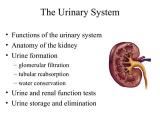
1. The Urinary System
• Functions of the urinary system
• Anatomy of the kidney
• Urine formation
– glomerular filtration
– tubular reabsorption
– water conservation
• Urine and renal function tests
• Urine storage and elimination
5. Excretion
• Separation of wastes from body fluids and
eliminating them
– respiratory system: CO2
– integumentary system: water, salts, lactic acid, urea
– digestive system: water, salts, CO2, lipids, bile
pigments, cholesterol
– urinary system: many metabolic wastes, toxins, drugs,
hormones, salts, H+ and water
6. Anatomy of Kidney
• Position, weight and size
– retroperitoneal about 160 g each
– about size of a bar of soap (12x6x3 cm)
• Shape
– lateral surface - convex; medial - concave
• CT coverings
– renal fascia: binds to abdominal wall
– adipose capsule: cushions kidney
– renal capsule: encloses kidney like cellophane wrap
7. Anatomy of Kidney
• Renal cortex: outer 1 cm
• Renal medulla: renal columns, pyramids - papilla
• Lobe of kidney: pyramid and it’s overlying cortex
12. Renal Corpuscle
Glomerular filtrate collects in capsular
space, flows into renal tubule
13. Renal (Uriniferous) Tubule
• Proximal convoluted tubule
(PCT)
– longest, most coiled, simple
cuboidal with brush border
• Nephron loop - U shaped;
descending + ascending limbs
– thick segment (simple cuboidal)
initial part of descending limb
and part or all of ascending limb,
active transport of salts
– thin segment (simple squamous)
very water permeable
• Distal convoluted tubule (DCT)
– cuboidal, minimal microvilli
21. Glomerular Filtration Rate (GFR)
• Filtrate formed per minute
• Filtration coefficient (Kf) depends on permeability
and surface area of filtration barrier
• GFR = NFP x Kf ≈ 125 ml/min or 180 L/day
• 99% of filtrate reabsorbed, 1 to 2 L urine excreted
22. Effects of GFR Abnormalities
∀ ↑GFR, urine output rises → dehydration,
electrolyte depletion
∀ ↓GFR → wastes reabsorbed (azotemia possible)
• GFR controlled by adjusting glomerular blood
pressure
– Auto regulation
– sympathetic control
– hormonal mechanism: renin and angiotensin
24. Renal Autoregulation of GFR
∀ ↑ BP → constrict afferent
arteriole, dilate efferent
• ↓ BP → dilate afferent
arteriole, constrict efferent
• Stable for BP range of 80 to
170 mmHg (systolic)
• Cannot compensate for
extreme BP
26. Sympathetic Control of GFR
• Strenuous exercise or acute conditions (circulatory
shock) stimulate afferent arterioles to constrict
∀ ↓ GFR and urine production, redirecting blood
flow to heart, brain and skeletal muscles
30. Peritubular Capillaries
• Blood has unusually high COP here, and BHP is
only 8 mm Hg (or lower when constricted by
angiotensin II); this favors reabsorption
• Water absorbed by osmosis and carries other
solutes with it (solvent drag)
31. Proximal Convoluted Tubules (PCT)
• Reabsorbs 65% of GF to peritubular capillaries
• Great length, prominent microvilli and abundant
mitochondria for active transport
• Reabsorbs greater variety of chemicals than other
parts of nephron
– transcellular route - through epithelial cells of PCT
– paracellular route - between epithelial cells of PCT
• Transport maximum: when transport proteins of plasma
membrane are saturated; glucose > 220 mg/dL remains in
urine (glycosuria)
32.
33. Tubular Secretion of PCT
and Nephron Loop
• Waste removal
– urea, uric acid, bile salts, ammonia, catecholamines,
many drugs
• Acid-base balance
– secretion of hydrogen and bicarbonate ions regulates
pH of body fluids
• Primary function of nephron loop
– water conservation, also involved in electrolyte
reabsorption
34. DCT and Collecting Duct
• Effect of aldosterone
↓ BP causes angiotensin II formation
– angiotensin II stimulates adrenal cortex
– adrenal cortex secretes aldosterone
– aldosterone promotes Na+ reabsorption
– Na+ reabsorption promotes water reabsorption
– water reabsorption ↓ urine volume
– BP drops less rapidly
35. DCT and Collecting Duct 2
• Effect of atrial natriuretic factor (ANF)
↑ BP stimulates right atrium
– atrium secretes ANF
– ANF promotes Na+ and water excretion
– BP drops
• Effect of ADH
– dehydration stimulates hypothalamus
– hypothalamus stimulates posterior pituitary
– posterior pituitary releases ADH
– ADH ↑ water reabsorption
– urine volume ↓
36. Collecting Duct Concentrates Urine
• Osmolarity 4x as high
deep in medulla
• Medullary portion of
CD is permeable to
water but not to NaCl
37. Control of Water Loss
• Producing hypotonic urine
– NaCl reabsorbed by cortical CD
– water remains in urine
• Producing hypertonic urine
– GFR drops
– tubular reabsorption ↑
– less NaCl remains in CD
– ADH ↑ CD’s water permeability
– more water is reabsorbed
– urine is more concentrated
38. Countercurrent Multiplier
• Recaptures NaCl and returns it to renal medulla
• Descending limb
– reabsorbs water but not salt
– concentrates tubular fluid
• Ascending limb
– reabsorbs Na+, K+, and Cl-
– maintains high osmolarity of renal medulla
– impermeable to water
– tubular fluid becomes hypotonic
• Recycling of urea: collecting duct-medulla
– urea accounts for 40% of high osmolarity of medulla
40. Countercurrent Exchange System
• Formed by vasa recta
– provide blood supply to medulla
– do not remove NaCl from medulla
• Descending capillaries
– water diffuses out of blood
– NaCl diffuses into blood
• Ascending capillaries
– water diffuses into blood
– NaCl diffuses out of blood
43. Composition and Properties of Urine
• Appearance
– almost colorless to deep amber; yellow color due to
urochrome, from breakdown of hemoglobin (RBC’s)
• Odor - as it stands bacteria degrade urea to ammonia
• Specific gravity
– density of urine ranges from 1.000 -1.035
• Osmolarity - (blood - 300 mOsm/L) ranges from
50 mOsm/L to 1,200 mOsm/L in dehydrated person
• pH - range: 4.5 - 8.2, usually 6.0
• Chemical composition: 95% water, 5% solutes
– urea, NaCl, KCl, creatinine, uric acid
44. Urine Volume
• Normal volume - 1 to 2 L/day
• Polyuria > 2L/day
• Oliguria < 500 mL/day
• Anuria - 0 to 100 mL
45. Diuretics
• Effects
↑ urine output
↓ blood volume
• Uses
– hypertension and congestive heart failure
• Mechanisms of action
↑ GFR
↓ tubular reabsorption
46. Renal Function Tests
• Renal clearance: volume of blood plasma cleared
of a waste in 1 minute
• Determine renal clearance (C) by assessing blood
and urine samples: C = UV/P
– U (waste concentration in urine)
– V (rate of urine output)
– P (waste concentration in plasma)
• Determine GFR: inulin is neither reabsorbed or
secreted so for this solute GFR = renal clearance
GFR = UV/P