Downloaded 344 times
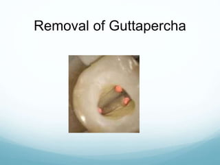
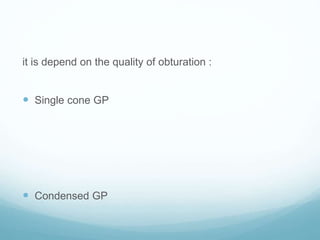
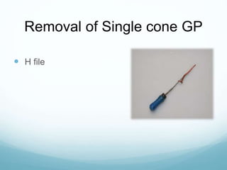
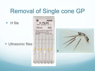
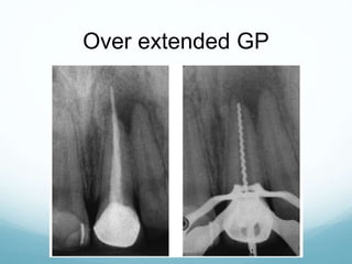
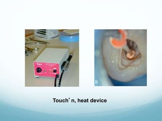
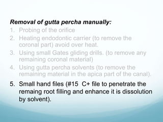
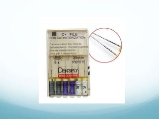
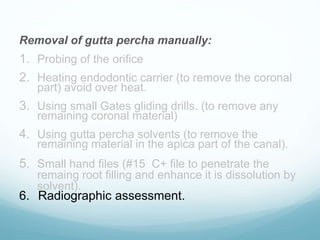
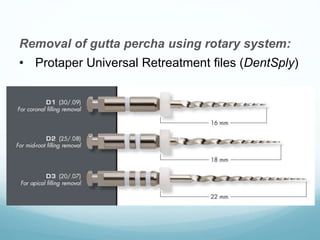
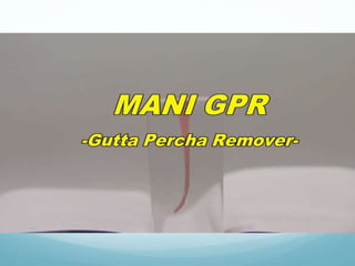
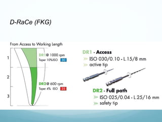
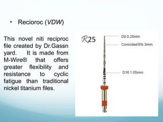
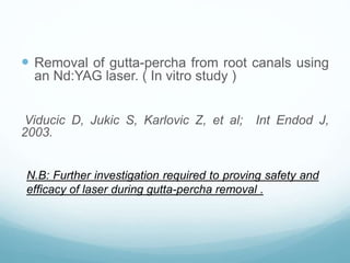
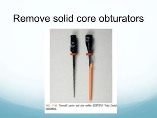
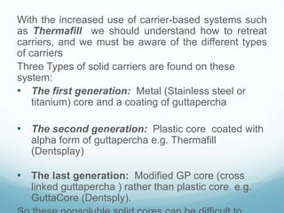
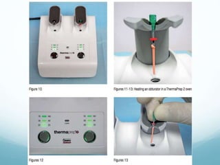
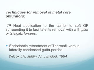
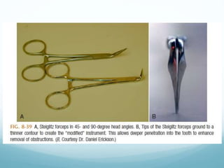
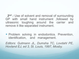
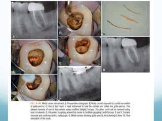
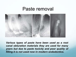
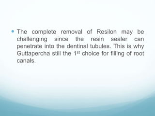
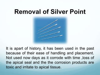
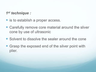
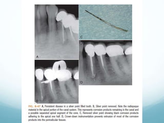
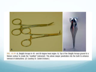
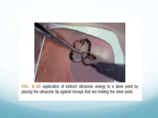
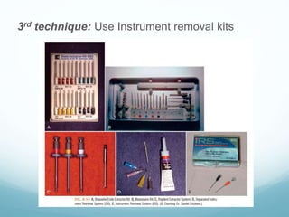

The document discusses various obturation materials and techniques for their removal during root canal retreatment. It describes the different types of gutta percha obturations including single cone, condensed, and overextended, and techniques for removing each using hand files, ultrasonic files, heat, and solvents. For other materials like solid core obturators, paste, Resilon, and silver points, it outlines techniques using rotary files, ultrasonics, solvents, and specialized removal kits. References are provided supporting the various methods described.


![ONFH[AVN HIP] -TRIPLE REGIME -A NOVAL SURGICAL CONCEPT .pptx](https://cdn.slidesharecdn.com/ss_thumbnails/onfhavnhip2026koaconcalicutdrgokuldevdrmashraf-260210064517-213ec005-thumbnail.jpg?width=640&height=640&fit=bounds)

![CTEV [ clubfoot] DR ARUN LAL ,DR MOHAMED ASHRAF travancore medical college k...](https://cdn.slidesharecdn.com/ss_thumbnails/ctevclubfootdrarunlaldrmohamedashraftravancoremedicalcollegekollamkeralaindia-260208063247-18fc466c-thumbnail.jpg?width=640&height=640&fit=bounds)


