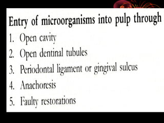
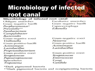
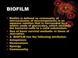
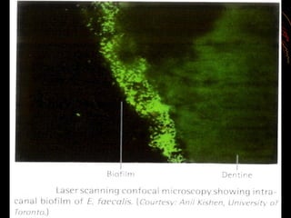
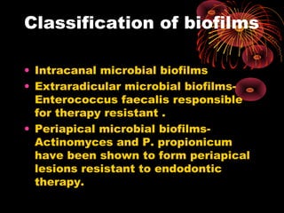
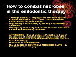
MICROBIOLOGY & 
IMMUNOLOGY IN 
ENDODONTICS 
DR.SADASIVA 
READER 
CHETTINAD HEALTH CITY
INTRODUCTION 
• Most changes in pulp and 
periradicular tissues are of 
bacteriological and must be dealt 
with as infectious processes. 
• Bacteria play a major role in 
pathogenesis of pulpo-periradicular 
lesion, a fundamental knowledge of 
endodontic microbiology needed to 
understand.
• 1. The role of bacteria in these diseases. 
• 2.Pathways of pulp and periradicular 
tissues. 
• 3. Response of pulpal and periradicular 
tissues to bacterial infection. 
• 4. Methods used to control and eradicate 
root canal infection during root canal 
treatment. 
• The intact hard tissues of the tooth 
normally protect the pulp by acting as 
physical barrier.
HISTORY 
• 1890- Miller – demonstrated 
presence of bacteria in necrotic pulp 
tissue. 
• Predominant species were 
streptococci, micrococci, and small 
% of anaerobic bacteria. 
• Kakehashi etal – experiment with 
germ free rats – failed to create 
lesions. 
• Poly microbial infections.
Flora of root canal and 
periradicular lesions 
• Most of them – multi bacterial. 
• Predominance of facultative 
organisms 
• Obligate anaerobic species 
• Gram negative cocci 
• Lactobacilli 
• spirochetes 
• Bacteroides- black pigmented 
species
MICROBIAL 
VIRULENCE&PATHOGENIC 
ITY 
• In 1965, Hobson gave an equation 
showing the relation of number of 
microorganisms , their virulence, 
resistance of host and severity of the 
disease. 
• Number of microorganisms x 
Virulence of microorganisms = severity of the disease 
--------------------------------------------------- 
resistance of host
VARIOUS VIRULENT 
FACTORS 
• 1. Lipopolysaccharides(lps) 
• 2. Extra cellular vesicles 
• 3. Enzymes 
• 4.Fatty acids 
• 5. Polyamines 
• 6. Capsule 
• 7.Pilli
Lipopolysaccharides(LP 
S) 
Present on the surface of gram 
negative bacteria. 
LPS HAVE NON SPECIFIC 
antigens which are not 
neutralized by antibodies. 
ENDOTOXINS 
Diffuse in to dentin . 
Studies have shown the 
relationship between endotoxins 
and the periradicular 
inflammation.
EXTRACELLULAR 
CELLULAR VESICLES 
• Produced by gram-ve bacteria in 
the form of endotoxins. 
• Vesicles contain various 
enzymes and toxic products-responsible 
for 
haemaggulutination, hemolysis 
and bacterial adhesion
ENZYMES 
• Helps in spread of infection 
• Neutralization of the immunoglobulin 
and complement components 
• PMN leucocytes release hydrolytic 
enzymes which degenerate and lyse 
to form purulent exudates and have 
adverse effects on the surrounding 
tissues.
FATTY 
ACIDS&POLYAMINES 
• ANAEROBIC BACTERIA –produces 
propionic acid, butryicacid. 
• They cause neutrophil chemotaxis, 
degranulation,and stimulate 
interleukin -1 production which 
causes bone resorptionand 
periradicular diseases. 
• Polyamines are active chemicals 
found in infected canals. Cadaverine, 
putrescine, spermidine are 
polyamines.
Factors influencing the 
growth and colonization of 
microorganisms 
• Influence of oxygen 
• Nutritional factors 
• Bacteriocins 
• Coaggregation 
• Bacterial relationships
Microbial ecosystem of 
the root canal system 
• NAIDORF summarized the few 
generalization in relation to organisms 
isolated freom the root canals. 
• 1. Mixed infections are more common than 
single organisms. 
• 2.Pulp contains flora almost similar to that 
of oral cavity. 
• 3. Approximately 25 % of isolated 
organisms are anaerobic. 
• 4. organisms isolated from flare-up as well 
as asymptomatic cases are almost similar.
Microbiology of infected 
root canal
BIOFILM 
• Biofilm is defined as community of 
microcolonies of microorganisms in an 
aqueous solution that is surrounded by a 
matrix made of glycocalyx, which attaches 
the bacterial cells to a solid substratum. 
• One of basic survival methods- in times of 
starvation. 
• A BIOFILM has the following attributes: 
• Autopoiesis 
• Homeostasis 
• Synergy 
• Communality
Classification of biofilms 
• Intracanal microbial biofilms 
• Extraradicular microbial biofilms- 
Enterococcus faecalis responsible 
for therapy resistant . 
• Periapical microbial biofilms- 
Actinomyces and P. propionicum 
have been shown to form periapical 
lesions resistant to endodontic 
therapy.
PATHOGENICITYOF 
BACTEROIDES 
• Related to the presence of LPS and Peptidoglycans 
• 1. induce hormones like cytokinins which play 
important role in inflammation. 
• Stimulate B-lymphocytes 
• Activate complement cascade 
• Release various enzymes like collgenase 
• Enhance production of various pain mediators like 
bradykinin,histamine and prostaglandins 
• LPS once released as ENDOTOXIN causes 
biological effects including inflammationand bone 
resorption.
Fungi& viruses 
• Using PCR, Candida albicans was 
detected in 21 % of infected root 
canals. 
• Bacteriocins are viruses that infect 
bacteria and carry DNA in to the 
genome of the bacteria. HIV , 
CYTOMEGALO VIRUS, and EPSTEIN 
BARR virus – associated with 
periapical disease.
Microbiology of 
periradicular endodontic 
infections 
• Biofilms are formed in the periapical region 
• Periapical lesions contains macrophages, 
lymphocytes(T- cells and B-cells), plasma cells, and 
neutrophils. 
• Their function is to prevent microorganisms from 
invading periradicular tissues. 
• The mosty common cause of persistent 
periradicular infections is incomplete debridement 
of the root canal system. Therefore three 
dimensional sealing of the rootcanal system is 
necessary for resolution of periapical pathologies
Microbiology of root 
canal failures 
• More facultative bacteria rather than 
strict anaerobes 
• Enterococcus fecalis. 
Propionbacterium alactolyticus, 
P.propionicum in filled root canal 
failure cases. 
• Vast majority of endodontic 
treatment failures are caused by 
intraradicular infections.
IDENTIFICATION OF 
THE BACTERIA 
• GRAMS STAIN- helps in 
differentiating Gram positive 
and Gram negative organisms. 
• CULTURE: 
• Molecular diagnostic methods 
• DNA- DNA hybridization method 
• Polymerase chain reaction
How to combat microbes 
in the endodontic therapy 
• Thorough cleaning & shaping of the root canal system-irrigants 
like NAOCL, H2O2, EDTA - excellent tissue 
dissolving and microbial agents. 
• Oxygenating a canal simply by opening is detrimental to 
anaerobes. 
• Drainage by canal or by soft tissues decrease discomfort 
caused by inflammatory mediators. 
• ANTIBIOITICS :Drug of choice is Penicillin vk bcos of 
its spectrum of microbial activity against most of the 
bacteria associated with endodontic infections and also 
of its low toxicity. 
• INTRACANAL MEDICAMENTS 
• Use of CAOH2, CHX2% TRIPLE ANTIBIOTIC PASTE - is 
beneficial in necrotic pulps
THANK U

Endo microbiology -key to success in endo

  • 1.
    MICROBIOLOGY & IMMUNOLOGYIN ENDODONTICS DR.SADASIVA READER CHETTINAD HEALTH CITY
  • 2.
    INTRODUCTION • Mostchanges in pulp and periradicular tissues are of bacteriological and must be dealt with as infectious processes. • Bacteria play a major role in pathogenesis of pulpo-periradicular lesion, a fundamental knowledge of endodontic microbiology needed to understand.
  • 3.
    • 1. Therole of bacteria in these diseases. • 2.Pathways of pulp and periradicular tissues. • 3. Response of pulpal and periradicular tissues to bacterial infection. • 4. Methods used to control and eradicate root canal infection during root canal treatment. • The intact hard tissues of the tooth normally protect the pulp by acting as physical barrier.
  • 5.
    HISTORY • 1890-Miller – demonstrated presence of bacteria in necrotic pulp tissue. • Predominant species were streptococci, micrococci, and small % of anaerobic bacteria. • Kakehashi etal – experiment with germ free rats – failed to create lesions. • Poly microbial infections.
  • 7.
    Flora of rootcanal and periradicular lesions • Most of them – multi bacterial. • Predominance of facultative organisms • Obligate anaerobic species • Gram negative cocci • Lactobacilli • spirochetes • Bacteroides- black pigmented species
  • 11.
    MICROBIAL VIRULENCE&PATHOGENIC ITY • In 1965, Hobson gave an equation showing the relation of number of microorganisms , their virulence, resistance of host and severity of the disease. • Number of microorganisms x Virulence of microorganisms = severity of the disease --------------------------------------------------- resistance of host
  • 13.
    VARIOUS VIRULENT FACTORS • 1. Lipopolysaccharides(lps) • 2. Extra cellular vesicles • 3. Enzymes • 4.Fatty acids • 5. Polyamines • 6. Capsule • 7.Pilli
  • 14.
    Lipopolysaccharides(LP S) Presenton the surface of gram negative bacteria. LPS HAVE NON SPECIFIC antigens which are not neutralized by antibodies. ENDOTOXINS Diffuse in to dentin . Studies have shown the relationship between endotoxins and the periradicular inflammation.
  • 15.
    EXTRACELLULAR CELLULAR VESICLES • Produced by gram-ve bacteria in the form of endotoxins. • Vesicles contain various enzymes and toxic products-responsible for haemaggulutination, hemolysis and bacterial adhesion
  • 16.
    ENZYMES • Helpsin spread of infection • Neutralization of the immunoglobulin and complement components • PMN leucocytes release hydrolytic enzymes which degenerate and lyse to form purulent exudates and have adverse effects on the surrounding tissues.
  • 17.
    FATTY ACIDS&POLYAMINES •ANAEROBIC BACTERIA –produces propionic acid, butryicacid. • They cause neutrophil chemotaxis, degranulation,and stimulate interleukin -1 production which causes bone resorptionand periradicular diseases. • Polyamines are active chemicals found in infected canals. Cadaverine, putrescine, spermidine are polyamines.
  • 18.
    Factors influencing the growth and colonization of microorganisms • Influence of oxygen • Nutritional factors • Bacteriocins • Coaggregation • Bacterial relationships
  • 19.
    Microbial ecosystem of the root canal system • NAIDORF summarized the few generalization in relation to organisms isolated freom the root canals. • 1. Mixed infections are more common than single organisms. • 2.Pulp contains flora almost similar to that of oral cavity. • 3. Approximately 25 % of isolated organisms are anaerobic. • 4. organisms isolated from flare-up as well as asymptomatic cases are almost similar.
  • 20.
  • 21.
    BIOFILM • Biofilmis defined as community of microcolonies of microorganisms in an aqueous solution that is surrounded by a matrix made of glycocalyx, which attaches the bacterial cells to a solid substratum. • One of basic survival methods- in times of starvation. • A BIOFILM has the following attributes: • Autopoiesis • Homeostasis • Synergy • Communality
  • 25.
    Classification of biofilms • Intracanal microbial biofilms • Extraradicular microbial biofilms- Enterococcus faecalis responsible for therapy resistant . • Periapical microbial biofilms- Actinomyces and P. propionicum have been shown to form periapical lesions resistant to endodontic therapy.
  • 26.
    PATHOGENICITYOF BACTEROIDES •Related to the presence of LPS and Peptidoglycans • 1. induce hormones like cytokinins which play important role in inflammation. • Stimulate B-lymphocytes • Activate complement cascade • Release various enzymes like collgenase • Enhance production of various pain mediators like bradykinin,histamine and prostaglandins • LPS once released as ENDOTOXIN causes biological effects including inflammationand bone resorption.
  • 27.
    Fungi& viruses •Using PCR, Candida albicans was detected in 21 % of infected root canals. • Bacteriocins are viruses that infect bacteria and carry DNA in to the genome of the bacteria. HIV , CYTOMEGALO VIRUS, and EPSTEIN BARR virus – associated with periapical disease.
  • 28.
    Microbiology of periradicularendodontic infections • Biofilms are formed in the periapical region • Periapical lesions contains macrophages, lymphocytes(T- cells and B-cells), plasma cells, and neutrophils. • Their function is to prevent microorganisms from invading periradicular tissues. • The mosty common cause of persistent periradicular infections is incomplete debridement of the root canal system. Therefore three dimensional sealing of the rootcanal system is necessary for resolution of periapical pathologies
  • 29.
    Microbiology of root canal failures • More facultative bacteria rather than strict anaerobes • Enterococcus fecalis. Propionbacterium alactolyticus, P.propionicum in filled root canal failure cases. • Vast majority of endodontic treatment failures are caused by intraradicular infections.
  • 30.
    IDENTIFICATION OF THEBACTERIA • GRAMS STAIN- helps in differentiating Gram positive and Gram negative organisms. • CULTURE: • Molecular diagnostic methods • DNA- DNA hybridization method • Polymerase chain reaction
  • 32.
    How to combatmicrobes in the endodontic therapy • Thorough cleaning & shaping of the root canal system-irrigants like NAOCL, H2O2, EDTA - excellent tissue dissolving and microbial agents. • Oxygenating a canal simply by opening is detrimental to anaerobes. • Drainage by canal or by soft tissues decrease discomfort caused by inflammatory mediators. • ANTIBIOITICS :Drug of choice is Penicillin vk bcos of its spectrum of microbial activity against most of the bacteria associated with endodontic infections and also of its low toxicity. • INTRACANAL MEDICAMENTS • Use of CAOH2, CHX2% TRIPLE ANTIBIOTIC PASTE - is beneficial in necrotic pulps
  • 33.

Editor's Notes