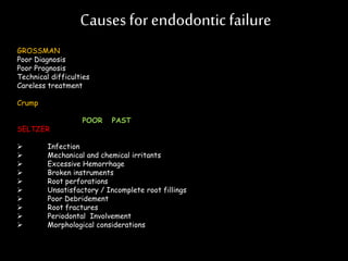
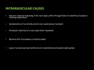
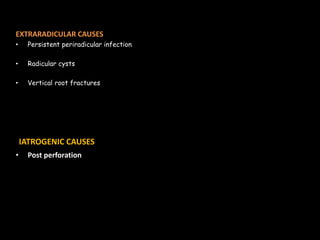
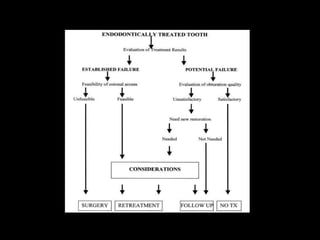
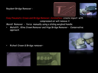
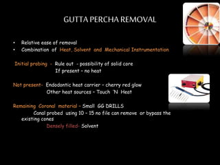
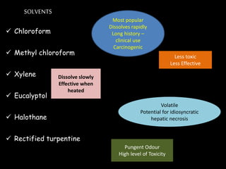
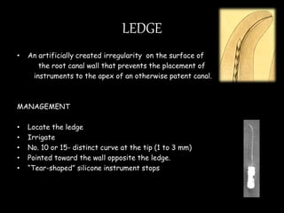
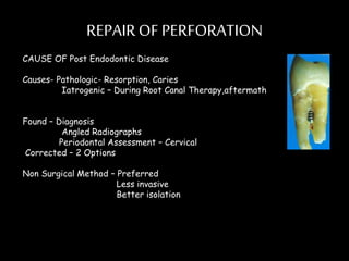
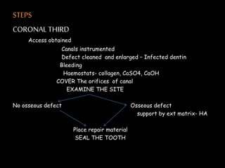
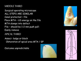
Endodontic retreatment involves removing previous root canal fillings and materials to address treatment failure. Success is defined as an asymptomatic tooth with healthy surrounding tissue and bone fill on radiographs. Causes of failure include missed canals, improper length determination, ledges, and perforations. The objectives of retreatment are to regain access, remove all materials, disinfect canals, and obtain proper seal length. Techniques include removing posts, solvents or heat to remove fillings, bypassing ledges or separated instruments, and repairing any perforations. Proper case selection, access, and following technical protocols are important for success.
Biomechanical preparation is the crucial step in endodontic procedure. Biological principles can only be preserved if the mechanical shaping of the perticular canal is completed with the cordial following of the endodontic priciples. This presentation is aimed to simplify the various endodontic techniques for root canal shaping in as conservative as possible manner.
This presentation is all about restoration of endodontically treated teeth, prefabricated post and core, cast post and core, direct and indirect technique.
About failures of root canal treatment and retreatment. This presentation describes about various techniques for gutta percha removal, posts removal, pastes removal, and removal of separated instrument
Cases of failed root canal treatments can now be successfully treated, thanks to modern technologies. A failed root canal treatment can be potentially recognised by pain and/or swelling.
New technologies have increased success rates of treating failed root canal treatments:
Magnification loupes/ Microscopes with fibre optic lighting system have improved a dentist’s vision and therefore the chances of the treatment’s success.
The Indian Dental Academy is the Leader in continuing dental education , training dentists in all aspects of dentistry and
offering a wide range of dental certified courses in different formats.
In this lecture I explain the basic concept of root canal filling or what called obturation. The lectures discuss different techniques used in that matter in step-by-step fashion and explanatory pictures.
It is directed to the level of undergraduate mind.
Management of non surgical root-canal treatment failureHamza Tahir
This presentation of mine is actually a brief overview of non surgical root canal re treatment procedure from removing permanent fillings to removal procedure of obturating material etc.
Advances in obturation system in endodontics /certified fixed orthodontic co...Indian dental academy
Welcome to Indian Dental Academy
The Indian Dental Academy is the Leader in continuing dental education , training dentists in all aspects of dentistry and offering a wide range of dental certified courses in different formats.
Indian dental academy has a unique training program & curriculum that provides students with exceptional clinical skills and enabling them to return to their office with high level confidence and start treating patients
State of the art comprehensive training-Faculty of world wide repute &Very affordable.
explained with Limited matter moreover I have included all the images. if you go through a standard textbook and referred to this PPT it will help you so much I hope It helps you. ask me for the books details.
Success in Endodontics was originally based on the triad of debridement, thorough disinfection and obturation with all the aspects equally important.
At present, successful root canal treatment is based on broader principles, these includes
Diagnosis and treatment planning
Knowledge of anatomy and morphology
Concepts of thorough debridement
Obturation of the root canal space
Finally the coronal seal / restoration.
A meta- analysis of factors influencing the root canal treatment found that the following four factors influenced success:
Absence of pre-treatment periapical lesion / Co-existing pathology
Root canal fillings with minimal or no voids
Obturation to within 2.0mm / or as close as possible to the radiographic apex
An adequate coronal restoration.
Ethanol (CH3CH2OH), or beverage alcohol, is a two-carbon alcohol
that is rapidly distributed in the body and brain. Ethanol alters many
neurochemical systems and has rewarding and addictive properties. It
is the oldest recreational drug and likely contributes to more morbidity,
mortality, and public health costs than all illicit drugs combined. The
5th edition of the Diagnostic and Statistical Manual of Mental Disorders
(DSM-5) integrates alcohol abuse and alcohol dependence into a single
disorder called alcohol use disorder (AUD), with mild, moderate,
and severe subclassifications (American Psychiatric Association, 2013).
In the DSM-5, all types of substance abuse and dependence have been
combined into a single substance use disorder (SUD) on a continuum
from mild to severe. A diagnosis of AUD requires that at least two of
the 11 DSM-5 behaviors be present within a 12-month period (mild
AUD: 2–3 criteria; moderate AUD: 4–5 criteria; severe AUD: 6–11 criteria).
The four main behavioral effects of AUD are impaired control over
drinking, negative social consequences, risky use, and altered physiological
effects (tolerance, withdrawal). This chapter presents an overview
of the prevalence and harmful consequences of AUD in the U.S.,
the systemic nature of the disease, neurocircuitry and stages of AUD,
comorbidities, fetal alcohol spectrum disorders, genetic risk factors, and
pharmacotherapies for AUD.
Flu Vaccine Alert in Bangalore Karnatakaaddon Scans
As flu season approaches, health officials in Bangalore, Karnataka, are urging residents to get their flu vaccinations. The seasonal flu, while common, can lead to severe health complications, particularly for vulnerable populations such as young children, the elderly, and those with underlying health conditions.
Dr. Vidisha Kumari, a leading epidemiologist in Bangalore, emphasizes the importance of getting vaccinated. "The flu vaccine is our best defense against the influenza virus. It not only protects individuals but also helps prevent the spread of the virus in our communities," he says.
This year, the flu season is expected to coincide with a potential increase in other respiratory illnesses. The Karnataka Health Department has launched an awareness campaign highlighting the significance of flu vaccinations. They have set up multiple vaccination centers across Bangalore, making it convenient for residents to receive their shots.
To encourage widespread vaccination, the government is also collaborating with local schools, workplaces, and community centers to facilitate vaccination drives. Special attention is being given to ensuring that the vaccine is accessible to all, including marginalized communities who may have limited access to healthcare.
Residents are reminded that the flu vaccine is safe and effective. Common side effects are mild and may include soreness at the injection site, mild fever, or muscle aches. These side effects are generally short-lived and far less severe than the flu itself.
Healthcare providers are also stressing the importance of continuing COVID-19 precautions. Wearing masks, practicing good hand hygiene, and maintaining social distancing are still crucial, especially in crowded places.
Protect yourself and your loved ones by getting vaccinated. Together, we can help keep Bangalore healthy and safe this flu season. For more information on vaccination centers and schedules, residents can visit the Karnataka Health Department’s official website or follow their social media pages.
Stay informed, stay safe, and get your flu shot today!
Report Back from SGO 2024: What’s the Latest in Cervical Cancer?bkling
Are you curious about what’s new in cervical cancer research or unsure what the findings mean? Join Dr. Emily Ko, a gynecologic oncologist at Penn Medicine, to learn about the latest updates from the Society of Gynecologic Oncology (SGO) 2024 Annual Meeting on Women’s Cancer. Dr. Ko will discuss what the research presented at the conference means for you and answer your questions about the new developments.
ARTIFICIAL INTELLIGENCE IN HEALTHCARE.pdfAnujkumaranit
Artificial intelligence (AI) refers to the simulation of human intelligence processes by machines, especially computer systems. It encompasses tasks such as learning, reasoning, problem-solving, perception, and language understanding. AI technologies are revolutionizing various fields, from healthcare to finance, by enabling machines to perform tasks that typically require human intelligence.
These simplified slides by Dr. Sidra Arshad present an overview of the non-respiratory functions of the respiratory tract.
Learning objectives:
1. Enlist the non-respiratory functions of the respiratory tract
2. Briefly explain how these functions are carried out
3. Discuss the significance of dead space
4. Differentiate between minute ventilation and alveolar ventilation
5. Describe the cough and sneeze reflexes
Study Resources:
1. Chapter 39, Guyton and Hall Textbook of Medical Physiology, 14th edition
2. Chapter 34, Ganong’s Review of Medical Physiology, 26th edition
3. Chapter 17, Human Physiology by Lauralee Sherwood, 9th edition
4. Non-respiratory functions of the lungs https://academic.oup.com/bjaed/article/13/3/98/278874
HOT NEW PRODUCT! BIG SALES FAST SHIPPING NOW FROM CHINA!! EU KU DB BK substit...GL Anaacs
Contact us if you are interested:
Email / Skype : kefaya1771@gmail.com
Threema: PXHY5PDH
New BATCH Ku !!! MUCH IN DEMAND FAST SALE EVERY BATCH HAPPY GOOD EFFECT BIG BATCH !
Contact me on Threema or skype to start big business!!
Hot-sale products:
NEW HOT EUTYLONE WHITE CRYSTAL!!
5cl-adba precursor (semi finished )
5cl-adba raw materials
ADBB precursor (semi finished )
ADBB raw materials
APVP powder
5fadb/4f-adb
Jwh018 / Jwh210
Eutylone crystal
Protonitazene (hydrochloride) CAS: 119276-01-6
Flubrotizolam CAS: 57801-95-3
Metonitazene CAS: 14680-51-4
Payment terms: Western Union,MoneyGram,Bitcoin or USDT.
Deliver Time: Usually 7-15days
Shipping method: FedEx, TNT, DHL,UPS etc.Our deliveries are 100% safe, fast, reliable and discreet.
Samples will be sent for your evaluation!If you are interested in, please contact me, let's talk details.
We specializes in exporting high quality Research chemical, medical intermediate, Pharmaceutical chemicals and so on. Products are exported to USA, Canada, France, Korea, Japan,Russia, Southeast Asia and other countries.
TEST BANK for Operations Management, 14th Edition by William J. Stevenson, Ve...kevinkariuki227
TEST BANK for Operations Management, 14th Edition by William J. Stevenson, Verified Chapters 1 - 19, Complete Newest Version.pdf
Recomendações da OMS sobre cuidados maternos e neonatais para uma experiência pós-natal positiva.
Em consonância com os ODS – Objetivos do Desenvolvimento Sustentável e a Estratégia Global para a Saúde das Mulheres, Crianças e Adolescentes, e aplicando uma abordagem baseada nos direitos humanos, os esforços de cuidados pós-natais devem expandir-se para além da cobertura e da simples sobrevivência, de modo a incluir cuidados de qualidade.
Estas diretrizes visam melhorar a qualidade dos cuidados pós-natais essenciais e de rotina prestados às mulheres e aos recém-nascidos, com o objetivo final de melhorar a saúde e o bem-estar materno e neonatal.
Uma “experiência pós-natal positiva” é um resultado importante para todas as mulheres que dão à luz e para os seus recém-nascidos, estabelecendo as bases para a melhoria da saúde e do bem-estar a curto e longo prazo. Uma experiência pós-natal positiva é definida como aquela em que as mulheres, pessoas que gestam, os recém-nascidos, os casais, os pais, os cuidadores e as famílias recebem informação consistente, garantia e apoio de profissionais de saúde motivados; e onde um sistema de saúde flexível e com recursos reconheça as necessidades das mulheres e dos bebês e respeite o seu contexto cultural.
Estas diretrizes consolidadas apresentam algumas recomendações novas e já bem fundamentadas sobre cuidados pós-natais de rotina para mulheres e neonatos que recebem cuidados no pós-parto em unidades de saúde ou na comunidade, independentemente dos recursos disponíveis.
É fornecido um conjunto abrangente de recomendações para cuidados durante o período puerperal, com ênfase nos cuidados essenciais que todas as mulheres e recém-nascidos devem receber, e com a devida atenção à qualidade dos cuidados; isto é, a entrega e a experiência do cuidado recebido. Estas diretrizes atualizam e ampliam as recomendações da OMS de 2014 sobre cuidados pós-natais da mãe e do recém-nascido e complementam as atuais diretrizes da OMS sobre a gestão de complicações pós-natais.
O estabelecimento da amamentação e o manejo das principais intercorrências é contemplada.
Recomendamos muito.
Vamos discutir essas recomendações no nosso curso de pós-graduação em Aleitamento no Instituto Ciclos.
Esta publicação só está disponível em inglês até o momento.
Prof. Marcus Renato de Carvalho
www.agostodourado.com
Title: Sense of Taste
Presenter: Dr. Faiza, Assistant Professor of Physiology
Qualifications:
MBBS (Best Graduate, AIMC Lahore)
FCPS Physiology
ICMT, CHPE, DHPE (STMU)
MPH (GC University, Faisalabad)
MBA (Virtual University of Pakistan)
Learning Objectives:
Describe the structure and function of taste buds.
Describe the relationship between the taste threshold and taste index of common substances.
Explain the chemical basis and signal transduction of taste perception for each type of primary taste sensation.
Recognize different abnormalities of taste perception and their causes.
Key Topics:
Significance of Taste Sensation:
Differentiation between pleasant and harmful food
Influence on behavior
Selection of food based on metabolic needs
Receptors of Taste:
Taste buds on the tongue
Influence of sense of smell, texture of food, and pain stimulation (e.g., by pepper)
Primary and Secondary Taste Sensations:
Primary taste sensations: Sweet, Sour, Salty, Bitter, Umami
Chemical basis and signal transduction mechanisms for each taste
Taste Threshold and Index:
Taste threshold values for Sweet (sucrose), Salty (NaCl), Sour (HCl), and Bitter (Quinine)
Taste index relationship: Inversely proportional to taste threshold
Taste Blindness:
Inability to taste certain substances, particularly thiourea compounds
Example: Phenylthiocarbamide
Structure and Function of Taste Buds:
Composition: Epithelial cells, Sustentacular/Supporting cells, Taste cells, Basal cells
Features: Taste pores, Taste hairs/microvilli, and Taste nerve fibers
Location of Taste Buds:
Found in papillae of the tongue (Fungiform, Circumvallate, Foliate)
Also present on the palate, tonsillar pillars, epiglottis, and proximal esophagus
Mechanism of Taste Stimulation:
Interaction of taste substances with receptors on microvilli
Signal transduction pathways for Umami, Sweet, Bitter, Sour, and Salty tastes
Taste Sensitivity and Adaptation:
Decrease in sensitivity with age
Rapid adaptation of taste sensation
Role of Saliva in Taste:
Dissolution of tastants to reach receptors
Washing away the stimulus
Taste Preferences and Aversions:
Mechanisms behind taste preference and aversion
Influence of receptors and neural pathways
Impact of Sensory Nerve Damage:
Degeneration of taste buds if the sensory nerve fiber is cut
Abnormalities of Taste Detection:
Conditions: Ageusia, Hypogeusia, Dysgeusia (parageusia)
Causes: Nerve damage, neurological disorders, infections, poor oral hygiene, adverse drug effects, deficiencies, aging, tobacco use, altered neurotransmitter levels
Neurotransmitters and Taste Threshold:
Effects of serotonin (5-HT) and norepinephrine (NE) on taste sensitivity
Supertasters:
25% of the population with heightened sensitivity to taste, especially bitterness
Increased number of fungiform papillae
2. CONTENTS
• Introduction
• Measuring The Success
• Causes Of Failure
• Definition
• Objectives
• Coronal Access Cavity Preparation
• Post Removal
• Regaining Access To The Apical Area
• Removal Of Separated Instruments
• Removal Technique
• Finishing Of Retreatment
• Repair Of Perforation
• Ten Commandments For Improved Success
• Conclusion
3. Success ……
• Asymptomatic
• Periodontium should be healthy
• Radiographs should demonstrate healing or progressive bone fill
overtime.
• Principles of restorative excellence should be satisfied.
5. INTRARADICULAR CAUSES
• Necrotic material remaining in the root canal, either through failure to identify all canals or
treating canals short.
• Contamination of an initially sterile root canal during treatment
• Persistent infection of a root canal after treatment
• Bacteria left in accessory or lateral canals
• Loss of coronal seal and reinfection of a disinfected and sealed canal system
7. DEFINED AS………….
A procedure to remove root canal filling materials from the tooth ,
revise the shape and obturate the canals ; usually accomplished
because the original treatment appears inadequate or has failed or
because the root canal has been contaminated by prolonged exposure
to oral environment.
-A.A. E
8. OBJECTIVES
To regain access to the apical area of the root canal space in the previously treated
tooth
Eliminate microorganisms that have either survived previous treatment or have re-
entered the root canal system.
Remove all necrotic material remaining in the root canal either due to missed main,
accessory , lateral canals.
Block all the portals of exit or achieve a three dimensional obturation upto the apex
of the tooth.
Treat any infection persisting after a treatment.
Regain proper coronal seal and establish proper sealed canals.
11. CORONAL ACCESS CAVITY
Synonym
Quality of coronal seal – assessed preoperatively
REMOVAL OF CROWN
Crown satisfactory - Retaining Adv- Isolation easy
Occlusion is preserved
Esthetics minimally changed
Cost effective
Disadv- Restricted visibility, Anatomy - Iatrogenic mishaps
Removal of canal obstructions - posts
Missing – canals ,hidden recurrent caries, fracture
12. Preserve
1.Access through Crown
Metal- Carbide fissure bur- # 1556
PFM- P- Round diamond copious coolant water spray
M- End cutting bur – Transmetal bur and Great White bur
2. Remove the crown
Chiesel , Flat Plastic and Coupland ‘s Chisel
Ultrasonics
Forceps
K.Y. Pliers- Small replacement Rubber tips and Emery powder
Wynman Crown Gripper , Trail Crown remover and
Trident Crown Placer/ remover
13. Roydent Bridge Remover -
Easy Pneumatic Crown and Bridge Remover, Coronaflex- create impact with
compressed air will remove it
Morell Remover - force manually using a sliding weighed handle
• Metalift , Kline Crown Remover and Higa Bridge Remover - Conservative
approach
• Richwil Crown & Bridge remover-
14. POST REMOVAL
Depends upon
• Type, Shape ,Design of the post
• Location of tooth
• Materials used to cement the post
• Techinques
“It is not only what is removed but also what is left behind that is important”.
First step-- Expose it properly
Bulk of core material-- High speed hand piece - Cylindrical or tapered
carbide or diamond burs
Last embedding core material – Less aggressive – Tapered , mid sized ultrasonic tip
Minimal restorative material remaining – Small sized ultrasonic instrument
15. Once well isolated and freed from all restorative material-
Retention of Post should be reduced
Medium sized US tips - AT THE INTERFACE
Care to be taken not push with too much force
If root is thin , smaller US tips are used –DRY - limit visibilty & accumulation of
debris
If rubber dam , Post flooded with Solvents like Chloroform and later
activated
Roto- Pro Bur – 3 shapes , Six sided , non cutting tapered burs in high speed
hand piece - decreases the retention
16. POST REMOVAL KITS
Gonon Post Removing System- Parallel or Tapered
Non active Preformed
Hollow Trephine bur
Specific extraction mandrel – create or tap a thread on to exposed milled
portion of post – Extraction forceps or vise is applied to tooth and post – Turning
the screw on the handle of the vise will create a coronal force
Drawbacks – Size of vise – makes the access diff- molars, crowded Mand incis.
Thomas Screw Post Removal Kit - Active or Screw post
Extraction mandrel are threaded in opposite direction
17. Ruddle Post Removal System – Combines the properties
JS Post Extractor – Advan – Smaller size- inaccessible areas
PULLING ACTION
Disadvantage- Large variety of trephine burs or
extraction mandrel
Post Puller or Eggler Post Remover – No trephine burs or extraction mandrels
2 sets of jaws – independent
High speed handpiece & bur tooth and post
Not recommended for Screw post
18. TOOTHCOLOUREDPOSTS
Ceramic , Zirconium, Various Type Of Fiber Reinforced Composite
LARGO BUR , PAESO DRILL
REMOVAL BURS IN THE KIT
Gyro tip- flutes, Plasma coated silicon carbide
POTENTIALCOMPLICATIONS OF POST REMOVAL
Fracture of tooth
Leaving the tooth non restorable
Root perforation
Inability to remove the post
Heat generated - ultrasonics
20. PASTES
N2 OR RC 2B- Formaldehyde , Heavy Metal Oxides
CAN BE
A. SOFT PASTES- Easy
Crown Down Instr . With copious Irrigation
B.HARD PASTES – Probed with endo explorer or file
BUR OR US tip in easily accessible
straight portions
Curvature- precurved , small files are inserted
Densely filled – solvents
21. Ultrasonics – Hard paste in curved apical area
Energy will break up the paste
Biocalex 6.9- EDTA CAN BE USED
22. GUTTA PERCHA REMOVAL
• Relative ease of removal
• Combination of Heat, Solvent and Mechanical Instrumentation
Initial probing - Rule out - possibility of solid core
If present – no heat
Not present- Endodontic heat carrier – cherry red glow
Other heat sources – Touch ’N Heat
Remaining Coronal material – Small GG DRILLS
Canal probed using 10 – 15 no file can remove or bypass the
existing cones
Densely filled- Solvent
23. SOLVENTS
Chloroform
Methyl chloroform
Xylene
Eucalyptol
Halothane
Rectified turpentine
Most popular
Dissolves rapidly
Long history –
clinical use
Carcinogenic
Less toxic
Less Effective
Dissolve slowly
Effective when
heated
Volatile
Potential for idiosyncratic
hepatic necrosis
Pungent Odour
High level of Toxicity
24. TECHNIQUE
Solvent introduced into coronal portions acts as reservior
Small files are used to penetrate the remaining root filling and SA
Precurved rigid files such as C+ files- more efficient
Radiograph – taken when estimated length is approached- avoid Overextending
Once WL reached – Progressive Larger diameter hand files - rotated in Passive, non binding
clockwise reaming motion – Remove bulk of GP
Frequent replenishment of solvent
When last loose fitting instrument – removed clean- solvent acts as Irrigant.
Solvent removed with paper points
Use kinked small files, probe the canal wall for irregularities
25. Glass ionomer assealer
• Insoluble In halothene and chloroform
• Done by removing GP – US to debride canal walls
OVEREXTENDINGFILLING
• H file -- extruded apical fragment of root filling – clockwise
rotation – withdrawn without rotation
• Should not be softened with Solvent
26. ROTARY SYSTEMS
Enhanced efficiency and effectiveness in removing
Risk of instrument separation
LASERS
Nd YAG – time is same
considerable amount was left
Root surface temperature increased
RESILON- Cone – Heat
Solvent- Endosolv -R
27. SOLID CORE OBTURATORS
More complex & difficult
Method depends upon-
Type- Plastic - smooth sided
Metal - fluted
Level at which carrier is cut- 2-3 mm above the pulp chamber
For post space prepa – nicked at the middle and inserted apically
28. TREATMENT STEPS
PREOPERATIVE RADIOGRAPH
CAREFUL ACCESS AND PROBING- metallic structure embedded in the GP mass
Black spot
Metallic carrier- Heat applied– soften GP- REMOVED – Peet silver point forceps or modified
Steiglitz forceps
If not enough space available--- Solvent application – using small hand instruments – followed by
ultrasonic activation-- removing it
Plastic Carrier – Heat should be avoided
Older – VECTRA- Insoluble in solvents
POLYSULFONE- soluble in Chloroform
Newer carriers not soluble
29. STEPS-
Access flooded –GP removed – Larger to smaller hand files
Solvent – replenished- 8 no. file - extend to apical area
When little GP remaining – large H File – inserted alongside plastic carrier –
gently turned clockwise to engage the flutes- pulled
Care to be taken not to overstress the instrument
Recently- System B Heat source- soften GP
AT 225 0 c
Rotary Instruments
Difficult to remove sealer and GP – alpha phase
Solvent –wicking with paper point
30. SILVERPOINTS
• Minimal Taper and smooth sided
Removal technique
Establish proper access
Coronal portion embedded in Core material – Carefully removed
with bur and US
Flood the access- for cement dissolution
Endo. Explorer and Small file carries solvent down the silver pt
Replenish the solvent
Grasp the exposed end with – Stieglitz pliers or some other
forceps
Gently pull it out
31. If no good purchase – cone held with forceps - that is held with hemostat or needle
driver - allow removal
If held in tight friction grip- Indirect US - can be used to loosen it
If not much exposure – Caufield silver point retrievers can be used
Spoon with groove in the tip
Available in three sizes – 25, 35 and 50.
Other techniques
1. H FILES- Requires some space in the coronal area
Sealer is dissolved
32. • If more exposure is required , Use of trephine bur and microtubes or
ultrasonics
• SEVERAL EXTRACTION DEVICES –
• Masserann kit
• Endoextractor
• Separated Instrument Retrieval System etc
• Once removed instrumentation – Crown down- prevent extrusion of
Corrosion Products
33. BROKEN INSTRUMENT
Types of Instrument
Can be seen during diagnosis
After removal of GP
Causes
“Stressed” instrument
Placing exaggerated bends
Forcing a file before canal opened
sufficiently + reaming motion.
Inadequate access
Anatomy
Manufacturing defects
The best antidote for a broken file is PREVENTION.
34. PROGNOSIS
Depends on
What stage
Preoperative Status
Whether file can be removed or bypassed
Removal depends on
Location
Root curvatures, External root concavities , Root thickness
Type of material – NiTi and Stainless Steel
35. RemovalTechniques
Headlamp and Magnifying loupes
Operative Microscope
Treatment Approach
Visible in Coronal Access – Grasp – Hemostat or Steiglitz Forceps
Technique- ACW
Deep – Visibility difficult
Create straight line Coronal – radicular access
Modified GG DRILLS – Create circumferential staging
platform
US tip – placed bet exposed file & wall and is vibrated around the
obstruction in CCW – Causes Unscrewing forces
Occasionally , file will jump out
36. Other methods
1. Microtube technique- SS TUBING
Small H file inserted
2.Wire Loop And Tube Method-25 Gauge injection needle
with 0.14 mm diameter steel ligature
3.ENDODONTIC EXTRACTOR KIT- 4 sizes of Trephine burs and extractors ,
Cyanoacrylate adhesive – Bonds hollow tube -exposed file
Imp factor- Snughly fit
Recommended overlap – 2mm
Disad- 1.Smaller separate instrum should be used
2. Very aggressive
37. • Masserann kit – Trephine burs + Extraction device
Cut in CCW Internal Stylus WEDGE The
file against the internal wall of mandrel
Disadv- Removes excess of tooth structure
38. • Extraction System From Roydent
1 Bur 3 Extraction Devices
Very conservative Small
Removes minimal amount of tooth Remove Smaller obstructions
Surround obstruction with six prongs
DISADV- Lack of variety of Instr
Possibility of separating obstruction with bur
Potential problem of breakage
42. LEDGE
• An artificially created irregularity on the surface of
the root canal wall that prevents the placement of
instruments to the apex of an otherwise patent canal.
MANAGEMENT
• Locate the ledge
• Irrigate
• No. 10 or 15- distinct curve at the tip (1 to 3 mm)
• Pointed toward the wall opposite the ledge.
• “Tear-shaped” silicone instrument stops
43. • Vaivén/watch-winding /stem-winding/twiddling
• Resistance - retract slightly, rotate, and advance again
• Until it bypasses; teased apically
• Radiograph
• Do not remove - circumferential filing
• Subsequent files used in the same manner to
maintain the true pathway
• Greater taper files can also used to reduce the
extent of the ledge while using minimum number
of files.
45. REPAIROF PERFORATION
CAUSE OF Post Endodontic Disease
Causes- Pathologic- Resorption, Caries
Iatrogenic – During Root Canal Therapy,aftermath
Found – Diagnosis
Angled Radiographs
Periodontal Assessment – Cervical
Corrected – 2 Options
Non Surgical Method – Preferred
Less invasive
Better isolation
46. • Prognosisdependson-
• Location- More coronal better
• Time elapsed - immediate better
• Previous contamination with Microbes
• Ability to seal the defect – Commonly used material- Amalgam, Super EBA
cements, bonded composite material
• Recently MTA-Seals well even in presence of Blood
Cementum like material has been shown to grow
47. STEPS
CORONAL THIRD
Access obtained
Canals instrumented
Defect cleaned and enlarged – Infected dentin
Bleeding
Haemostats- collagen, CaSO4, CaOH
COVER The orifices of canal
EXAMINE THE SITE
No osseous defect Osseous defect
support by ext matrix- HA
Place repair material
SEAL THE TOOTH
48. MIDDLE THIRD
Surgical operating microscope
ALL STEPS ARE SIMILAR
Canal protected – file
Place MTA – US energy on the file
MTA slumps into defect
File – should be 1-2 mm push pull-
Easily remove
APICAL THIRD
Associ- ledge or block
Obturation of apical area MTA / GP
Outcome unpredictable
49. Prognosis of Retreatment
• Proper diagnosis and all technical aspects
• Largely depends on apical periodontitis proir to
treatment
50. TEN COMMANDMENTS
1. Use great care in case selection
2.Use greater care in treatment
3.Establish adequate cavity preparation
4.Determine the exact length of tooth to the foramen
5.Always use curved, sharp instruments in curved canals
6.Use great care in fitting the primary filling point
7.Use periradicular surgery only in those cases for which surgery is definitely indicated.
8.Always check the apical density of the completed root canal filling
9.Properly restore each treated pulpless tooth to prevent coronal fracture and
microleakage
10.Practice endodontic techniques