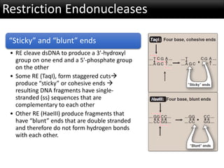
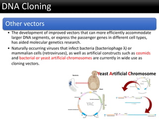
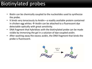
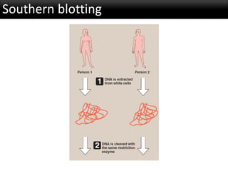
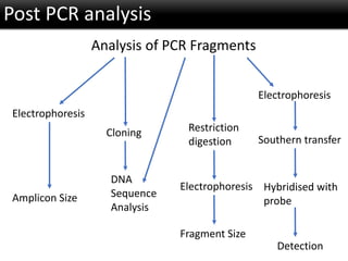
r-DNA Technology
Class outline
Restriction endonucleases
• Permit the dissection of huge DNA molecules into defined
fragments.
Cloning techniques
• Mechanism for amplification of specific nucleotide
sequences.
Specific Probes
• Allowed the identification and manipulation of nucleotide
sequences of interest.
Introduction
The human genome contains approximately three billion (109) base
pairs (bp) that encode 20,000 to 30,000 protein coding genes
located on 23 pairs of chromosomes.
Restriction Endonucleases
Specificity of restriction
endonucleases (RE)
• RE recognize short stretches of DNA
(four to eight bp) that contain
specific nucleotide sequences, which
differ for each restriction enzyme
• These sequences are palindromes
(exhibit twofold rotational
symmetry)  Within a short region
of the double helix, the nucleotide
sequence on the two strands is
identical if each is read in the 5'→3'
direction
Restriction Endonucleases
“Sticky” and “blunt” ends
• RE cleave dsDNA to produce a 3'-hydroxyl
group on one end and a 5'-phosphate group
on the other
• Some RE (TaqI), form staggered cuts
produce “sticky” or cohesive ends 
resulting DNA fragments have single-
stranded (ss) sequences that are
complementary to each other
• Other RE (HaeIII) produce fragments that
have “blunt” ends that are double stranded
and therefore do not form hydrogen bonds
with each other.
Restriction Endonucleases
“Sticky” and “blunt” ends
• Using the enzyme DNA ligase, sticky ends of a DNA
fragment of interest can be covalently joined with
other DNA fragments that have sticky ends
produced by cleavage with the same restriction
endonuclease
• Another ligase, encoded by bacteriophage T4, can
covalently join blunt-ended fragments.
Restriction sites
• A DNA sequence that is recognized and cut by a RE
• RE cleave dsDNA into fragments of different sizes.
• RE that recognizes a specific four-base-pair
sequence produces many cuts in the DNA
molecule, one every 44 bp.
• In contrast, RE requiring a unique sequence of six
base pairs produces fewer cuts (one every 46 bp)
and, hence, longer pieces.
DNA Cloning
Introduction of a foreign DNA molecule into a replicating cell permits the cloning
or amplification of that DNA. (In some cases, a single DNA fragment can be
isolated and purified prior to cloning).
Cloning a nucleotide sequence of interest
• Cellular DNA is cleaved with a specific RE, creating hundreds of thousands of
fragments  Each of the resulting DNA fragments is joined to a DNA vector
molecule (referred to as a cloning vector) to form a hybrid or recombinant
molecule  Each recombinant DNA molecule conveys its inserted DNA
fragment into a single host cell, for example, a bacterium, where it is replicated.
• As the host cell multiplies, it forms a clone in which every bacterium carries
copies of the same inserted DNA fragment, hence the name “cloning.”  The
cloned DNA is eventually released from its vector by cleavage (using the
appropriate restriction endonuclease) and is isolated  Production of many
identical copies of DNA of interest.
DNA Cloning
Vectors
• A vector is a molecule of DNA to which the fragment of DNA to be cloned is joined.
• Common vectors - Plasmids and viruses.
Essential properties of a vector include:
• It must be capable of autonomous replication within a host cell
• It must contain at least one specific nucleotide sequence recognized by a restriction
endonuclease
• It must carry at least one gene that confers the ability to select for the vector, such as
an antibiotic resistance gene
DNA Cloning
Prokaryotic plasmids
• Plasmid DNA undergoes replication that may or may not be
synchronized to chromosomal division.
• Plasmids may carry genes that convey antibiotic resistance to the host
bacterium, and may facilitate the transfer of genetic information from
one bacterium to another.
• Plasmids can be readily isolated from bacterial cells, their circular DNA
cleaved at specific sites by restriction endonucleases, and up to 10 kb
of foreign DNA (cut with the same restriction enzyme) inserted.
• The recombinant plasmid can be introduced into a bacterium, and large
numbers of copies of the plasmid produced.
• The bacteria are grown in the presence of antibiotics, thus selecting for
cells containing the hybrid plasmids, which provide antibiotic
resistance.
DNA Cloning
Other vectors
• The development of improved vectors that can more efficiently accommodate
larger DNA segments, or express the passenger genes in different cell types,
has aided molecular genetics research.
• Naturally occurring viruses that infect bacteria (bacteriophage λ) or
mammalian cells (retroviruses), as well as artificial constructs such as cosmids
and bacterial or yeast artificial chromosomes are currently in wide use as
cloning vectors.
DNA libraries
A DNA library is a collection of cloned restriction fragments of the DNA of
an organism.
Two kinds of libraries are commonly used: genomic libraries &
complementary DNA (cDNA) libraries.
• Genomic libraries ideally contain a copy of every DNA nucleotide sequence in the genome.
• cDNA libraries contain those DNA sequences that only appear as processed mRNA molecules,
and these differ from one cell type to another.
Genomic DNA libraries
A genomic library is created by digestion of the total DNA of the organism with
a RE and subsequent ligation to an appropriate vector  Recombinant DNA
molecules replicate within host bacteria  Amplified DNA fragments thus
represent the entire genome of the organism
Regardless of the RE used, the chances are rather good that the gene of
interest contains more than one restriction site recognized by that enzyme.
• If this is the case, and if the digestion is allowed to go to completion, the gene of interest is
fragmented—that is, it is not contained in any one clone in the library.
• To avoid this usually undesirable result, a partial digestion is performed in which either the amount
or the time of action of the enzyme is limited.
• This results in cleavage occurring at only a fraction of the restriction sites on any one DNA molecule,
thus producing fragments of about 20 kb.
• Enzymes that cut very frequently (that is, those that recognize 4 bp sequences) are generally used
for this purpose so that the result is an almost random collection of fragments.
• This ensures a high degree of probability that the gene of interest is contained, intact, in some
fragment.
cDNA libraries
If a protein-coding gene of interest is expressed
at a high level in a particular tissue, it is likely that
mRNA transcribed from that gene is also present
at high concentrations in the cell
mRNA can be used as a template to make a
complementary DNA (cDNA) molecule using the
enzyme reverse transcriptase
The resulting cDNA is thus a double-stranded
copy of mRNA. cDNA can be amplified by cloning
or by the polymerase chain reaction.
cDNA libraries
It can be used as a probe to locate the gene that coded for
the original mRNA (or fragments of the gene) in mixtures
containing many unrelated DNA fragments. If the mRNA
used as a template is a mixture of many different size
species, the resulting cDNA is heterogeneous.
These mixtures can be cloned to form a cDNA library.
Because cDNA has no intervening sequences, it can be
cloned into an expression vector for the synthesis of
eukaryotic proteins by bacteria
These special plasmids contain a bacterial promoter for
transcription of the cDNA, and a Shine-Dalgarno sequence
that allows the bacterial ribosome to initiate translation of
the resulting mRNA molecule
Sequencing of cloned DNA fragments
The ssDNA to be sequenced is used as the template
for DNA synthesis by DNA polymerase.
A radiolabeled primer complementary to the 3'-end of
the target DNA is added, along with the four dNTPs
Sample is divided into four reaction tubes, and a small
amount of one of the four ddNTP is added to each
tube  As it contains no 3'-OH group, incorporation of
a ddNMP terminates elongation at that point
The products of this reaction, then, consist of a
mixture of DNA strands of different lengths, each
terminating at a specific base
Sequencing of cloned DNA fragments
Probes
Cleavage of large DNA molecules by restriction enzymes produces a
bewildering array of fragments. How can the DNA sequence of interest
be picked out of a mixture of thousands or even millions of irrelevant
DNA fragments?
Probe—
• A short piece of ssDNA, labeled with a radioisotope, such
as 32P, or with a nonradioactive molecule, such as biotin.
• The sequence of a probe is complementary to a sequence
in the DNA of interest, called the target DNA.
• Probes are used to identify which band on a gel or which
clone in a library contains the target DNA, a process called
screening.
Probes
Hybridization of a probe to DNA fragments
• The utility of probes hinges on the phenomenon of
hybridization (or annealing) in which a probe containing a
complementary sequence binds a single-stranded sequence of
a target DNA.
• Alkaline denaturation of dsDNA  ssDNA  Bound to a solid
support (nitrocellulose membrane)  The immobilized DNA
strands are prevented from self-annealing, but are available for
hybridization to an exogenous, radiolabeled, ssDNA probe.
• The extent of hybridization is measured by the retention of
radioactivity on the membrane. Excess probe molecules that do
not hybridize are removed by washing the filter.
Probes
Synthetic oligonucleotide probes
• If the sequence of all or part of the target DNA is known, ss-
oligonucleotide probes of 20–30 nucleotides can be synthesized
that are complementary to a small region of the gene of
interest.
• If the sequence of the gene is unknown, the amino acid
sequence of the protein— the final gene product—may be used
to construct a probe.
• Short, ssDNA sequences (15–30 nucleotides) are synthesized,
using the genetic code as a guide  Because of the degeneracy
of the genetic code, it is necessary to synthesize several
oligonucleotides.
Biotinylated probes
• Biotin can be chemically coupled to the nucleotides used to synthesize
the probe.
• It binds very tenaciously to Avidin—a readily available protein contained
in chicken egg whites  Avidin can be attached to a fluorescent dye
detectable optically with great sensitivity.
• DNA fragment that hybridizes with the biotinylated probe can be made
visible by immersing the gel in a solution of dye-coupled avidin.
• After washing away the excess avidin, the DNA fragment that binds the
probe is fluorescent.
Southern blotting
Southern blotting
Southern blotting
Introduction
PCR is an exponentially progressing synthesis of the
defined target DNA sequences in vitro.
Polymerase
Chain
Reaction
Invented by Dr. Kary Mullis (1983)
 Nobel Prize in Chemistry (1993)
Introduction
Requirements for PCR
dGTP
dTTP
dCTP
dATP
Taq
Template DNA (Genomic DNA,
cDNA, or cell lysates)
Primers - forward and reverse
Each of the four deoxynucleotide
triphosphates (dNTPs)
Thermostable DNA polymerase
(Taq polymerase)
Reaction buffer
The story inside
Step 1: Denaturation
• At 950C, DNA is
denatured
Step 2: Annealing
• At 400C- 650C, Primers
anneal to their
complementary
sequences
Step 3: Extension
• At 720C, DNA Polymerase
extends the DNA chain
PCR protocol
• Temperature Protocol
• Initial Melt: 94ºC for 2 minutes
• Melt: 94ºC for 30 seconds
• Anneal: 55ºC for 30 seconds
• Extend: 72ºC for 1 minute
• Final Extension: 72ºC for 6 minutes
• Hold: 4ºC
30-35
cycles
5’
5’
3’
3’
d.NTPs
Thermal Stable
DNA Polymerase
Primers
Denaturation
Annealing
Add to Reaction Tube
The story inside
Extension
5’ 3’
5’3’
Extension Continued
5’ 3’
5’3’
Taq
Taq
3’
5’3’
Taq
Taq
Repeat
The story inside
5’3’
3’
3’
3’
5’3’
3’
5’3’
3’
Cycle 2
4 Copies
Cycle 3
8 Copies
3’
3’
5’3’
3’
5’3’
3’
5’3’
3’
5’3’
3’
5’3’
3’
5’3’
3’
5’3’
3’
The story inside
Post PCR analysis
Analysis of PCR Fragments
Electrophoresis
Amplicon Size
Electrophoresis
Southern transfer
Hybridised with
probe
Detection
Electrophoresis
Restriction
digestion
Fragment Size
Cloning
DNA
Sequence
Analysis
Electrophoresis
39
Inverse PCR
Overlap extension PCR
 Assemble PCR
 Helicase dependent amplication
 Intersequence-specific PCR(ISSR)
 Ligation-mediated PCR
 Methylation –specific PCR
 Miniprimer PCR
 Multiplex PCR
 Nested PCR
 Solid phase PCR
 Touch down PCR
Other types of PCR
• Poor Precision
• Low sensitivity
• Short dynamic range < 2 logs
• Low resolution
• Non - Automated
• Size-based discrimination only
• Results are not expressed as
numbers
• Ethidium bromide for staining is
not very quantitative
• Post PCR processing
Limitations of PCR
Real Time PCR
Real-Time PCR is a specialized technique that allows a PCR
reaction to be visualized “in real time” as the reaction
progresses.
• Components:
• Thermal Cycler (PCR
machine)
• Optical Module (to detect
fluorescence in the tubes
during the run)
Detection in Real Time PCR
0
500000
1000000
1500000
2000000
2500000
3000000
3500000
4000000
4500000
5000000
0 5 10 15 20 25 30 35 40
Cycle Sample A Sample B
23 250,000 1,000,000
24 500,000 2,000,000
25 1,000,000 4,000,000
0
500000
1000000
1500000
2000000
2500000
3000000
3500000
4000000
4500000
5000000
0 5 10 15 20 25 30 35 40
Restriction Fragment Length Polymorphism (RFLP)
• RFLP is a genetic variant that can be observed by cleaving the DNA into fragments
(restriction fragments) with a restriction enzyme.
• The length of the restriction fragments is altered if the genetic variant alters the DNA
so as to create or abolish a site of restriction endonuclease cleavage
• RFLP can be used to detect human genetic variations, e.g., in prospective parents or
in fetal tissue.
Genome variations are differences in the sequence of DNA among individuals.
It has been estimated that the genomes of nonrelated people differ at about one
in 1,200 DNA bases, or about 0.1% of the genome.
These genome variations include both mutations and polymorphisms.
A mutation refers to an
infrequent, but
potentially harmful,
genome variation that is
associated with a specific
human disease.
• A polymorphism is a clinically harmless DNA
variation that does not affect the phenotype.
• It is traditionally defined as a sequence variation at a
given locus (allele) in more than 1% of a population.
• Polymorphisms primarily occur in regions of the
genome that do not encode proteins, that is, in
introns and intergenic regions.
Restriction Fragment Length Polymorphism (RFLP)
DNA variations resulting in RFLP
Single base changes in DNA
• About 90% of human genome variation comes in
the form of single-nucleotide polymorphisms,
(SNPs), that is, variations that involve just one
base.
• The substitution of one nucleotide at a restriction
site can render the site unrecognizable by a
particular restriction endonuclease.
• A new restriction site can also be created by the
same mechanism. In either case, cleavage with an
endonuclease results in fragments of lengths
differing from the normal, which can be detected
by DNA hybridization
Restriction Fragment Length Polymorphism (RFLP)
DNA variations resulting in RFLP
Variable Number Tandem repeats (VNTR)
• These are short sequences of DNA at scattered locations in the genome, repeated in
tandem (one after another).
• The number of these repeat units varies from person to person, but is unique for any given
individual and, therefore, serves as a molecular fingerprint.
• Cleavage by restriction enzymes yields fragments that vary in length depending on how
many repeated segments are contained in the fragment.
• Variation in the number of tandem repeats can lead to polymorphisms.
• Many different VNTR loci have been identified, and are extremely useful for DNA
fingerprint analysis, such as in forensic and paternity identity cases.
Restriction Fragment Length Polymorphism (RFLP)
Analysis of Gene Expression
1. Northern blots:
• Northern blots are very similar to
Southern blots, except that the
original sample contains a mixture of
mRNA molecules that are separated
by electrophoresis, then transferred
to a membrane and hybridized to a
radiolabeled probe.
• The bands obtained by
autoradiography give a measure of
the amount and size of particular
mRNA molecules in the sample.
Determination of mRNA levels
Messenger RNA levels are usually determined by the hybridization of labeled probes to either
mRNA itself or to cDNA produced from mRNA.
2. Microarrays:
• DNA microarrays contain thousands of
immobilized DNA sequences organized
in an area no larger than a microscope
slide.
• These microarrays are used to analyze
a sample for the presence of gene
variations or mutations (genotyping),
or to determine the patterns of mRNA
production (gene expression analysis),
analyzing thousands of genes at the
same time.
Analysis of Gene Expression
Determination of mRNA levels
• Microarray
• For genotyping analysis, the cellular sample
is genomic DNA.
• For expression analysis, the population of
mRNA molecules from a particular cell type
is converted to cDNA and labeled with a
fluorescent tag
Analysis of Gene Expression
Determination of mRNA levels
• Microarray
• This mixture is then exposed to a gene
chip, which is a glass slide or
membrane containing thousands of
tiny spots of DNA, each corresponding
to a different gene.
• The amount of fluorescence bound to
each spot is a measure of the amount
of that particular mRNA in the sample.
• DNA microarrays are often used to
determine the differing patterns of
gene expression in two different types
of cell—for example, normal and
cancer cells.
Analysis of Gene Expression
Analysis of Protein levels
1. Enzyme-linked immunosorbent
assays (ELISA)
2. Western Blots
3. Proteomics
Gene therapy
• Aim - To insert the normal, cloned DNA for a gene into the somatic cells
of a patient who has a defect in that gene as a result of some disease-
causing mutation.
• Mechanism - DNA must become permanently integrated into the
patient’s chromosomes in such a way as to be properly expressed to
produce the correct protein.
• Example - Patients with SCID have an immune deficiency as a result of
mutations in either the gene for adenosine deaminase or a gene coding
for an interleukin receptor subunit (X-linked severe combined
immunodeficiency, or SCIDX1).
• Patients with both kinds of SCID have been successfully treated by
incorporating functional copies of the appropriate gene into their
cells
• Risks - Retrovirus-mediated gene transfer was able to correct SCID-X1 in
nine of ten patients  leukemia's developed in several of the patients,
presumably because of activation of a hematopoietic oncogene.
Summary
Recombinant DNA Technology

Recombinant DNA Technology

  • 1.
  • 2.
    Class outline Restriction endonucleases •Permit the dissection of huge DNA molecules into defined fragments. Cloning techniques • Mechanism for amplification of specific nucleotide sequences. Specific Probes • Allowed the identification and manipulation of nucleotide sequences of interest.
  • 3.
    Introduction The human genomecontains approximately three billion (109) base pairs (bp) that encode 20,000 to 30,000 protein coding genes located on 23 pairs of chromosomes.
  • 4.
    Restriction Endonucleases Specificity ofrestriction endonucleases (RE) • RE recognize short stretches of DNA (four to eight bp) that contain specific nucleotide sequences, which differ for each restriction enzyme • These sequences are palindromes (exhibit twofold rotational symmetry)  Within a short region of the double helix, the nucleotide sequence on the two strands is identical if each is read in the 5'→3' direction
  • 5.
    Restriction Endonucleases “Sticky” and“blunt” ends • RE cleave dsDNA to produce a 3'-hydroxyl group on one end and a 5'-phosphate group on the other • Some RE (TaqI), form staggered cuts produce “sticky” or cohesive ends  resulting DNA fragments have single- stranded (ss) sequences that are complementary to each other • Other RE (HaeIII) produce fragments that have “blunt” ends that are double stranded and therefore do not form hydrogen bonds with each other.
  • 6.
    Restriction Endonucleases “Sticky” and“blunt” ends • Using the enzyme DNA ligase, sticky ends of a DNA fragment of interest can be covalently joined with other DNA fragments that have sticky ends produced by cleavage with the same restriction endonuclease • Another ligase, encoded by bacteriophage T4, can covalently join blunt-ended fragments. Restriction sites • A DNA sequence that is recognized and cut by a RE • RE cleave dsDNA into fragments of different sizes. • RE that recognizes a specific four-base-pair sequence produces many cuts in the DNA molecule, one every 44 bp. • In contrast, RE requiring a unique sequence of six base pairs produces fewer cuts (one every 46 bp) and, hence, longer pieces.
  • 7.
    DNA Cloning Introduction ofa foreign DNA molecule into a replicating cell permits the cloning or amplification of that DNA. (In some cases, a single DNA fragment can be isolated and purified prior to cloning). Cloning a nucleotide sequence of interest • Cellular DNA is cleaved with a specific RE, creating hundreds of thousands of fragments  Each of the resulting DNA fragments is joined to a DNA vector molecule (referred to as a cloning vector) to form a hybrid or recombinant molecule  Each recombinant DNA molecule conveys its inserted DNA fragment into a single host cell, for example, a bacterium, where it is replicated. • As the host cell multiplies, it forms a clone in which every bacterium carries copies of the same inserted DNA fragment, hence the name “cloning.”  The cloned DNA is eventually released from its vector by cleavage (using the appropriate restriction endonuclease) and is isolated  Production of many identical copies of DNA of interest.
  • 8.
    DNA Cloning Vectors • Avector is a molecule of DNA to which the fragment of DNA to be cloned is joined. • Common vectors - Plasmids and viruses. Essential properties of a vector include: • It must be capable of autonomous replication within a host cell • It must contain at least one specific nucleotide sequence recognized by a restriction endonuclease • It must carry at least one gene that confers the ability to select for the vector, such as an antibiotic resistance gene
  • 9.
    DNA Cloning Prokaryotic plasmids •Plasmid DNA undergoes replication that may or may not be synchronized to chromosomal division. • Plasmids may carry genes that convey antibiotic resistance to the host bacterium, and may facilitate the transfer of genetic information from one bacterium to another. • Plasmids can be readily isolated from bacterial cells, their circular DNA cleaved at specific sites by restriction endonucleases, and up to 10 kb of foreign DNA (cut with the same restriction enzyme) inserted. • The recombinant plasmid can be introduced into a bacterium, and large numbers of copies of the plasmid produced. • The bacteria are grown in the presence of antibiotics, thus selecting for cells containing the hybrid plasmids, which provide antibiotic resistance.
  • 11.
    DNA Cloning Other vectors •The development of improved vectors that can more efficiently accommodate larger DNA segments, or express the passenger genes in different cell types, has aided molecular genetics research. • Naturally occurring viruses that infect bacteria (bacteriophage λ) or mammalian cells (retroviruses), as well as artificial constructs such as cosmids and bacterial or yeast artificial chromosomes are currently in wide use as cloning vectors.
  • 12.
    DNA libraries A DNAlibrary is a collection of cloned restriction fragments of the DNA of an organism. Two kinds of libraries are commonly used: genomic libraries & complementary DNA (cDNA) libraries. • Genomic libraries ideally contain a copy of every DNA nucleotide sequence in the genome. • cDNA libraries contain those DNA sequences that only appear as processed mRNA molecules, and these differ from one cell type to another.
  • 13.
    Genomic DNA libraries Agenomic library is created by digestion of the total DNA of the organism with a RE and subsequent ligation to an appropriate vector  Recombinant DNA molecules replicate within host bacteria  Amplified DNA fragments thus represent the entire genome of the organism Regardless of the RE used, the chances are rather good that the gene of interest contains more than one restriction site recognized by that enzyme. • If this is the case, and if the digestion is allowed to go to completion, the gene of interest is fragmented—that is, it is not contained in any one clone in the library. • To avoid this usually undesirable result, a partial digestion is performed in which either the amount or the time of action of the enzyme is limited. • This results in cleavage occurring at only a fraction of the restriction sites on any one DNA molecule, thus producing fragments of about 20 kb. • Enzymes that cut very frequently (that is, those that recognize 4 bp sequences) are generally used for this purpose so that the result is an almost random collection of fragments. • This ensures a high degree of probability that the gene of interest is contained, intact, in some fragment.
  • 14.
    cDNA libraries If aprotein-coding gene of interest is expressed at a high level in a particular tissue, it is likely that mRNA transcribed from that gene is also present at high concentrations in the cell mRNA can be used as a template to make a complementary DNA (cDNA) molecule using the enzyme reverse transcriptase The resulting cDNA is thus a double-stranded copy of mRNA. cDNA can be amplified by cloning or by the polymerase chain reaction.
  • 15.
    cDNA libraries It canbe used as a probe to locate the gene that coded for the original mRNA (or fragments of the gene) in mixtures containing many unrelated DNA fragments. If the mRNA used as a template is a mixture of many different size species, the resulting cDNA is heterogeneous. These mixtures can be cloned to form a cDNA library. Because cDNA has no intervening sequences, it can be cloned into an expression vector for the synthesis of eukaryotic proteins by bacteria These special plasmids contain a bacterial promoter for transcription of the cDNA, and a Shine-Dalgarno sequence that allows the bacterial ribosome to initiate translation of the resulting mRNA molecule
  • 16.
    Sequencing of clonedDNA fragments The ssDNA to be sequenced is used as the template for DNA synthesis by DNA polymerase. A radiolabeled primer complementary to the 3'-end of the target DNA is added, along with the four dNTPs Sample is divided into four reaction tubes, and a small amount of one of the four ddNTP is added to each tube  As it contains no 3'-OH group, incorporation of a ddNMP terminates elongation at that point The products of this reaction, then, consist of a mixture of DNA strands of different lengths, each terminating at a specific base
  • 17.
    Sequencing of clonedDNA fragments
  • 18.
    Probes Cleavage of largeDNA molecules by restriction enzymes produces a bewildering array of fragments. How can the DNA sequence of interest be picked out of a mixture of thousands or even millions of irrelevant DNA fragments? Probe— • A short piece of ssDNA, labeled with a radioisotope, such as 32P, or with a nonradioactive molecule, such as biotin. • The sequence of a probe is complementary to a sequence in the DNA of interest, called the target DNA. • Probes are used to identify which band on a gel or which clone in a library contains the target DNA, a process called screening.
  • 19.
    Probes Hybridization of aprobe to DNA fragments • The utility of probes hinges on the phenomenon of hybridization (or annealing) in which a probe containing a complementary sequence binds a single-stranded sequence of a target DNA. • Alkaline denaturation of dsDNA  ssDNA  Bound to a solid support (nitrocellulose membrane)  The immobilized DNA strands are prevented from self-annealing, but are available for hybridization to an exogenous, radiolabeled, ssDNA probe. • The extent of hybridization is measured by the retention of radioactivity on the membrane. Excess probe molecules that do not hybridize are removed by washing the filter.
  • 20.
    Probes Synthetic oligonucleotide probes •If the sequence of all or part of the target DNA is known, ss- oligonucleotide probes of 20–30 nucleotides can be synthesized that are complementary to a small region of the gene of interest. • If the sequence of the gene is unknown, the amino acid sequence of the protein— the final gene product—may be used to construct a probe. • Short, ssDNA sequences (15–30 nucleotides) are synthesized, using the genetic code as a guide  Because of the degeneracy of the genetic code, it is necessary to synthesize several oligonucleotides.
  • 21.
    Biotinylated probes • Biotincan be chemically coupled to the nucleotides used to synthesize the probe. • It binds very tenaciously to Avidin—a readily available protein contained in chicken egg whites  Avidin can be attached to a fluorescent dye detectable optically with great sensitivity. • DNA fragment that hybridizes with the biotinylated probe can be made visible by immersing the gel in a solution of dye-coupled avidin. • After washing away the excess avidin, the DNA fragment that binds the probe is fluorescent.
  • 22.
  • 23.
  • 24.
  • 25.
    Introduction PCR is anexponentially progressing synthesis of the defined target DNA sequences in vitro. Polymerase Chain Reaction Invented by Dr. Kary Mullis (1983)  Nobel Prize in Chemistry (1993)
  • 26.
  • 27.
    Requirements for PCR dGTP dTTP dCTP dATP Taq TemplateDNA (Genomic DNA, cDNA, or cell lysates) Primers - forward and reverse Each of the four deoxynucleotide triphosphates (dNTPs) Thermostable DNA polymerase (Taq polymerase) Reaction buffer
  • 28.
    The story inside Step1: Denaturation • At 950C, DNA is denatured Step 2: Annealing • At 400C- 650C, Primers anneal to their complementary sequences Step 3: Extension • At 720C, DNA Polymerase extends the DNA chain
  • 29.
    PCR protocol • TemperatureProtocol • Initial Melt: 94ºC for 2 minutes • Melt: 94ºC for 30 seconds • Anneal: 55ºC for 30 seconds • Extend: 72ºC for 1 minute • Final Extension: 72ºC for 6 minutes • Hold: 4ºC 30-35 cycles
  • 30.
  • 31.
    Extension 5’ 3’ 5’3’ Extension Continued 5’3’ 5’3’ Taq Taq 3’ 5’3’ Taq Taq Repeat The story inside
  • 32.
    5’3’ 3’ 3’ 3’ 5’3’ 3’ 5’3’ 3’ Cycle 2 4 Copies Cycle3 8 Copies 3’ 3’ 5’3’ 3’ 5’3’ 3’ 5’3’ 3’ 5’3’ 3’ 5’3’ 3’ 5’3’ 3’ 5’3’ 3’ The story inside
  • 33.
    Post PCR analysis Analysisof PCR Fragments Electrophoresis Amplicon Size Electrophoresis Southern transfer Hybridised with probe Detection Electrophoresis Restriction digestion Fragment Size Cloning DNA Sequence Analysis
  • 34.
  • 35.
    39 Inverse PCR Overlap extensionPCR  Assemble PCR  Helicase dependent amplication  Intersequence-specific PCR(ISSR)  Ligation-mediated PCR  Methylation –specific PCR  Miniprimer PCR  Multiplex PCR  Nested PCR  Solid phase PCR  Touch down PCR Other types of PCR
  • 36.
    • Poor Precision •Low sensitivity • Short dynamic range < 2 logs • Low resolution • Non - Automated • Size-based discrimination only • Results are not expressed as numbers • Ethidium bromide for staining is not very quantitative • Post PCR processing Limitations of PCR
  • 37.
    Real Time PCR Real-TimePCR is a specialized technique that allows a PCR reaction to be visualized “in real time” as the reaction progresses. • Components: • Thermal Cycler (PCR machine) • Optical Module (to detect fluorescence in the tubes during the run)
  • 38.
    Detection in RealTime PCR 0 500000 1000000 1500000 2000000 2500000 3000000 3500000 4000000 4500000 5000000 0 5 10 15 20 25 30 35 40 Cycle Sample A Sample B 23 250,000 1,000,000 24 500,000 2,000,000 25 1,000,000 4,000,000 0 500000 1000000 1500000 2000000 2500000 3000000 3500000 4000000 4500000 5000000 0 5 10 15 20 25 30 35 40
  • 39.
    Restriction Fragment LengthPolymorphism (RFLP) • RFLP is a genetic variant that can be observed by cleaving the DNA into fragments (restriction fragments) with a restriction enzyme. • The length of the restriction fragments is altered if the genetic variant alters the DNA so as to create or abolish a site of restriction endonuclease cleavage • RFLP can be used to detect human genetic variations, e.g., in prospective parents or in fetal tissue. Genome variations are differences in the sequence of DNA among individuals. It has been estimated that the genomes of nonrelated people differ at about one in 1,200 DNA bases, or about 0.1% of the genome. These genome variations include both mutations and polymorphisms. A mutation refers to an infrequent, but potentially harmful, genome variation that is associated with a specific human disease. • A polymorphism is a clinically harmless DNA variation that does not affect the phenotype. • It is traditionally defined as a sequence variation at a given locus (allele) in more than 1% of a population. • Polymorphisms primarily occur in regions of the genome that do not encode proteins, that is, in introns and intergenic regions.
  • 40.
    Restriction Fragment LengthPolymorphism (RFLP) DNA variations resulting in RFLP Single base changes in DNA • About 90% of human genome variation comes in the form of single-nucleotide polymorphisms, (SNPs), that is, variations that involve just one base. • The substitution of one nucleotide at a restriction site can render the site unrecognizable by a particular restriction endonuclease. • A new restriction site can also be created by the same mechanism. In either case, cleavage with an endonuclease results in fragments of lengths differing from the normal, which can be detected by DNA hybridization
  • 41.
    Restriction Fragment LengthPolymorphism (RFLP) DNA variations resulting in RFLP Variable Number Tandem repeats (VNTR) • These are short sequences of DNA at scattered locations in the genome, repeated in tandem (one after another). • The number of these repeat units varies from person to person, but is unique for any given individual and, therefore, serves as a molecular fingerprint. • Cleavage by restriction enzymes yields fragments that vary in length depending on how many repeated segments are contained in the fragment. • Variation in the number of tandem repeats can lead to polymorphisms. • Many different VNTR loci have been identified, and are extremely useful for DNA fingerprint analysis, such as in forensic and paternity identity cases.
  • 42.
    Restriction Fragment LengthPolymorphism (RFLP)
  • 43.
    Analysis of GeneExpression 1. Northern blots: • Northern blots are very similar to Southern blots, except that the original sample contains a mixture of mRNA molecules that are separated by electrophoresis, then transferred to a membrane and hybridized to a radiolabeled probe. • The bands obtained by autoradiography give a measure of the amount and size of particular mRNA molecules in the sample. Determination of mRNA levels Messenger RNA levels are usually determined by the hybridization of labeled probes to either mRNA itself or to cDNA produced from mRNA. 2. Microarrays: • DNA microarrays contain thousands of immobilized DNA sequences organized in an area no larger than a microscope slide. • These microarrays are used to analyze a sample for the presence of gene variations or mutations (genotyping), or to determine the patterns of mRNA production (gene expression analysis), analyzing thousands of genes at the same time.
  • 44.
    Analysis of GeneExpression Determination of mRNA levels • Microarray • For genotyping analysis, the cellular sample is genomic DNA. • For expression analysis, the population of mRNA molecules from a particular cell type is converted to cDNA and labeled with a fluorescent tag
  • 45.
    Analysis of GeneExpression Determination of mRNA levels • Microarray • This mixture is then exposed to a gene chip, which is a glass slide or membrane containing thousands of tiny spots of DNA, each corresponding to a different gene. • The amount of fluorescence bound to each spot is a measure of the amount of that particular mRNA in the sample. • DNA microarrays are often used to determine the differing patterns of gene expression in two different types of cell—for example, normal and cancer cells.
  • 46.
    Analysis of GeneExpression Analysis of Protein levels 1. Enzyme-linked immunosorbent assays (ELISA) 2. Western Blots 3. Proteomics
  • 47.
    Gene therapy • Aim- To insert the normal, cloned DNA for a gene into the somatic cells of a patient who has a defect in that gene as a result of some disease- causing mutation. • Mechanism - DNA must become permanently integrated into the patient’s chromosomes in such a way as to be properly expressed to produce the correct protein. • Example - Patients with SCID have an immune deficiency as a result of mutations in either the gene for adenosine deaminase or a gene coding for an interleukin receptor subunit (X-linked severe combined immunodeficiency, or SCIDX1). • Patients with both kinds of SCID have been successfully treated by incorporating functional copies of the appropriate gene into their cells • Risks - Retrovirus-mediated gene transfer was able to correct SCID-X1 in nine of ten patients  leukemia's developed in several of the patients, presumably because of activation of a hematopoietic oncogene.
  • 48.