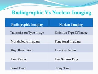
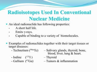
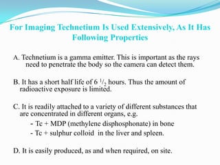
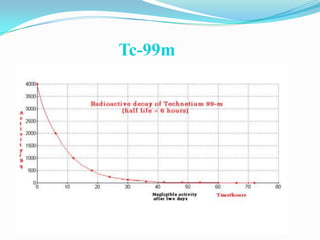
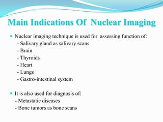
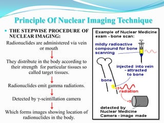
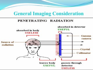
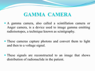
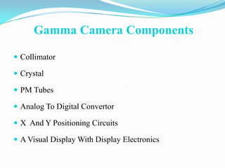
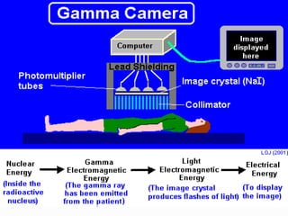
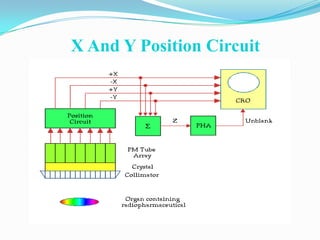
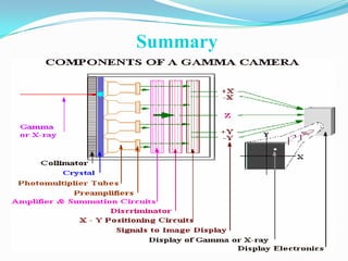
NUCLEAR IMAGING
   Prepared By:
   Muhammad Yaseen
   Trainee Med. Physicist
   Radiology Department
   Mail: myaseena@hotmail.com
Contents
 Introduction
 Advantages Of Nuclear Imaging
 Radiography Vs Nuclear Imaging
 Radioisotopes used in NM
 Radiopharmaceuticals
 Gamma Camera
 SPECT
 Safety
 Summary
INTRODUCTION
 DEFINITION:           Nuclear imaging is a method of producing
  images by detecting radiation from different parts of the body
  after a radioactive tracer material is administered.

 The images are recorded on computer and on film.

 The nuclear imaging physician interprets the images to make a
  diagnosis.

 Radioactive tracers used in nuclear medicine are, in most cases,
  injected into a vein.
Advantages Of Nuclear Medicine

 Target tissue function is investigated.

 All similar target tissues can be examined during one
  investigation, e.g. the whole skeleton can be imaged
  during one bone scan.

 Computer analysis and enhancement of results are
  available.
Radiographic Vs Nuclear Imaging

Radiographic Imaging         Nuclear Imaging

Transmission Type Image   Emission Type Of Image

Morphologic Imaging       Functional Imaging

High Resolution           Low Resolution

Use X-rays                Use Gamma Rays

Short Time                Long Time
Radioisotopes Used In Conventional
          Nuclear Medicine
 An ideal radionuclide has following properties:
  -      A short half life.
  -      Emits γ-rays.
  -      Capable of binding to a variety of biomolecules.

 Examples of radionuclides together with their target tissues or
  target diseases:
    - Technetium (99mTc)      – Salivary glands, thyroid, bone,
                                 blood, liver, lung & heart.
      - Iodine (131I )        – Thyroid
      - Gallium (67Ga)        – Tumors & inflammation
For Imaging Technetium Is Used Extensively, As It Has
                Following Properties

A. Technetium is a gamma emitter. This is important as the rays
    need to penetrate the body so the camera can detect them.

B. It has a short half life of 6 1/2 hours. Thus the amount of
  radioactive exposure is limited.

C. It is readily attached to a variety of different substances that
  are concentrated in different organs, e.g.
       - Tc + MDP (methylene disphosphonate) in bone
       - Tc + sulphur colloid in the liver and spleen.

D. It is easily produced, as and when required, on site.
Tc-99m
Main Indications Of Nuclear Imaging
 Nuclear imaging technique is used for assessing function of:
   - Salivary gland as salivary scans
   - Brain
   - Thyroids
   - Heart
   - Lungs
   - Gastro-intestinal system

 It is also used for diagnosis of:
  - Metastatic diseases
  - Bone tumors as bone scans
Principle Of Nuclear Imaging Technique
 THE STEPWISE PROCEDURE OF
 NUCLEAR IMAGING:
Radionuclides are administered via vein
                or mouth

They distribute in the body according to
  their strength for particular tissues so
            called target tissues.

 Radionuclides emit gamma radiations.

   Detected by γ-scintillation camera

Which forms images showing location of
        radionuclides in the body.
Bone Scan & Thyroid Scan Images
Pharmaceutical
Organ                      Pharmaceutical
          Tc-99m pertechnetate
 Brain    Tc-99m DTPA
          Tc-99m glucoheptonate
Cardiac   Tc-99m pyrophosphate

 Liver    Tc-99m sulfur colloid

          Tc-99m DTPA
Kidney    Tc-99m DMSA
          Tc-99m glucoheptonate
          I-131
Thyroid   I-131 Hippuran
General Imaging Consideration
GAMMA CAMERA
 A gamma camera, also called a scintillation camera or
 Anger camera, is a device used to image gamma emitting
 radioisotopes, a technique known as scintigraphy.

 These cameras capture photons and convert them to light
 and then to a voltage signal.

 These signals are reconstructed to an image that shows
 distribution of radionuclide in the patient.
Gamma Camera Components

 Collimator

 Crystal

 PM Tubes

 Analog To Digital Convertor

 X And Y Positioning Circuits

 A Visual Display With Display Electronics
Collimator
 The collimator can be made from lead foil.

 The collimator stops about 99.9% of the available
 photons.

 The walls of each channel in the collimator are called
 septa, and if a photon manages to penetrate the wall, it
 is called septal penetration.
Types Of Collimator

 Parallel Hole Collimator

 Converging Collimator

 Diverging Collimator

 Pin Hole Collimator
Parallel Hole Collimator
 LEGP (low energy general
  purpose) or LEAP (low
  energy all purpose).

 To increase the resolution,
  smaller diameter holes are
  needed.

 To increase sensitivity, the
  holes need to be wider.
Parallel Hole Collimator
 To image higher energy
  isotopes such as Ga-67 or
 I-131, the collimator needs
 to have thicker septa in
 order to stop penetration.

 This produces a heavier
 collimator with lower
 sensitivity.
Pin Hole ,Converging &Diverging
           Collimators


 Converging
Crystal (NaI)
 The γ-rays that pass through the collimator then strike
 scintillation crystal.
 Made up of sodium iodide with trace amount of
 thallium.
 This crystal shows florescence when it absorbs γ-rays.
 These flashes of light are detected by photomultiplier
 tubes coupled to the crystal.
Photo Multiplier Tube
 Extremely sensitive detector of light in the ultraviolet,
  visible and near infrared
 Multiplies the signal produced by incident light by as
  much as 108
   • single photons can be resolved
 High gain, low noise, high frequency response, and large
  area of collection
 A tiny and normally undetectable current becomes a
  much larger and easily measurable current
Components Of PMT

 Made of a glass

  vacuum tube

 Photocathode

 Several dynodes

 One anode
How It Works
Analog To Digital Convertor
 The signals from photomultiplier tubes go through an analog to
  digital converter (ADC)

 This component is used to convert the analogue information
  produced by the imaging system so that it is coded in the form
  of binary numbers.

 In this way the analog signal is digitalized & used to produce
  image by computer
X And Y Position Circuit
Further Developments in Radioisotope
     Imaging Techniques Include

 SPECT (single photon emission computed tomography)

                 And

 PET (Positron emission tomography)
Single Photon Emission Computed
           Tomography
 SPECT is a method of acquiring tomographic slices
  through a patient.

 Most gamma cameras have SPECT capability.

 In this technique either a single or multiple gamma
  cameras is rotated 360 degrees about the patient.

 Image acquisition takes about 30 – 45 min.
Applications of SPECT

 Heart Imaging

 Brain Imaging

 Tumor detection SPECT can be used to detect tumors in

 cancer patients in the early stages.

 Bone Scans
Advantages of SPECT

 Better detailed resolution

 Enhanced contrast

 Localization of defects is more precise and more
 clearly seen.

 Extend and size of defects is better defined.
Limitation Of Nuclear Medicine
 Poor image resolution – only minimal information of target
  tissue is obtained.

 The radiation dose to the whole body can be relatively high.


 Images are not usually disease-specific.


 Difficult to localize exact anatomical site of source of emission.


 Facilities are not widely available.
Safety Precautions
 Injected patient should avoid to keep away from everyone

  but specially from children and pregnant women

 Only authorized users are allowed to handle the source.

 Do not look directly into bore hole of the source holder or
  cover it with any part of your body.

 Use Lead Apron, lead goggles and lead thyroid shield
Summary
References
 Medical instrumentation by :

  Jennifer Prekeges

 The essential physics of medical imaging by:

  Bush Berg
Muhammad yaseen

Muhammad yaseen

  • 1.
    NUCLEAR IMAGING Prepared By: Muhammad Yaseen Trainee Med. Physicist Radiology Department Mail: myaseena@hotmail.com
  • 2.
    Contents  Introduction  AdvantagesOf Nuclear Imaging  Radiography Vs Nuclear Imaging  Radioisotopes used in NM  Radiopharmaceuticals  Gamma Camera  SPECT  Safety  Summary
  • 3.
    INTRODUCTION  DEFINITION: Nuclear imaging is a method of producing images by detecting radiation from different parts of the body after a radioactive tracer material is administered.  The images are recorded on computer and on film.  The nuclear imaging physician interprets the images to make a diagnosis.  Radioactive tracers used in nuclear medicine are, in most cases, injected into a vein.
  • 4.
    Advantages Of NuclearMedicine  Target tissue function is investigated.  All similar target tissues can be examined during one investigation, e.g. the whole skeleton can be imaged during one bone scan.  Computer analysis and enhancement of results are available.
  • 5.
    Radiographic Vs NuclearImaging Radiographic Imaging Nuclear Imaging Transmission Type Image Emission Type Of Image Morphologic Imaging Functional Imaging High Resolution Low Resolution Use X-rays Use Gamma Rays Short Time Long Time
  • 6.
    Radioisotopes Used InConventional Nuclear Medicine  An ideal radionuclide has following properties: - A short half life. - Emits γ-rays. - Capable of binding to a variety of biomolecules.  Examples of radionuclides together with their target tissues or target diseases: - Technetium (99mTc) – Salivary glands, thyroid, bone, blood, liver, lung & heart. - Iodine (131I ) – Thyroid - Gallium (67Ga) – Tumors & inflammation
  • 7.
    For Imaging TechnetiumIs Used Extensively, As It Has Following Properties A. Technetium is a gamma emitter. This is important as the rays need to penetrate the body so the camera can detect them. B. It has a short half life of 6 1/2 hours. Thus the amount of radioactive exposure is limited. C. It is readily attached to a variety of different substances that are concentrated in different organs, e.g. - Tc + MDP (methylene disphosphonate) in bone - Tc + sulphur colloid in the liver and spleen. D. It is easily produced, as and when required, on site.
  • 8.
  • 9.
    Main Indications OfNuclear Imaging  Nuclear imaging technique is used for assessing function of: - Salivary gland as salivary scans - Brain - Thyroids - Heart - Lungs - Gastro-intestinal system  It is also used for diagnosis of: - Metastatic diseases - Bone tumors as bone scans
  • 10.
    Principle Of NuclearImaging Technique  THE STEPWISE PROCEDURE OF NUCLEAR IMAGING: Radionuclides are administered via vein or mouth They distribute in the body according to their strength for particular tissues so called target tissues. Radionuclides emit gamma radiations. Detected by γ-scintillation camera Which forms images showing location of radionuclides in the body.
  • 11.
    Bone Scan &Thyroid Scan Images
  • 12.
    Pharmaceutical Organ Pharmaceutical Tc-99m pertechnetate Brain Tc-99m DTPA Tc-99m glucoheptonate Cardiac Tc-99m pyrophosphate Liver Tc-99m sulfur colloid Tc-99m DTPA Kidney Tc-99m DMSA Tc-99m glucoheptonate I-131 Thyroid I-131 Hippuran
  • 13.
  • 14.
    GAMMA CAMERA  Agamma camera, also called a scintillation camera or Anger camera, is a device used to image gamma emitting radioisotopes, a technique known as scintigraphy.  These cameras capture photons and convert them to light and then to a voltage signal.  These signals are reconstructed to an image that shows distribution of radionuclide in the patient.
  • 15.
    Gamma Camera Components Collimator  Crystal  PM Tubes  Analog To Digital Convertor  X And Y Positioning Circuits  A Visual Display With Display Electronics
  • 17.
    Collimator  The collimatorcan be made from lead foil.  The collimator stops about 99.9% of the available photons.  The walls of each channel in the collimator are called septa, and if a photon manages to penetrate the wall, it is called septal penetration.
  • 18.
    Types Of Collimator Parallel Hole Collimator  Converging Collimator  Diverging Collimator  Pin Hole Collimator
  • 19.
    Parallel Hole Collimator LEGP (low energy general purpose) or LEAP (low energy all purpose).  To increase the resolution, smaller diameter holes are needed.  To increase sensitivity, the holes need to be wider.
  • 20.
    Parallel Hole Collimator To image higher energy isotopes such as Ga-67 or I-131, the collimator needs to have thicker septa in order to stop penetration.  This produces a heavier collimator with lower sensitivity.
  • 21.
    Pin Hole ,Converging&Diverging Collimators Converging
  • 22.
    Crystal (NaI)  Theγ-rays that pass through the collimator then strike scintillation crystal.  Made up of sodium iodide with trace amount of thallium.  This crystal shows florescence when it absorbs γ-rays.  These flashes of light are detected by photomultiplier tubes coupled to the crystal.
  • 23.
    Photo Multiplier Tube Extremely sensitive detector of light in the ultraviolet, visible and near infrared  Multiplies the signal produced by incident light by as much as 108 • single photons can be resolved  High gain, low noise, high frequency response, and large area of collection  A tiny and normally undetectable current becomes a much larger and easily measurable current
  • 24.
    Components Of PMT Made of a glass vacuum tube  Photocathode  Several dynodes  One anode
  • 25.
  • 26.
    Analog To DigitalConvertor  The signals from photomultiplier tubes go through an analog to digital converter (ADC)  This component is used to convert the analogue information produced by the imaging system so that it is coded in the form of binary numbers.  In this way the analog signal is digitalized & used to produce image by computer
  • 27.
    X And YPosition Circuit
  • 28.
    Further Developments inRadioisotope Imaging Techniques Include  SPECT (single photon emission computed tomography) And  PET (Positron emission tomography)
  • 29.
    Single Photon EmissionComputed Tomography  SPECT is a method of acquiring tomographic slices through a patient.  Most gamma cameras have SPECT capability.  In this technique either a single or multiple gamma cameras is rotated 360 degrees about the patient.  Image acquisition takes about 30 – 45 min.
  • 30.
    Applications of SPECT Heart Imaging  Brain Imaging  Tumor detection SPECT can be used to detect tumors in cancer patients in the early stages.  Bone Scans
  • 31.
    Advantages of SPECT Better detailed resolution  Enhanced contrast  Localization of defects is more precise and more clearly seen.  Extend and size of defects is better defined.
  • 32.
    Limitation Of NuclearMedicine  Poor image resolution – only minimal information of target tissue is obtained.  The radiation dose to the whole body can be relatively high.  Images are not usually disease-specific.  Difficult to localize exact anatomical site of source of emission.  Facilities are not widely available.
  • 33.
    Safety Precautions  Injectedpatient should avoid to keep away from everyone but specially from children and pregnant women  Only authorized users are allowed to handle the source.  Do not look directly into bore hole of the source holder or cover it with any part of your body.  Use Lead Apron, lead goggles and lead thyroid shield
  • 34.
  • 35.
    References  Medical instrumentationby : Jennifer Prekeges  The essential physics of medical imaging by: Bush Berg