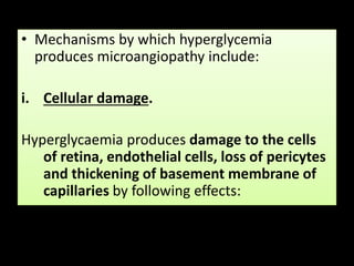
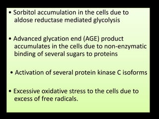
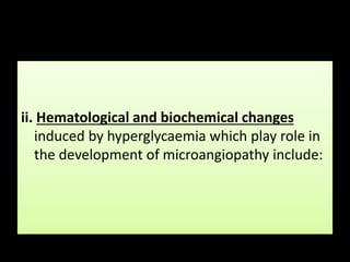
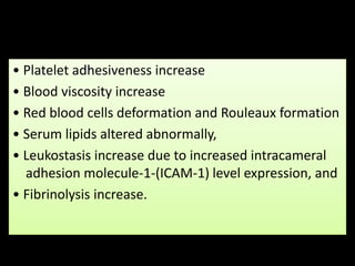
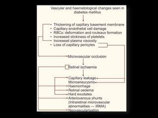
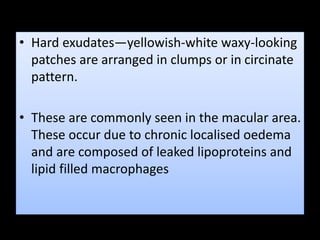
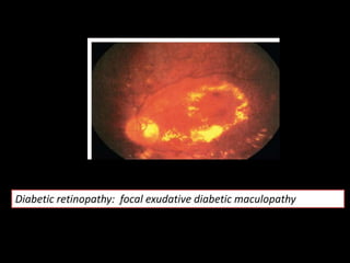
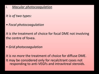
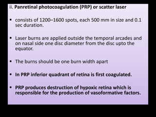
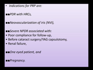
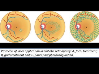
Diabetic Retinopathy
-By Priyanka ( MBBS Student )
• Diabetic retinopathy (DR) refers to retinal
changes seen in patients with diabetes mellitus.
• Diabetic retinopathy is a leading cause of
blindness
Risk Factor
• 1. Duration of diabetes is the most important
determining factor.
• After 10 years, 20% of type I and 25% of type II
diabetics develop retinopathy.
• After 20 years, 90% of type I and 60% of type II
diabetics develop retinopathy.
• After 30 years, 95% of both type I and type II diabetics
develop retinopathy.
• 2. Age of onset of diabetes also act as a risk
factor.
• The risk of retinopathy in a child with onset of
diabetes at the age of 2 years is negligible for
the first 10 years.
• After onset of puberty, age of onset is not a
risk factor.
3. Sex.
• Incidence is more in females than males (4:3).
4. Poor metabolic control
5. Heredity.
• It is transmitted as a recessive trait without sex
linkage. The effect of heredity is more on the
proliferative retinopathy.
6. Pregnancy may accelerate the changes of
diabetic retinopathy.
7. Hypertension, when associated, may also
accentuate the changes of diabetic retinopathy.
8. Other risk factors include smoking, obesity,
anaemia and hyperlipidaemia.
Pathogenesis
• Hypergylcemia, in uncontrolled diabetes
mellitus, is the starting point for development
of DR.
• Microangiopathy, affecting retinal pre-
capillary arterioles, capillaries and venules,
produced by hyperglycaemia is the basic
pathology in diabetic retinopathy.
• Mechanisms by which hyperglycemia
produces microangiopathy include:
i. Cellular damage.
Hyperglycaemia produces damage to the cells
of retina, endothelial cells, loss of pericytes
and thickening of basement membrane of
capillaries by following effects:
• Sorbitol accumulation in the cells due to
aldose reductase mediated glycolysis
• Advanced glycation end (AGE) product
accumulates in the cells due to non-enzymatic
binding of several sugars to proteins
• Activation of several protein kinase C isoforms
• Excessive oxidative stress to the cells due to
excess of free radicals.
ii. Hematological and biochemical changes
induced by hyperglycaemia which play role in
the development of microangiopathy include:
• Platelet adhesiveness increase
• Blood viscosity increase
• Red blood cells deformation and Rouleaux formation
• Serum lipids altered abnormally,
• Leukostasis increase due to increased intracameral
adhesion molecule-1-(ICAM-1) level expression, and
• Fibrinolysis increase.
Effects of microangiopathy
• Breakdown of blood-retinal barrier leads to
retinal oedema, haemorrhages, and leakage of
lipids (hard exudates).
• Weakened capillary wall produces
microaneurysms, and haemorrhages.
• Microvascular occlusions produce ischaemia
and its effects, and arteriovenous shunts, i.e.,
intraretinal microvascular abnormalities
(IRMAs).
Neovascularization of retina is induced by:
• Proangiogenic factors such as vasculoendothelial
growth factors (VEGFs), platelet derived growth factor
(PDGF), and hepatocyte growth factor (HGF) which are
released as a result of ischaemia produced by
microvascular occlusions.
• Release of angiogenic factors is also mediated by
hyperglycemia-induced oxidative stress, activation of
protein kinase C and cytokines.
• Deletion of anti–angiogenic factors such as endostatin,
angiostatin, pigment epithelial derived factor (PEDF),
thrombospondin-1 and platelet factor 4, also play role
in causing neovascularization.
• Classification of Diabetic Retinopathy Diabetic
retinopathy has been variously classified. Presently
followed classification is as follows:
I. Non-proliferative diabetic retinopathy (NPDR)
• Mild NPDR
• Moderate NPDR
• Severe NPDR
• Very severe NPDR
II. Proliferative diabetic retinopathy (PDR)
III. Diabetic maculopathy
IV. Advanced diabetic eye disease (ADED)
Non-proliferative diabetic retinopathy
(NPDR)
Ophthalmoscopic features of NPDR
include:
• Microaneurysms are seen in the macular area
(the earliest detectable lesion) and elsewhere
in relation to area of capillary non perfusion.
• These are formed due to focal dilation (out
pouching) of capillary wall following loss of
pericytes.
• These appear as red dots and leak fluid,
proteins, lipids and also fluorescein dye on FFA
• Retinal haemorrhages. Both deep (dot and
blot haemorrhages which are more common)
and superficial haemorrhages (flame-shaped),
occur from capillary leakage.
• Retinal oedema characterized by retinal
thickening is caused by capillary leakage.
• Hard exudates—yellowish-white waxy-looking
patches are arranged in clumps or in circinate
pattern.
• These are commonly seen in the macular area.
These occur due to chronic localised oedema
and are composed of leaked lipoproteins and
lipid filled macrophages
• Cotton-wool spots, are small whitish fluffy
superficial lesions.
• These represent areas of nerve fibre infarcts.
• Venous abnormalities (beading, looping and
dilatation) occur adjacent to area of capillary
non-perfusion.
• Intraretinal microvascular abnormalities
(IRMA) seen as fine irregular red lines
connecting arterioles with venules, represent
arteriovenular shunts.
• Dark-blot haemorrhages representing
haemorrhagic retinal infarcts.
MHOHECVID
1. Mild NPDR
• At least one microaneurysm must be present.
2. Moderate NPDR
• Microaneurysms/intraretinal haemorrhage in 2 or 3 quadrants.
• Early mild IRMA.
• Hard/soft exudates may or may not present.
3. Severe NPDR. Any one of the following (4–2–1 Rule)
• Four quadrants of microaneurysms and extensive intraretinal haemorrhages.
• Two quadrants of venous beading.
• One quadrant of IRMA changes.
4. Very severe NPDR. Any two of the following (4–2–1 Rule)
• Four quadrants of microaneurysms and extensive intraretinal haemorrhages.
• Two quadrants of venous beading.
• One quadrant of IRMA changes
Diabetic retinopathy: A, mild NPDR; B, moderate NPDR; C, severe NPDR; D, very
severe NPDR
• Risk factors for progression to PDR, observed
in ETDRS include presence of:
• IRMAs,
• Multiple increasing intraretinal haemorrhages,
• Venous beading and loops, and
• Wide spread capillary non-perfusion (CNP)
areas.
II. Proliferative diabetic retinopathy (PDR)
• Proliferative diabetic retinopathy develops in more
than 50% of cases after about 25 years of the onset of
disease. Therefore, it is more common in patients with
juvenile onset diabetes.
■Occurrence of neovascularization over the changes of
very severe non-proliferative diabetic retinopathy is
hallmark of PDR.
• It is characterised by proliferation of new vessels from
the capillaries, in the form of neovascularization at the
optic disc (NVD) and/or elsewhere (NVE) in the
fundus, usually along the course of the major temporal
retinal vessels.
• New vessels may proliferate in the plane of
retina or spread into the vitreous as vascular
fronds.
• Later on results in formation of:
• Fibrovascular epiretinal membrane formed
due to condensation of connective tissue
around the new vessels.
• Vitreous detachment and vitreous
haemorrhage may occur in this stage.
On the basis of high-risk characteristics (HRCs) described by
diabetic retinopathy study (DRS) group, the PDR can be
further classified as below:
• 1. Early NVD or NVE PDR without HRCs (Early PDR)
• 2. PDR with HRCs. High-risk characteristics (HRC) of
PDR are as follows
• NVD 1/4 to 1/3 of disc area with or without vitreous
haemorrhage (VH) or preretinal haemorrhage (PRH)
• NVD 1/2 disc area with VH or PRH
E, early PDR; F, high-risk PDR
III. Diabetic Maculopathy
• Changes in macular region need special mention,
due to their effect on vision.
• These changes may be associated with non-
proliferative diabetic retinopathy (NPDR) or
proliferative diabetic retinopathy (PDR).
• The diabetic macular oedema (DME) occurs due
to increased permeability of the retinal
capillaries.
Clinically significant macular oedema (CSME)
• CSME is the term coined during early
treatment diabetic retinopathy study (ETDRS).
It is diagnosed if one of the following three
criteria are present on slit-lamp examination
with 90D lens:
• Thickening of the retina at or within 500 micron of
the centre of the fovea.
• Hard exudates at or within 500 micron of the
centre of fovea associated with adjacent retinal
thickening.
• Development of a zone of retinal thickening one
disc diameter or larger in size, at least a part of
which is within one disc diameter of the foveal
centre.
• Clinico-angiographic classification of diabetic
maculopathy
• 1. Focal exudative maculopathy
• It is characterised by microaneurysms,
haemorrhages, well-circumscribed macular
oedema and hard exudates which are usually
arranged in a circinate pattern.
• Fluorescein angiography reveals focal leakage
with adequate macular perfusion.
Diabetic retinopathy: focal exudative diabetic maculopathy
• 2. Diffuse exudative maculopathy.
• It is characterised by diffuse retinal oedema
and thickening throughout the posterior pole,
with relatively few hard exudates.
• Fluorescein angiography reveals diffuse
leakage at the posterior pole.
• 3. Ischaemic maculopathy.
• It occurs due to microvascular blockage.
• Clinically, it is characterised by marked visual loss with
microaneurysms, haemorrhages, mild or no macular
oedema and a few hard exudates.
• Fluorescein angiography shows areas of non-perfusion
which in early cases are in the form of enlargement of
foveal avascular zone (FAZ), later on areas of capillary
dropouts are seen and in advanced cases precapillary
arterioles are blocked.
• 4. Mixed maculopathy. In it combined features
of ischaemic and exudative maculopathy are
present
OCT classification of diabetic macular oedema.
On the basis of OCT examination the diabetic macular oedema
(DME) has been classified as below:
1. Non-tractional DME. It may be of following types:
• a. Spongy thickness of macula (>250 µ),
• b. Cystoid macular oedema (CME)
• c. Neurosensory detachment with or without (a)
or (b) above.
2. Tractional DME. It may be of following types:
• a. Vitreo-foveal traction (VFT)
• b. Taut/thickened posterior hyaloid membrane
IV. Advanced diabetic eye disease
• It is the end result of uncontrolled proliferative
diabetic retinopathy. It is marked by
complications such as:
• Persistent vitreous haemorrhage,
• Tractional retinal detachment, and
• Neovascular glaucoma.
MANAGEMENT
SCREENING
To prevent visual loss occurring from diabetic retinopathy a periodic
follow-up is very important for a timely intervention. The
recommendations for periodic fundus examination are as follows:
• First examination, 5 years after diagnosis of type 1 DM and at the
time of diagnosis in type 2 DM.
• Every year, till there is no diabetic retinopathy or there is mild
NPDR.
• Every 6 months, in moderate NPDR.
• Every 3 months, in severe NPDR.
• Every 2 months, in PDR with no high-risk characteristics.
INVESTIGATION
• Urine examination
• Blood sugar estimation
• 24 hour urinary protein
• Renal function tests
• Lipid profile
• Haemogram
• Glycosylated haemoglobin (HbA1C)
• Fundus fluorescein angiography should be carried out to
elucidate areas of neovascularization, leakage and capillary
nonperfusion.
• Optical coherence tomography (OCT) to study detailed
structural changes in diabetic maculopathy.
TREATMENT
• FIRST IS METABOLIC CONTROL OF DM AND
ASSOCIATED RISK FACTORS
• INTRAVITREAL ANTI-VEGFS
• INTRAVITREAL STEROID
• LASER THERAPY
• SURGERY
• Control of glycaemia. Target blood glucose level: fasting <120 mg%,
post-prandial <180 mg%, and HbA1c (glycosylated haemoglobin) <7%.
• Control of dyslipidaemia. Target lipid profile (fasting): Cholesterol <200
mg%, Triglycerides <150 mg%, HDL >50 mg%, and LDL <150 mg%.
• Renal function tests. Target level are serum creatinine 1.0 mg%, blood
urea 20–40 mg%, and 24-hour urinary protein <200 mg%.
• Control of associated anaemia. Target hemoglobin >10 mg%.
• Control of associated hypertension. Target blood pressure
levels:130/80 mm Hg.
• Life style changes. Patients should be counselled to prohibit smoking
and alcohol consumption, and take regular exercises.
Anti-VEGFs, e.g., Bevacizumab (1.25 mg) and
Ranibizumab (0.5 mg) when given intravitrealy in 0.1
ml vehicle lead to improvement in vision in >40% cases
and stabilize vision in another >40% cases. These drugs
should be preferred over laser therapy particularly in
patients with:
• Focal CME involving centre of fovea,
• Diffuse DME,
• Diabetic CME, and
• DME with neurosensory detachment
• Anti-VEGFs are also recommended before panretinal
photocoagulation (PRP) in patients with PDR and diffuse
DME.
• Intravitreal triamcinolone acetonide (IVTA) (20
mg) is another drug which is being tried. It
restores inner retinal barrier and has some anti-
VEGF effects as well.
• However, risk of glaucoma, steroid induced
cataract, and increased vulnerability to
endophthalmitis restrict its use.
• Hence, anti-VEGFs are preferred over IVTA these
days. However, in recalcitrant cases IVTA may be
given along with anti-VEGFs.
• ETDRS had recommended focal laser for focal
DME and grid laser for diffuse DME.
• Laser helps possibly by stimulating the RPE pump
mechanism and by inhibiting VEGF release.
• However, with the introduction of anti-VEGF
drugs, which also improve vision, the role of laser
therapy has become limited.
• Laser therapy is performed using double
frequency YAG laser 532 nm or argon green
laser, or diode laser.
i. Macular photocoagulation
It is of two types:
• Focal photocoagulation
It is the treatment of choice for focal DME not involving
the centre of fovea.
• Grid photocoagulation
It is no more the treatment of choice for diffuse DME.
It may be considered only for recalcitrant cases not
responding to anti-VEGFs and intravitreal steroids.
ii. Panretinal photocoagulation (PRP) or scatter laser
 consists of 1200–1600 spots, each 500 mm in size and 0.1
sec duration.
 Laser burns are applied outside the temporal arcades and
on nasal side one disc diameter from the disc upto the
equator.
 The burns should be one burn width apart
 In PRP inferior quadrant of retina is first coagulated.
 PRP produces destruction of hypoxic retina which is
responsible for the production of vasoformative factors.
• Indications for PRP are:
■■PDR with HRCs,
■■Neovascularization of iris (NVI),
■■Severe NPDR associated with:
• Poor compliance for follow-up,
• Before cataract surgery/YAG capsulotomy,
• Renal failure,
■■One eyed patient, and
■■Pregnancy.
Protocols of laser application in diabetic retinopathy: A, focal treatment;
B, grid treatment and; C, panretinal photocoagulation
• Surgical treatment is indicated in following cases:
• Tractional DME with NPDR. Treatment of choice is pars
plana vitrectomy (PPV) with removal of posterior
hyaloid.
• Advanced PDR with dense vitreous haemorrhage. PPV
along with removal of opaque vitreous gel and
endophotocoagulation should be done at an early
stage.
• Advanced PDR with extensive fibrovascular epiretinal
membrane should be treated by PPV along with
removal of fibrovascular epiretinal membrane and
endophotocoagulation.
• Advanced PDR with tractional retinal
detachment should be treated by PPV with
endophotocoagulation and reattachment of
detached retina along with other methods like
scleral buckling and internal tamponade using
intravitreal silicone oil or gases like sulphur
hexafluoride (SF6) or perfluoropropane
(C3F8).
THANK YOU!
ACKNOWLEDGEMENT
• A K KHURANA- COMPREHENSIVE
OPTHALMOLOGY

Quick Review on Diabetic retinopathy

  • 1.
  • 2.
    • Diabetic retinopathy(DR) refers to retinal changes seen in patients with diabetes mellitus. • Diabetic retinopathy is a leading cause of blindness
  • 3.
  • 4.
    • 1. Durationof diabetes is the most important determining factor. • After 10 years, 20% of type I and 25% of type II diabetics develop retinopathy. • After 20 years, 90% of type I and 60% of type II diabetics develop retinopathy. • After 30 years, 95% of both type I and type II diabetics develop retinopathy.
  • 5.
    • 2. Ageof onset of diabetes also act as a risk factor. • The risk of retinopathy in a child with onset of diabetes at the age of 2 years is negligible for the first 10 years. • After onset of puberty, age of onset is not a risk factor.
  • 6.
    3. Sex. • Incidenceis more in females than males (4:3). 4. Poor metabolic control 5. Heredity. • It is transmitted as a recessive trait without sex linkage. The effect of heredity is more on the proliferative retinopathy.
  • 7.
    6. Pregnancy mayaccelerate the changes of diabetic retinopathy. 7. Hypertension, when associated, may also accentuate the changes of diabetic retinopathy. 8. Other risk factors include smoking, obesity, anaemia and hyperlipidaemia.
  • 8.
  • 9.
    • Hypergylcemia, inuncontrolled diabetes mellitus, is the starting point for development of DR.
  • 10.
    • Microangiopathy, affectingretinal pre- capillary arterioles, capillaries and venules, produced by hyperglycaemia is the basic pathology in diabetic retinopathy.
  • 11.
    • Mechanisms bywhich hyperglycemia produces microangiopathy include: i. Cellular damage. Hyperglycaemia produces damage to the cells of retina, endothelial cells, loss of pericytes and thickening of basement membrane of capillaries by following effects:
  • 12.
    • Sorbitol accumulationin the cells due to aldose reductase mediated glycolysis • Advanced glycation end (AGE) product accumulates in the cells due to non-enzymatic binding of several sugars to proteins • Activation of several protein kinase C isoforms • Excessive oxidative stress to the cells due to excess of free radicals.
  • 13.
    ii. Hematological andbiochemical changes induced by hyperglycaemia which play role in the development of microangiopathy include:
  • 14.
    • Platelet adhesivenessincrease • Blood viscosity increase • Red blood cells deformation and Rouleaux formation • Serum lipids altered abnormally, • Leukostasis increase due to increased intracameral adhesion molecule-1-(ICAM-1) level expression, and • Fibrinolysis increase.
  • 16.
  • 17.
    • Breakdown ofblood-retinal barrier leads to retinal oedema, haemorrhages, and leakage of lipids (hard exudates). • Weakened capillary wall produces microaneurysms, and haemorrhages. • Microvascular occlusions produce ischaemia and its effects, and arteriovenous shunts, i.e., intraretinal microvascular abnormalities (IRMAs).
  • 18.
    Neovascularization of retinais induced by: • Proangiogenic factors such as vasculoendothelial growth factors (VEGFs), platelet derived growth factor (PDGF), and hepatocyte growth factor (HGF) which are released as a result of ischaemia produced by microvascular occlusions. • Release of angiogenic factors is also mediated by hyperglycemia-induced oxidative stress, activation of protein kinase C and cytokines. • Deletion of anti–angiogenic factors such as endostatin, angiostatin, pigment epithelial derived factor (PEDF), thrombospondin-1 and platelet factor 4, also play role in causing neovascularization.
  • 19.
    • Classification ofDiabetic Retinopathy Diabetic retinopathy has been variously classified. Presently followed classification is as follows: I. Non-proliferative diabetic retinopathy (NPDR) • Mild NPDR • Moderate NPDR • Severe NPDR • Very severe NPDR II. Proliferative diabetic retinopathy (PDR) III. Diabetic maculopathy IV. Advanced diabetic eye disease (ADED)
  • 20.
  • 21.
    Ophthalmoscopic features ofNPDR include: • Microaneurysms are seen in the macular area (the earliest detectable lesion) and elsewhere in relation to area of capillary non perfusion. • These are formed due to focal dilation (out pouching) of capillary wall following loss of pericytes. • These appear as red dots and leak fluid, proteins, lipids and also fluorescein dye on FFA
  • 22.
    • Retinal haemorrhages.Both deep (dot and blot haemorrhages which are more common) and superficial haemorrhages (flame-shaped), occur from capillary leakage.
  • 23.
    • Retinal oedemacharacterized by retinal thickening is caused by capillary leakage.
  • 24.
    • Hard exudates—yellowish-whitewaxy-looking patches are arranged in clumps or in circinate pattern. • These are commonly seen in the macular area. These occur due to chronic localised oedema and are composed of leaked lipoproteins and lipid filled macrophages
  • 25.
    • Cotton-wool spots,are small whitish fluffy superficial lesions. • These represent areas of nerve fibre infarcts.
  • 26.
    • Venous abnormalities(beading, looping and dilatation) occur adjacent to area of capillary non-perfusion. • Intraretinal microvascular abnormalities (IRMA) seen as fine irregular red lines connecting arterioles with venules, represent arteriovenular shunts.
  • 27.
    • Dark-blot haemorrhagesrepresenting haemorrhagic retinal infarcts. MHOHECVID
  • 28.
    1. Mild NPDR •At least one microaneurysm must be present. 2. Moderate NPDR • Microaneurysms/intraretinal haemorrhage in 2 or 3 quadrants. • Early mild IRMA. • Hard/soft exudates may or may not present. 3. Severe NPDR. Any one of the following (4–2–1 Rule) • Four quadrants of microaneurysms and extensive intraretinal haemorrhages. • Two quadrants of venous beading. • One quadrant of IRMA changes. 4. Very severe NPDR. Any two of the following (4–2–1 Rule) • Four quadrants of microaneurysms and extensive intraretinal haemorrhages. • Two quadrants of venous beading. • One quadrant of IRMA changes
  • 29.
    Diabetic retinopathy: A,mild NPDR; B, moderate NPDR; C, severe NPDR; D, very severe NPDR
  • 30.
    • Risk factorsfor progression to PDR, observed in ETDRS include presence of: • IRMAs, • Multiple increasing intraretinal haemorrhages, • Venous beading and loops, and • Wide spread capillary non-perfusion (CNP) areas.
  • 31.
    II. Proliferative diabeticretinopathy (PDR)
  • 32.
    • Proliferative diabeticretinopathy develops in more than 50% of cases after about 25 years of the onset of disease. Therefore, it is more common in patients with juvenile onset diabetes. ■Occurrence of neovascularization over the changes of very severe non-proliferative diabetic retinopathy is hallmark of PDR. • It is characterised by proliferation of new vessels from the capillaries, in the form of neovascularization at the optic disc (NVD) and/or elsewhere (NVE) in the fundus, usually along the course of the major temporal retinal vessels.
  • 33.
    • New vesselsmay proliferate in the plane of retina or spread into the vitreous as vascular fronds. • Later on results in formation of: • Fibrovascular epiretinal membrane formed due to condensation of connective tissue around the new vessels. • Vitreous detachment and vitreous haemorrhage may occur in this stage.
  • 34.
    On the basisof high-risk characteristics (HRCs) described by diabetic retinopathy study (DRS) group, the PDR can be further classified as below: • 1. Early NVD or NVE PDR without HRCs (Early PDR) • 2. PDR with HRCs. High-risk characteristics (HRC) of PDR are as follows • NVD 1/4 to 1/3 of disc area with or without vitreous haemorrhage (VH) or preretinal haemorrhage (PRH) • NVD 1/2 disc area with VH or PRH
  • 35.
    E, early PDR;F, high-risk PDR
  • 36.
  • 37.
    • Changes inmacular region need special mention, due to their effect on vision. • These changes may be associated with non- proliferative diabetic retinopathy (NPDR) or proliferative diabetic retinopathy (PDR). • The diabetic macular oedema (DME) occurs due to increased permeability of the retinal capillaries.
  • 38.
    Clinically significant macularoedema (CSME) • CSME is the term coined during early treatment diabetic retinopathy study (ETDRS). It is diagnosed if one of the following three criteria are present on slit-lamp examination with 90D lens:
  • 39.
    • Thickening ofthe retina at or within 500 micron of the centre of the fovea. • Hard exudates at or within 500 micron of the centre of fovea associated with adjacent retinal thickening. • Development of a zone of retinal thickening one disc diameter or larger in size, at least a part of which is within one disc diameter of the foveal centre.
  • 40.
  • 41.
    • 1. Focalexudative maculopathy • It is characterised by microaneurysms, haemorrhages, well-circumscribed macular oedema and hard exudates which are usually arranged in a circinate pattern. • Fluorescein angiography reveals focal leakage with adequate macular perfusion.
  • 42.
    Diabetic retinopathy: focalexudative diabetic maculopathy
  • 43.
    • 2. Diffuseexudative maculopathy. • It is characterised by diffuse retinal oedema and thickening throughout the posterior pole, with relatively few hard exudates. • Fluorescein angiography reveals diffuse leakage at the posterior pole.
  • 44.
    • 3. Ischaemicmaculopathy. • It occurs due to microvascular blockage. • Clinically, it is characterised by marked visual loss with microaneurysms, haemorrhages, mild or no macular oedema and a few hard exudates. • Fluorescein angiography shows areas of non-perfusion which in early cases are in the form of enlargement of foveal avascular zone (FAZ), later on areas of capillary dropouts are seen and in advanced cases precapillary arterioles are blocked.
  • 45.
    • 4. Mixedmaculopathy. In it combined features of ischaemic and exudative maculopathy are present
  • 46.
    OCT classification ofdiabetic macular oedema. On the basis of OCT examination the diabetic macular oedema (DME) has been classified as below: 1. Non-tractional DME. It may be of following types: • a. Spongy thickness of macula (>250 µ), • b. Cystoid macular oedema (CME) • c. Neurosensory detachment with or without (a) or (b) above. 2. Tractional DME. It may be of following types: • a. Vitreo-foveal traction (VFT) • b. Taut/thickened posterior hyaloid membrane
  • 47.
  • 48.
    • It isthe end result of uncontrolled proliferative diabetic retinopathy. It is marked by complications such as: • Persistent vitreous haemorrhage, • Tractional retinal detachment, and • Neovascular glaucoma.
  • 49.
  • 50.
    SCREENING To prevent visualloss occurring from diabetic retinopathy a periodic follow-up is very important for a timely intervention. The recommendations for periodic fundus examination are as follows: • First examination, 5 years after diagnosis of type 1 DM and at the time of diagnosis in type 2 DM. • Every year, till there is no diabetic retinopathy or there is mild NPDR. • Every 6 months, in moderate NPDR. • Every 3 months, in severe NPDR. • Every 2 months, in PDR with no high-risk characteristics.
  • 51.
    INVESTIGATION • Urine examination •Blood sugar estimation • 24 hour urinary protein • Renal function tests • Lipid profile • Haemogram • Glycosylated haemoglobin (HbA1C) • Fundus fluorescein angiography should be carried out to elucidate areas of neovascularization, leakage and capillary nonperfusion. • Optical coherence tomography (OCT) to study detailed structural changes in diabetic maculopathy.
  • 52.
    TREATMENT • FIRST ISMETABOLIC CONTROL OF DM AND ASSOCIATED RISK FACTORS • INTRAVITREAL ANTI-VEGFS • INTRAVITREAL STEROID • LASER THERAPY • SURGERY
  • 53.
    • Control ofglycaemia. Target blood glucose level: fasting <120 mg%, post-prandial <180 mg%, and HbA1c (glycosylated haemoglobin) <7%. • Control of dyslipidaemia. Target lipid profile (fasting): Cholesterol <200 mg%, Triglycerides <150 mg%, HDL >50 mg%, and LDL <150 mg%. • Renal function tests. Target level are serum creatinine 1.0 mg%, blood urea 20–40 mg%, and 24-hour urinary protein <200 mg%. • Control of associated anaemia. Target hemoglobin >10 mg%. • Control of associated hypertension. Target blood pressure levels:130/80 mm Hg. • Life style changes. Patients should be counselled to prohibit smoking and alcohol consumption, and take regular exercises.
  • 54.
    Anti-VEGFs, e.g., Bevacizumab(1.25 mg) and Ranibizumab (0.5 mg) when given intravitrealy in 0.1 ml vehicle lead to improvement in vision in >40% cases and stabilize vision in another >40% cases. These drugs should be preferred over laser therapy particularly in patients with: • Focal CME involving centre of fovea, • Diffuse DME, • Diabetic CME, and • DME with neurosensory detachment • Anti-VEGFs are also recommended before panretinal photocoagulation (PRP) in patients with PDR and diffuse DME.
  • 55.
    • Intravitreal triamcinoloneacetonide (IVTA) (20 mg) is another drug which is being tried. It restores inner retinal barrier and has some anti- VEGF effects as well. • However, risk of glaucoma, steroid induced cataract, and increased vulnerability to endophthalmitis restrict its use. • Hence, anti-VEGFs are preferred over IVTA these days. However, in recalcitrant cases IVTA may be given along with anti-VEGFs.
  • 56.
    • ETDRS hadrecommended focal laser for focal DME and grid laser for diffuse DME. • Laser helps possibly by stimulating the RPE pump mechanism and by inhibiting VEGF release. • However, with the introduction of anti-VEGF drugs, which also improve vision, the role of laser therapy has become limited.
  • 57.
    • Laser therapyis performed using double frequency YAG laser 532 nm or argon green laser, or diode laser.
  • 58.
    i. Macular photocoagulation Itis of two types: • Focal photocoagulation It is the treatment of choice for focal DME not involving the centre of fovea. • Grid photocoagulation It is no more the treatment of choice for diffuse DME. It may be considered only for recalcitrant cases not responding to anti-VEGFs and intravitreal steroids.
  • 59.
    ii. Panretinal photocoagulation(PRP) or scatter laser  consists of 1200–1600 spots, each 500 mm in size and 0.1 sec duration.  Laser burns are applied outside the temporal arcades and on nasal side one disc diameter from the disc upto the equator.  The burns should be one burn width apart  In PRP inferior quadrant of retina is first coagulated.  PRP produces destruction of hypoxic retina which is responsible for the production of vasoformative factors.
  • 60.
    • Indications forPRP are: ■■PDR with HRCs, ■■Neovascularization of iris (NVI), ■■Severe NPDR associated with: • Poor compliance for follow-up, • Before cataract surgery/YAG capsulotomy, • Renal failure, ■■One eyed patient, and ■■Pregnancy.
  • 61.
    Protocols of laserapplication in diabetic retinopathy: A, focal treatment; B, grid treatment and; C, panretinal photocoagulation
  • 62.
    • Surgical treatmentis indicated in following cases: • Tractional DME with NPDR. Treatment of choice is pars plana vitrectomy (PPV) with removal of posterior hyaloid. • Advanced PDR with dense vitreous haemorrhage. PPV along with removal of opaque vitreous gel and endophotocoagulation should be done at an early stage. • Advanced PDR with extensive fibrovascular epiretinal membrane should be treated by PPV along with removal of fibrovascular epiretinal membrane and endophotocoagulation.
  • 63.
    • Advanced PDRwith tractional retinal detachment should be treated by PPV with endophotocoagulation and reattachment of detached retina along with other methods like scleral buckling and internal tamponade using intravitreal silicone oil or gases like sulphur hexafluoride (SF6) or perfluoropropane (C3F8).
  • 64.
  • 65.
    ACKNOWLEDGEMENT • A KKHURANA- COMPREHENSIVE OPTHALMOLOGY