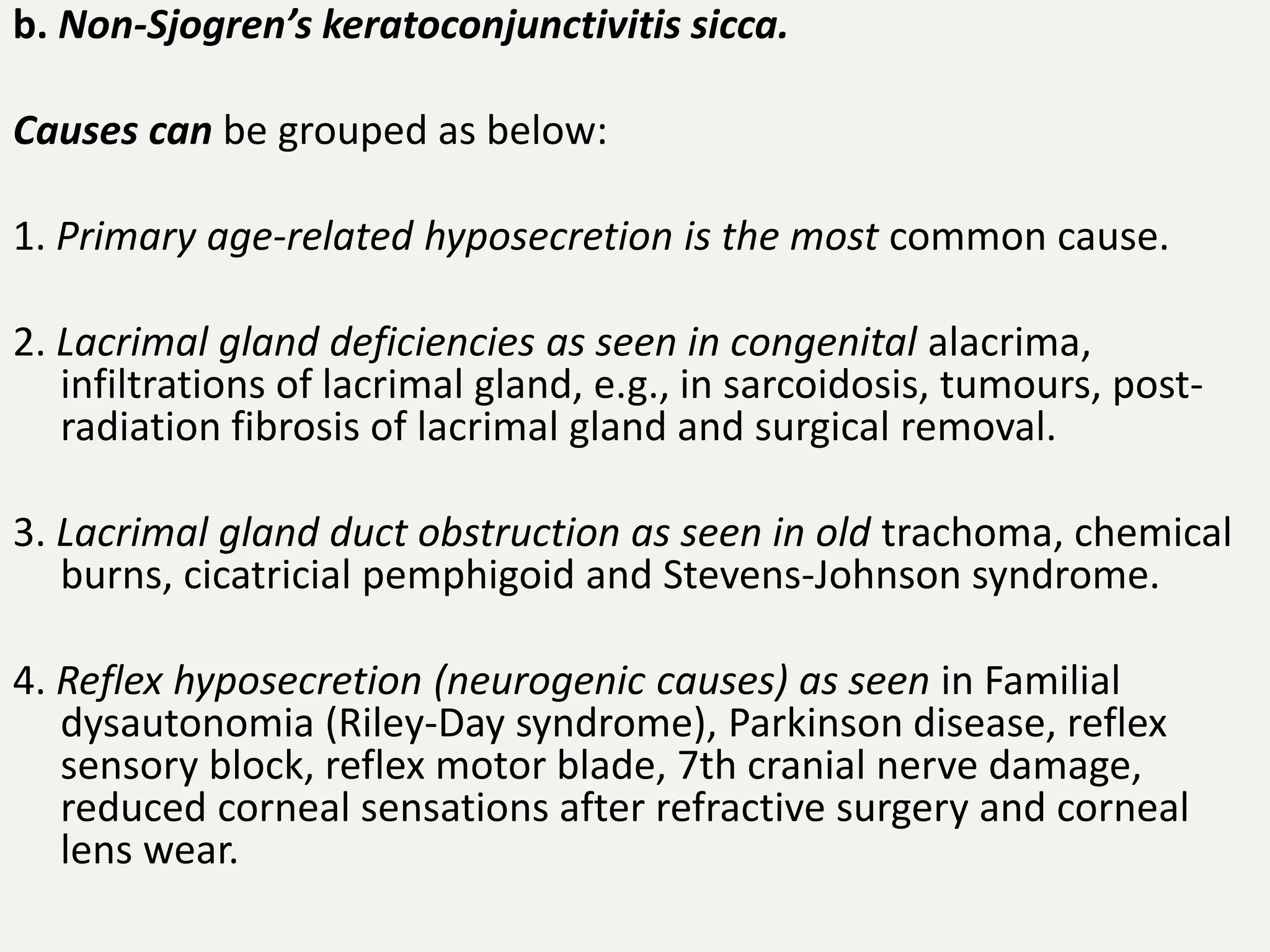
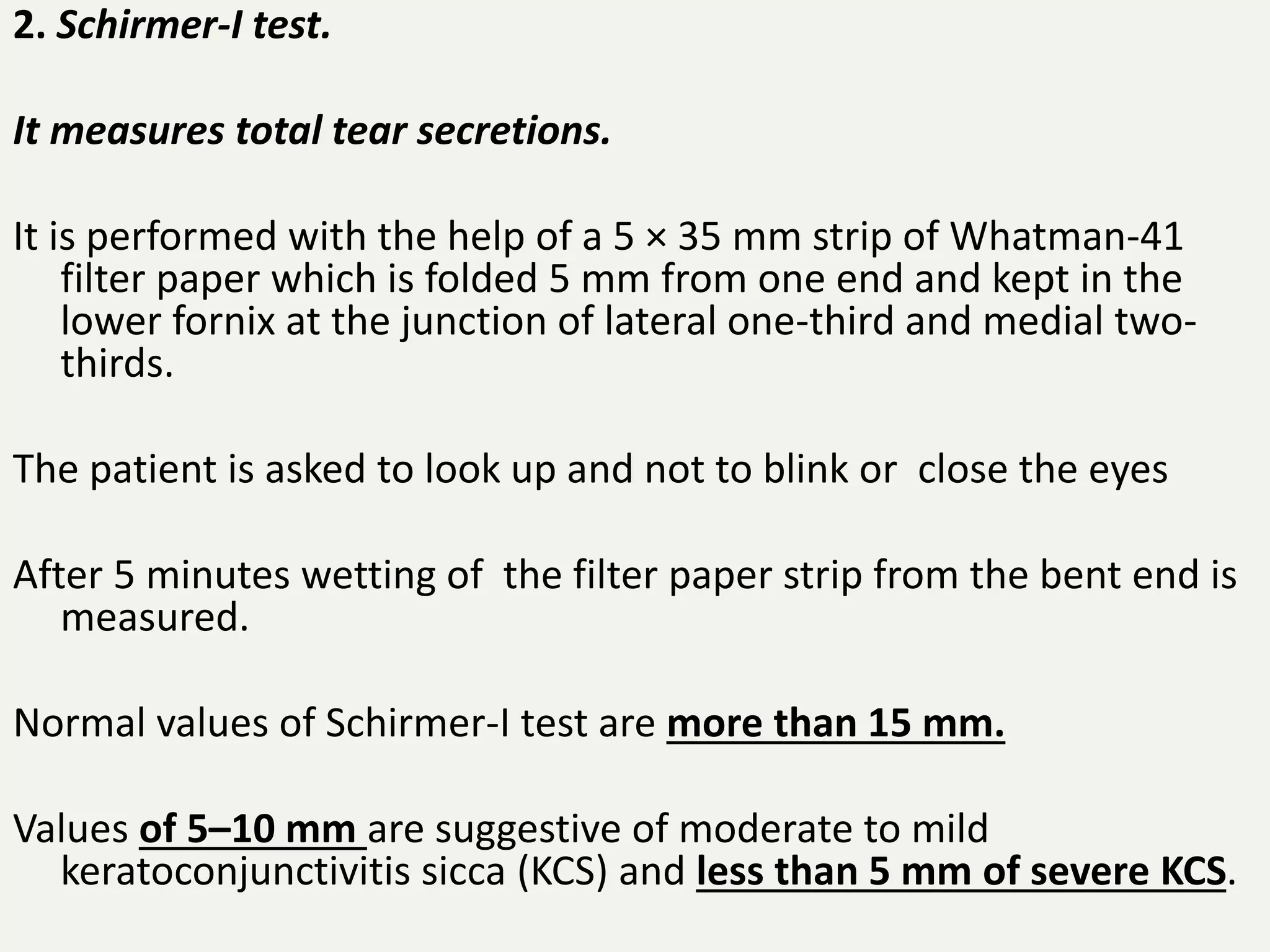
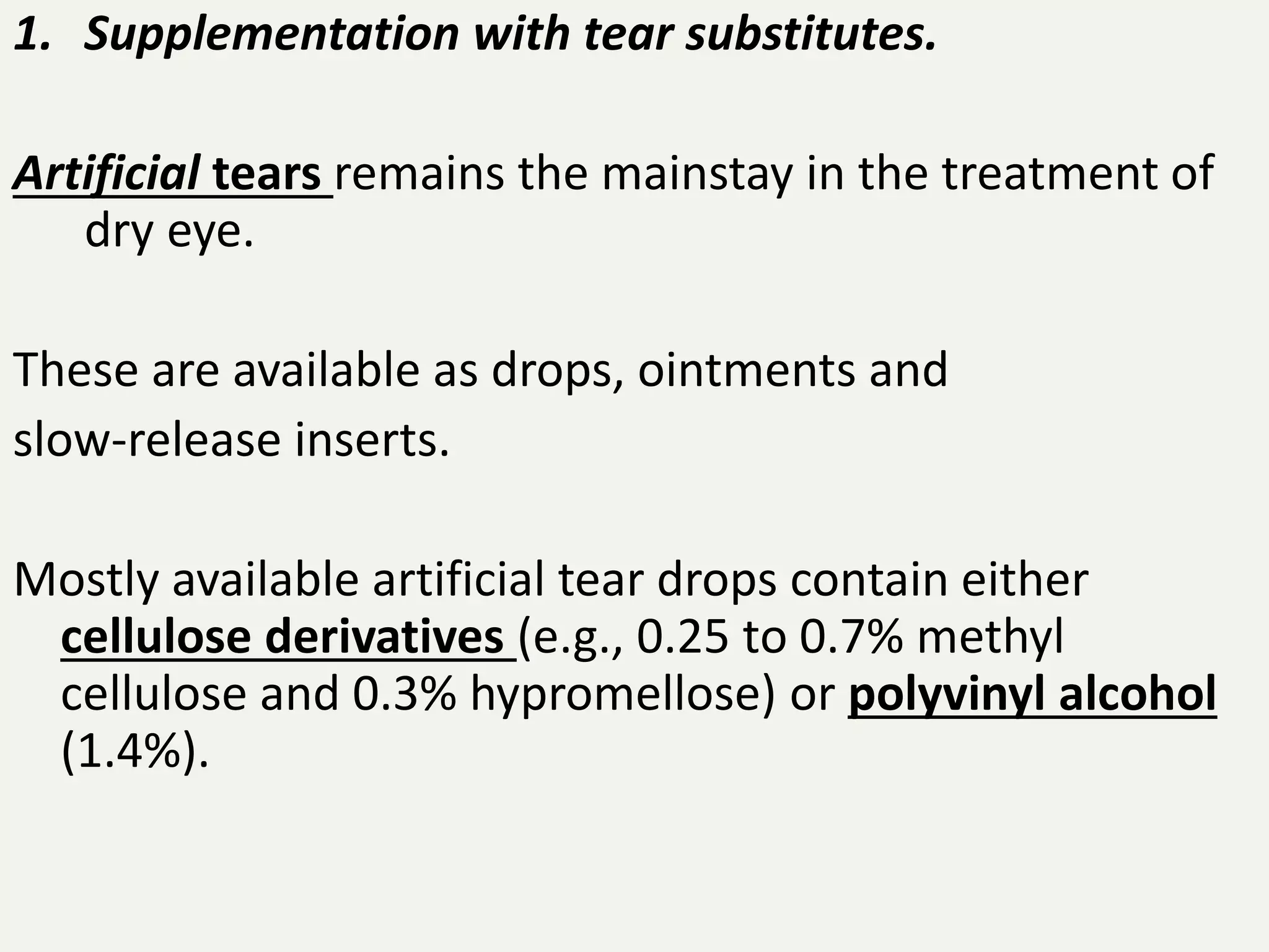
DRY EYE
By Priyanka ( MBBS Student )
The dry eye is not a disease entity, but a
symptom complex occurring as a sequelae to
deficiency or abnormalities of the tear film.
Aqueous deficiency dry eye also known as
keratoconjunctivitis sicca (KCS).
Its causes include:
a. Sjogren’s syndrome (Primary keratoconjunctivitis
sicca).
b. Non-Sjogren’s keratoconjunctivitis sicca.
Causes can be grouped as below:
1. Primary age-related hyposecretion is the most common cause.
2. Lacrimal gland deficiencies as seen in congenital alacrima,
infiltrations of lacrimal gland, e.g., in sarcoidosis, tumours, post-
radiation fibrosis of lacrimal gland and surgical removal.
3. Lacrimal gland duct obstruction as seen in old trachoma, chemical
burns, cicatricial pemphigoid and Stevens-Johnson syndrome.
4. Reflex hyposecretion (neurogenic causes) as seen in Familial
dysautonomia (Riley-Day syndrome), Parkinson disease, reflex
sensory block, reflex motor blade, 7th cranial nerve damage,
reduced corneal sensations after refractive surgery and corneal
lens wear.
II. Evaporative dry eye
It is caused by the conditions which decrease tear film stability and thus
increase evaporation.
Causes can be grouped as:
1. Meibomian gland dysfunction as seen in chronic posterior blepharitis,
rosacea, and congenital absence of meibomian glands.
2. Lagophthalmos as seen in facial nerve palsy, severe proptosis,
symblepharon and eyelid scarring.
3. Defective blinking such as low blink rate as seen in prolonged
computer users and other causes.
4. Vitamin A deficiency and other factors affecting ocular surface, e.g.,
topical drugs, preservatives, contact lens wear, ocular surface allergic
disease and scarring disorders.
Symptoms suggestive of dry eye include
irritation,
foreign body (sandy) sensation,
feeling of dryness,
itching,
nonspecific ocular discomfort
chronically sore eyes not responding to a variety of
drops instilled earlier.
Signs of dry eye are as below:
• Tear film signs. It may show presence of stingy mucous and
particulate matter. Marginal tear strip is reduced or absent
(normal height is 1 mm). Froth in the tears along the lid
margin is a sign of meibomian gland dysfunction.
• Conjunctival signs. It becomes lustureless, mildly congested,
conjunctival xerosis and keratinization may occur.
• Corneal signs. It may show punctate epithelial erosions,
filaments and mucus plaques. Cornea may loose lusture.
•
Signs of causative disease such as posterior blepharitis,
conjunctival scarring diseases (trachoma, Stevens-Johnson
syndrome, chemical burns, ocular pemphigoid) and
lagophthalmos may be depicted.
1. Tear film break-up (BUT).
It is the interval between
a complete blink and appearance of first randomly
distributed dry spot on the cornea.
It is noted after instilling a drop of fluorescein and examining in
a cobalt-blue light of a slit-lamp.
BUT is an indicator of adequacy of mucin component of tears.
Its normal values range from 15 to 35 seconds. Values less than
10 seconds imply an unstable tear film.
2. Schirmer-I test.
It measures total tear secretions.
It is performed with the help of a 5 × 35 mm strip of Whatman-41
filter paper which is folded 5 mm from one end and kept in the
lower fornix at the junction of lateral one-third and medial two-
thirds.
The patient is asked to look up and not to blink or close the eyes
After 5 minutes wetting of the filter paper strip from the bent end is
measured.
Normal values of Schirmer-I test are more than 15 mm.
Values of 5–10 mm are suggestive of moderate to mild
keratoconjunctivitis sicca (KCS) and less than 5 mm of severe KCS.
Schirmer’s Test
3. Rose Bengal staining.
It is a very useful test for detecting even mild cases of KCS.
Depending upon the severity of KCS three staining patterns
A, B and C have been described:
‘C’ pattern represents mild or early cases with fine punctate
stains in the interpalpebral area
‘B’ the moderate cases with extensive staining
‘A’ the severe cases with confluent staining of conjunctiva and
cornea.
Recently accepted system based on
severity of signs and tear film tests recommended
by Dry Eye Workshop (DEWS) Report (2007) grades
the severity of dry eye into 4 levels:
• Level 1 (mild dry eye),
• Level 2 (moderate dry eye),
• Level 3 (severe dry eye), and
• Level 4 (very severe dry eye).
Treatment modalities
1. Supplementation with tear substitutes.
Artificial tears remains the mainstay in the treatment of
dry eye.
These are available as drops, ointments and
slow-release inserts.
Mostly available artificial tear drops contain either
cellulose derivatives (e.g., 0.25 to 0.7% methyl
cellulose and 0.3% hypromellose) or polyvinyl alcohol
(1.4%).
2. Topical cyclosporine (0.05%, 0.1%) is reported to be
very effective drug for dry eye in many recent studies
It helps by reducing the cell-mediated inflammation
of the lacrimal tissue.
3. Mucolytics, such as 5% acetylcystine used 4 times
a day helps by dispersing the mucus threads and
decreasing tear viscosity.
4. Preservation of existing tears by reducing evaporation and
decreasing drainage.
• Evaporation can be reduced by
 decreasing room temperature,
 use of moist chambers and protective glasses.
• Punctal occlusion to decrease drainage can be
carried out by
 collagen implants,
 Cynoacrylate tissue adhesives,
 electrocauterisation,
 argon laser occlusion and surgical occlusion
to decrease the drainage of tears in patients with very severe dry eye.
• Permanent lateral tarsorrhaphy may be required
in very severe cases.
5. Treatment of the causative disease of dry eye
when discovered is very useful e.g.:
• Systemic tetracyclines and lid hygiene in patients
with chronic posterior blepharitis
• Vitamin A supplement for the deficiency
• Treat the cause of lagophthalmos.
Thank You!
• Acknowledgement – A K Khurana Comprehensive
Ophthalomology

quick review on Dry eye

  • 1.
    DRY EYE By Priyanka( MBBS Student )
  • 2.
    The dry eyeis not a disease entity, but a symptom complex occurring as a sequelae to deficiency or abnormalities of the tear film.
  • 3.
    Aqueous deficiency dryeye also known as keratoconjunctivitis sicca (KCS). Its causes include: a. Sjogren’s syndrome (Primary keratoconjunctivitis sicca).
  • 4.
    b. Non-Sjogren’s keratoconjunctivitissicca. Causes can be grouped as below: 1. Primary age-related hyposecretion is the most common cause. 2. Lacrimal gland deficiencies as seen in congenital alacrima, infiltrations of lacrimal gland, e.g., in sarcoidosis, tumours, post- radiation fibrosis of lacrimal gland and surgical removal. 3. Lacrimal gland duct obstruction as seen in old trachoma, chemical burns, cicatricial pemphigoid and Stevens-Johnson syndrome. 4. Reflex hyposecretion (neurogenic causes) as seen in Familial dysautonomia (Riley-Day syndrome), Parkinson disease, reflex sensory block, reflex motor blade, 7th cranial nerve damage, reduced corneal sensations after refractive surgery and corneal lens wear.
  • 5.
    II. Evaporative dryeye It is caused by the conditions which decrease tear film stability and thus increase evaporation. Causes can be grouped as: 1. Meibomian gland dysfunction as seen in chronic posterior blepharitis, rosacea, and congenital absence of meibomian glands. 2. Lagophthalmos as seen in facial nerve palsy, severe proptosis, symblepharon and eyelid scarring. 3. Defective blinking such as low blink rate as seen in prolonged computer users and other causes. 4. Vitamin A deficiency and other factors affecting ocular surface, e.g., topical drugs, preservatives, contact lens wear, ocular surface allergic disease and scarring disorders.
  • 6.
    Symptoms suggestive ofdry eye include irritation, foreign body (sandy) sensation, feeling of dryness, itching, nonspecific ocular discomfort chronically sore eyes not responding to a variety of drops instilled earlier.
  • 7.
    Signs of dryeye are as below: • Tear film signs. It may show presence of stingy mucous and particulate matter. Marginal tear strip is reduced or absent (normal height is 1 mm). Froth in the tears along the lid margin is a sign of meibomian gland dysfunction. • Conjunctival signs. It becomes lustureless, mildly congested, conjunctival xerosis and keratinization may occur. • Corneal signs. It may show punctate epithelial erosions, filaments and mucus plaques. Cornea may loose lusture. • Signs of causative disease such as posterior blepharitis, conjunctival scarring diseases (trachoma, Stevens-Johnson syndrome, chemical burns, ocular pemphigoid) and lagophthalmos may be depicted.
  • 8.
    1. Tear filmbreak-up (BUT). It is the interval between a complete blink and appearance of first randomly distributed dry spot on the cornea. It is noted after instilling a drop of fluorescein and examining in a cobalt-blue light of a slit-lamp. BUT is an indicator of adequacy of mucin component of tears. Its normal values range from 15 to 35 seconds. Values less than 10 seconds imply an unstable tear film.
  • 9.
    2. Schirmer-I test. Itmeasures total tear secretions. It is performed with the help of a 5 × 35 mm strip of Whatman-41 filter paper which is folded 5 mm from one end and kept in the lower fornix at the junction of lateral one-third and medial two- thirds. The patient is asked to look up and not to blink or close the eyes After 5 minutes wetting of the filter paper strip from the bent end is measured. Normal values of Schirmer-I test are more than 15 mm. Values of 5–10 mm are suggestive of moderate to mild keratoconjunctivitis sicca (KCS) and less than 5 mm of severe KCS.
  • 10.
  • 11.
    3. Rose Bengalstaining. It is a very useful test for detecting even mild cases of KCS. Depending upon the severity of KCS three staining patterns A, B and C have been described: ‘C’ pattern represents mild or early cases with fine punctate stains in the interpalpebral area ‘B’ the moderate cases with extensive staining ‘A’ the severe cases with confluent staining of conjunctiva and cornea.
  • 12.
    Recently accepted systembased on severity of signs and tear film tests recommended by Dry Eye Workshop (DEWS) Report (2007) grades the severity of dry eye into 4 levels: • Level 1 (mild dry eye), • Level 2 (moderate dry eye), • Level 3 (severe dry eye), and • Level 4 (very severe dry eye).
  • 13.
  • 14.
    1. Supplementation withtear substitutes. Artificial tears remains the mainstay in the treatment of dry eye. These are available as drops, ointments and slow-release inserts. Mostly available artificial tear drops contain either cellulose derivatives (e.g., 0.25 to 0.7% methyl cellulose and 0.3% hypromellose) or polyvinyl alcohol (1.4%).
  • 15.
    2. Topical cyclosporine(0.05%, 0.1%) is reported to be very effective drug for dry eye in many recent studies It helps by reducing the cell-mediated inflammation of the lacrimal tissue. 3. Mucolytics, such as 5% acetylcystine used 4 times a day helps by dispersing the mucus threads and decreasing tear viscosity.
  • 16.
    4. Preservation ofexisting tears by reducing evaporation and decreasing drainage. • Evaporation can be reduced by  decreasing room temperature,  use of moist chambers and protective glasses. • Punctal occlusion to decrease drainage can be carried out by  collagen implants,  Cynoacrylate tissue adhesives,  electrocauterisation,  argon laser occlusion and surgical occlusion to decrease the drainage of tears in patients with very severe dry eye. • Permanent lateral tarsorrhaphy may be required in very severe cases.
  • 17.
    5. Treatment ofthe causative disease of dry eye when discovered is very useful e.g.: • Systemic tetracyclines and lid hygiene in patients with chronic posterior blepharitis • Vitamin A supplement for the deficiency • Treat the cause of lagophthalmos.
  • 18.
    Thank You! • Acknowledgement– A K Khurana Comprehensive Ophthalomology