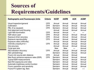
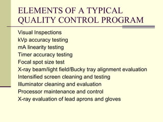
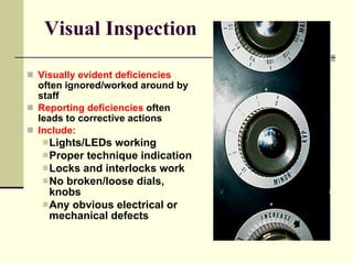
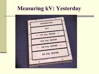
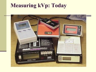
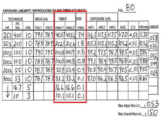
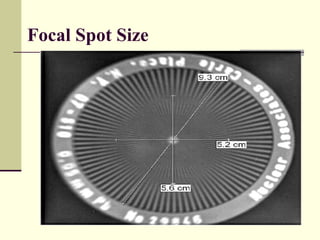
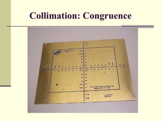
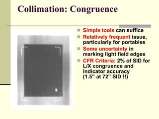
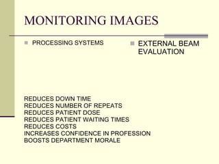
Quality Management A broad term which encompasses both quality assurance and quality improvement, describing a program of evaluating the quality of care using a variety of methodologies and techniques This may entail going beyond commonly accepted standards to striving for stricter yet generally achievable performance levels
Reasons for Inspections State regulatory requirement 3rd party payer requirement Employer expectations  Standards of good practice It is not uncommon that “inspections” include the minimum set of tests and evaluations needed to fulfill the expectation or legal requirement   (perhaps due to time constraints and priorities)
Philosophy of Inspections The goal of radiographic and fluoroscopic (R/F) inspections should be to provide value by evaluating and (if necessary) improving : radiation safety image quality  image consistency
Quality Assurance A planned and systematic pattern of all actions necessary to provide adequate confidence that the item or product conforms to established technical requirements. Identifies problems Monitors problems Resolves problems
Quality Control The operational techniques and the activities used to fulfill and verify requirements of quality. Inspection, analysis and action required to ensure quality of output. A procedure for keeping quality of inputs or outputs to specifications. Techniques ensuring that high quality is maintained through various stages of a process. For example, quality control during image capture might include comparing the scanned image to the original. The control of variation of workmanship, processes, and materials in order to produce a consistent, uniform product.
Sources of Requirements/Guidelines
GUIDELINES NCRP Report No 102 Manufactureres Radiographers Oregon Board of Radiologic Technology ARRT
WHO MAKE IT WORK RADIOGRAPHERS QC TECHNOLOGISTS SERVICE ENGINEERS MEDICAL PHYSICIST
ELEMENTS OF A TYPICAL QUALITY CONTROL PROGRAM Visual Inspections kVp accuracy testing mA linearity testing Timer accuracy testing Focal spot size test X-ray beam/light field/Bucky tray alignment evaluation Intensified screen cleaning and testing Illuminator cleaning and evaluation Processor maintenance and control X-ray evaluation of lead aprons and gloves
Visual Inspection Visually evident deficiencies  often ignored/worked around by staff Reporting deficiencies  often leads to corrective actions Include: Lights/LEDs working Proper technique indication  Locks and interlocks work No broken/loose dials, knobs Any obvious electrical or  mechanical defects
Measuring kV: Yesterday
Measuring kVp: Today
mA Linearity
 
TIMER ACCURACY
 
Timer Accuracy
Focal Spot Size
Collimation: Congruence
Collimation: Congruence Simple tools   can suffice Relatively frequent   issue, particularly for portables Some uncertainty   in marking light field edges CFR Criteria:   2% of SID for L/X congruence and indicator accuracy  (1.5” at 72” SID !!)
OTHER CASSETTES: EXTERIOR SCREENS VISUALLY INSPECTED ILLUMINATION LIGHT METER LEAD APRONS AND GLOVES REPEAT RATES
CR/DR QC Daily Density Check CR Cassettes IP plates
FOCUS – PDCA METHOD F ind and define a problem O rganize a team to work on the improvement C larify the problem with current knowledge U nderstand the problem and its causes S elect a method to improve the process P lan implementation D o the implementation and measure change. C heck the results A ct to continue improvements
MONITORING IMAGES PROCESSING SYSTEMS REDUCES DOWN TIME REDUCES NUMBER OF REPEATS REDUCES PATIENT DOSE REDUCES PATIENT WAITING TIMES REDUCES COSTS INCREASES CONFIDENCE IN PROFESSION BOOSTS DEPARTMENT MORALE EXTERNAL BEAM EVALUATION

Quality Management for Diagnostic Imaging

  • 1.
    Quality Management Abroad term which encompasses both quality assurance and quality improvement, describing a program of evaluating the quality of care using a variety of methodologies and techniques This may entail going beyond commonly accepted standards to striving for stricter yet generally achievable performance levels
  • 2.
    Reasons for InspectionsState regulatory requirement 3rd party payer requirement Employer expectations Standards of good practice It is not uncommon that “inspections” include the minimum set of tests and evaluations needed to fulfill the expectation or legal requirement (perhaps due to time constraints and priorities)
  • 3.
    Philosophy of InspectionsThe goal of radiographic and fluoroscopic (R/F) inspections should be to provide value by evaluating and (if necessary) improving : radiation safety image quality image consistency
  • 4.
    Quality Assurance Aplanned and systematic pattern of all actions necessary to provide adequate confidence that the item or product conforms to established technical requirements. Identifies problems Monitors problems Resolves problems
  • 5.
    Quality Control Theoperational techniques and the activities used to fulfill and verify requirements of quality. Inspection, analysis and action required to ensure quality of output. A procedure for keeping quality of inputs or outputs to specifications. Techniques ensuring that high quality is maintained through various stages of a process. For example, quality control during image capture might include comparing the scanned image to the original. The control of variation of workmanship, processes, and materials in order to produce a consistent, uniform product.
  • 6.
  • 7.
    GUIDELINES NCRP ReportNo 102 Manufactureres Radiographers Oregon Board of Radiologic Technology ARRT
  • 8.
    WHO MAKE ITWORK RADIOGRAPHERS QC TECHNOLOGISTS SERVICE ENGINEERS MEDICAL PHYSICIST
  • 9.
    ELEMENTS OF ATYPICAL QUALITY CONTROL PROGRAM Visual Inspections kVp accuracy testing mA linearity testing Timer accuracy testing Focal spot size test X-ray beam/light field/Bucky tray alignment evaluation Intensified screen cleaning and testing Illuminator cleaning and evaluation Processor maintenance and control X-ray evaluation of lead aprons and gloves
  • 10.
    Visual Inspection Visuallyevident deficiencies often ignored/worked around by staff Reporting deficiencies often leads to corrective actions Include: Lights/LEDs working Proper technique indication Locks and interlocks work No broken/loose dials, knobs Any obvious electrical or mechanical defects
  • 11.
  • 12.
  • 13.
  • 14.
  • 15.
  • 16.
  • 17.
  • 18.
  • 19.
  • 20.
    Collimation: Congruence Simpletools can suffice Relatively frequent issue, particularly for portables Some uncertainty in marking light field edges CFR Criteria: 2% of SID for L/X congruence and indicator accuracy (1.5” at 72” SID !!)
  • 21.
    OTHER CASSETTES: EXTERIORSCREENS VISUALLY INSPECTED ILLUMINATION LIGHT METER LEAD APRONS AND GLOVES REPEAT RATES
  • 22.
    CR/DR QC DailyDensity Check CR Cassettes IP plates
  • 23.
    FOCUS – PDCAMETHOD F ind and define a problem O rganize a team to work on the improvement C larify the problem with current knowledge U nderstand the problem and its causes S elect a method to improve the process P lan implementation D o the implementation and measure change. C heck the results A ct to continue improvements
  • 24.
    MONITORING IMAGES PROCESSINGSYSTEMS REDUCES DOWN TIME REDUCES NUMBER OF REPEATS REDUCES PATIENT DOSE REDUCES PATIENT WAITING TIMES REDUCES COSTS INCREASES CONFIDENCE IN PROFESSION BOOSTS DEPARTMENT MORALE EXTERNAL BEAM EVALUATION

Editor's Notes

  • #4 This may entail going beyond commonly accepted standards to striving for stricter yet generally achievable performance levels
  • #5 ACTIVITIES THAT PROVIDE CONFIDENCE THAT A RADIOLOGY SERVICE RENDERS CONSISTENTLY HIGH QUALITY IMAGES AND SERVICES. EVOLUTIONARY PROCESS THAT ASSESSES EVEYTHING THAT AFFECTS PATIETN CARE. ENSURES THAT REATKESA RE NOTREQUIRED AS THE RESULT OF EQUIPMENT MALFUNCTION EMPLOYEES RECEIVE OREINTATION ON NEW EQUIPMENT
  • #6 THE ASPECT OF QUALITY ASSURANCE THAT MONITORS THE TECHNICAL EQUIPMENT TO MAINTAIN STANDARDS MONITORING AND REGULATING THE VARIABLES ASSOCIAOTED WITH IMAGE PRODUCTION AND PATIENT CARE. EVERY FACILITY MUST ESTABLISH QUAILITY CONTROL GUIDELINES AND CONDUCT QC PROGRAMS TO PROVIDE A CONSISTENT STANDARD OF CARE. PROPERLY DOCUMENTED, ONGOING, AND EFFECTIVE QC PROGRAMS ARE REQUIRED BY HOSPITAL ACCREDITING AGENCIES AND STATE DEPARTMENTSO FO HEALTH INCOMING LINE CURRENT X-RAY PRODUCTION PROCESSING ERRATIC EQUIPMENT PERFORMANCE REPEAT RADIOGRAPHS UNNECESSARY PATIENT EXPOSURE
  • #8 RADIOGRAPHIC QC IS AN ORGANIZED AND METHODICAL EVALUATION OF IMAGING COMPONENTS – FREQUENCY OF TESTING RANGES FROM DAILY TO SEMI ANNUAL OR ANNUAL. 102: SERVES A A GUIDE TO GOOD MEDICAL RADIATION PRACTICES BY DESCRIBING THE FEDERAL REGULATIONS ON EQUIPMENT DESIGN, PERFORMANCE AND USE MANUFACTURERS: MUST FOLLOW GUIDELINES THAT STATE MAXIMUM X-RAY OUTPOUT AS SPECIFIC DISTANCES TOTAL QUANTITIES OF FILTRATION POSITIVE BEAM LIMITAION RADIOGRAPHERS: PRACTICE SAFE PINCIPLES OF OPERATION PREVENATTIVE MAINTENANCE QUALITY CONTROL CHECKS AT SPECIFIC INTERVALS TO ENSURE CONTINUED SAFE EQUIPMENT.
  • #12 FORMERLY EVALUATED USING A WSCONSION TEST TOOL AND CASSETTE
  • #13 DIGITAL: MORE CONVENIENT AND SIMPLE Measurements at many settings practical--allows comprehensive eval of accuracy & reproducibility VARIOUS KV’S ARE TESTED IN NORMAL DIAGNOSTIC IMAGING RANGE: 40 -150 KVP SELECTED KV AND ACTUAL Kv VALUE SHOULD NOT DIFFER BY MOR THAN + KvP FOR GENERAL AND BY 5% OF NOMICAL KvP FOR MAMMO EXAMPLE: 30 KvP A MARGIN OF ERROR SHOULD NOT EXCEED 1.5 KvP (30 *5% = 1.5 KvP)
  • #14 ACCURACY OF INDIVIDUAL mA STATIONS IS RELATED TO PATIENT DOSE AND IS ESSENTIAL TO PRODUCTION OF EXPECTED DENSITY LEVELS NO FILM OLD WAY: AN ALUMINUM STEP WEDGE (PENTROMETER0 CAN BE USED TO EVELAUTE EACH MA STATION A SERIES OF EXPOSURES IS MADE AT A PARTICULAR KvP USING THE mAs value at each m STSTAION, WITH EXPOSURE TIME ADJUSTED TO MAINTAIN A CONSTANT MaS. GOOD EXAMPLE OF THE RECRIPROCITY LAW: NEW WAY: DIGITAL DOSIMETER 9IONIZATION CHAMBER) EXPOSURES ARE MADE AT A PARTICUALR mAs with cvarious combinations of mA and time; x-ray output is measured in mr/mAs and should be accurate to within 10%
  • #16 TIMER ACCURACY IS RELATED TO PATEIENT DOSE AND THE PRODUCTION OF EXPECTED RADIOGRAPHIC DENSITY SHOULD BE TESTED ANNUALLY SPINNING TOP: CIRCULAR STEEL OR LEAD DISC WITH SMALL HOLES IN THE PERIPHERY
  • #17 SINGLE PHASE FULL WAVE: : 120 USEFUL X-RAY IMPULSES PER SECOND EXAMPLE: IF THE X-RAY TIMER IS SET TO USE SOME PORTION OF THE IMPULSES OR ¼ OF A SECOND, THEN 1/4 OF 120 = 30 DOTS MULTIPLY THE NUMBER OF IMPULSES PER SECOND BY THE EXPOSURE TIME. ½ THE CORRECT NUMBER OF DOTS = RECITIFER PROBLEM THREE PHASE OR HIGH FREQUENCY: PRODUCES A RIPPLE WAVE OF ALMOST CONSTANT POTENTIAL: DEMONSTRATES AN ARC EXAMPLE: IF THE EXPOSURE WAS 1 SECOND THEN 360 DEGREE ARC IF THE EXPOSURE WAS ¼ OF A SECOND; THEN A 90 DEGREE ARC.
  • #18 DIGITAL: SELECTED EXPOSURE TIME SHOULD BE WITHIN 5% OF THE ACTUAL EXPOSURE TIME.
  • #19 FOCAL SPOT SIZE IS REALTED TO DEGREE OF GEOMETRIC BLUR MANUFACTURER TOLERANCE FRO NEW FOCAL SPOTS IS 50%; THAT IS 0.3 MM FOCALSPOT MAY BE .45 MM FOCAL SPOT INCREASES AS TUBE AGES; THEREFORE ANNUAL TESTING IS IMPORTANT MEASURED WITH A SLIT CAMERA, OR STAR PATTEN TYPE.
  • #22 VISUALLY INSPECT SCREENS WIRE MESH TEST ILLUMINATION : USE LIGHT METER HELD AT SAME DISTANCE FROM EACH ILLUMINATOR (3 FEET) CLEANED TO REMOVE DUST AND GRIME CHANGE ALL BULBS IN A BANK IF 1 GOES OUT.
  • #23 DENSITY CHECK: ON THE PRINTER ACCORDING TO MANUFACTUERERS RECOMMENDATIONS CR CASSETTES: VISUALLY CHECKED IP PLATE SHOULD BE REMOVED BEFORE LEANING THE CASSETTE NEVER PUT A DAMP CR CASSETTE IN THE CR READER IP: CLEANED MONTHYL: USE ONLY ANHYDROUS ETHANOL CHECKED FOR SCRATCHES: APPEAR AS CLEAR AREAS ON THE RESULTING IMAGE LIFE OF AN IP PLATE: 10,000 EXPOSURES