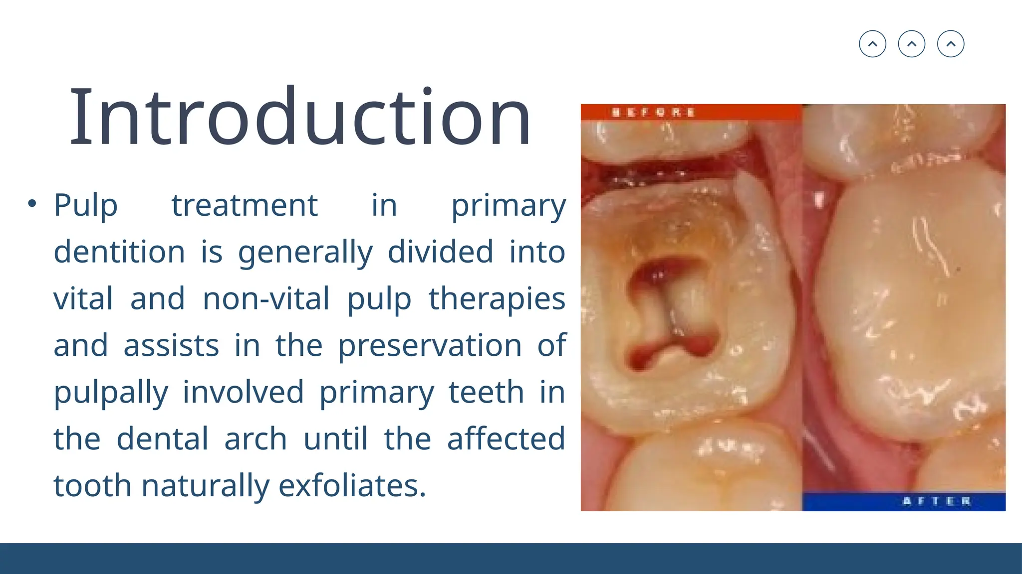
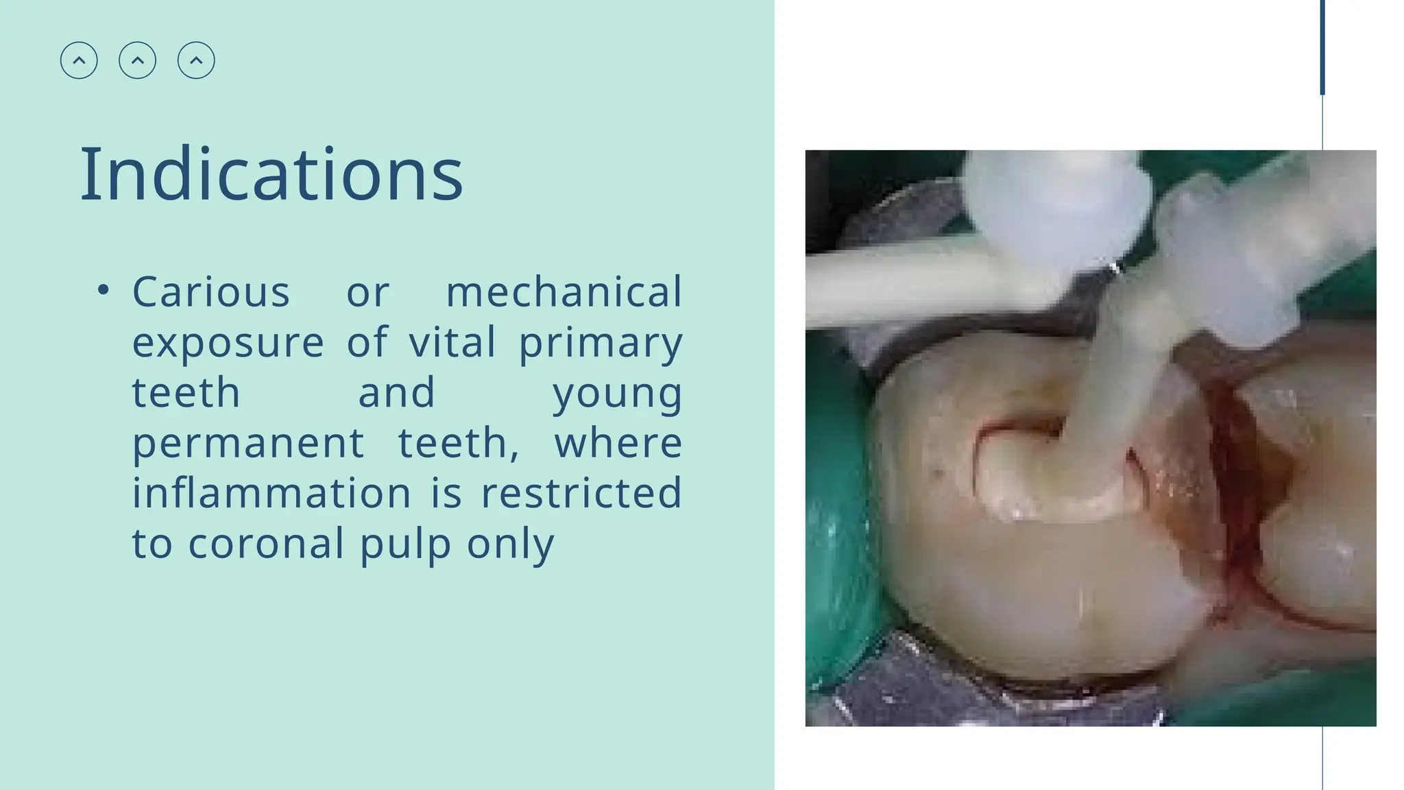
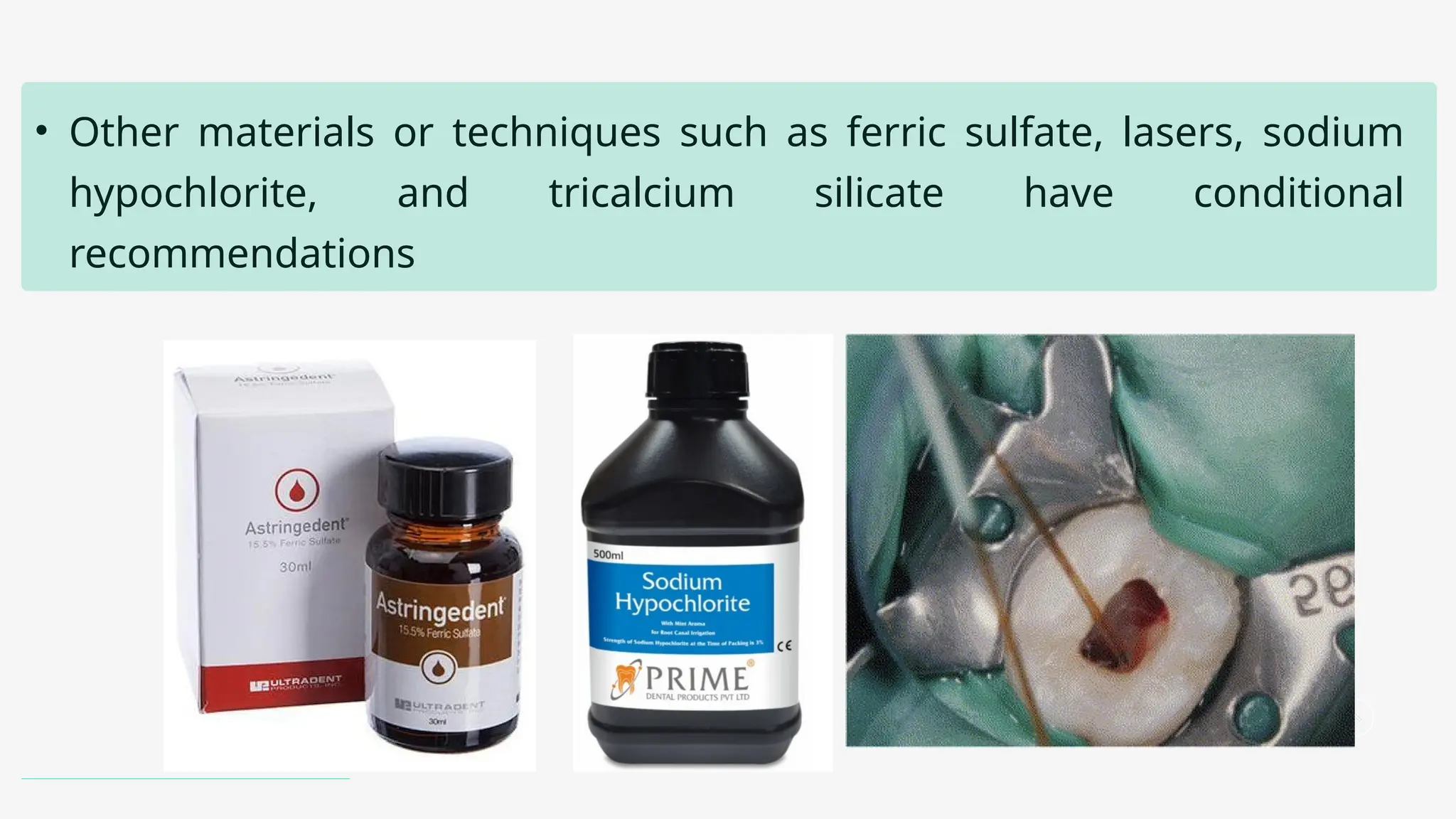
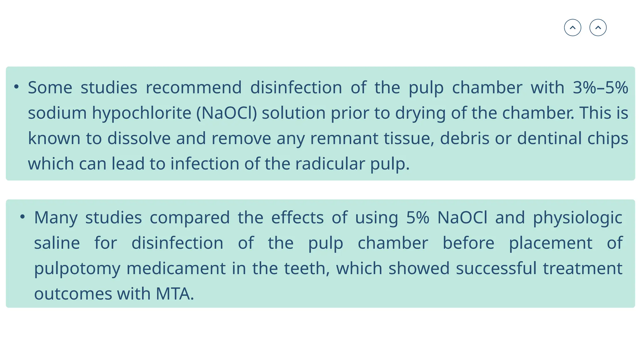
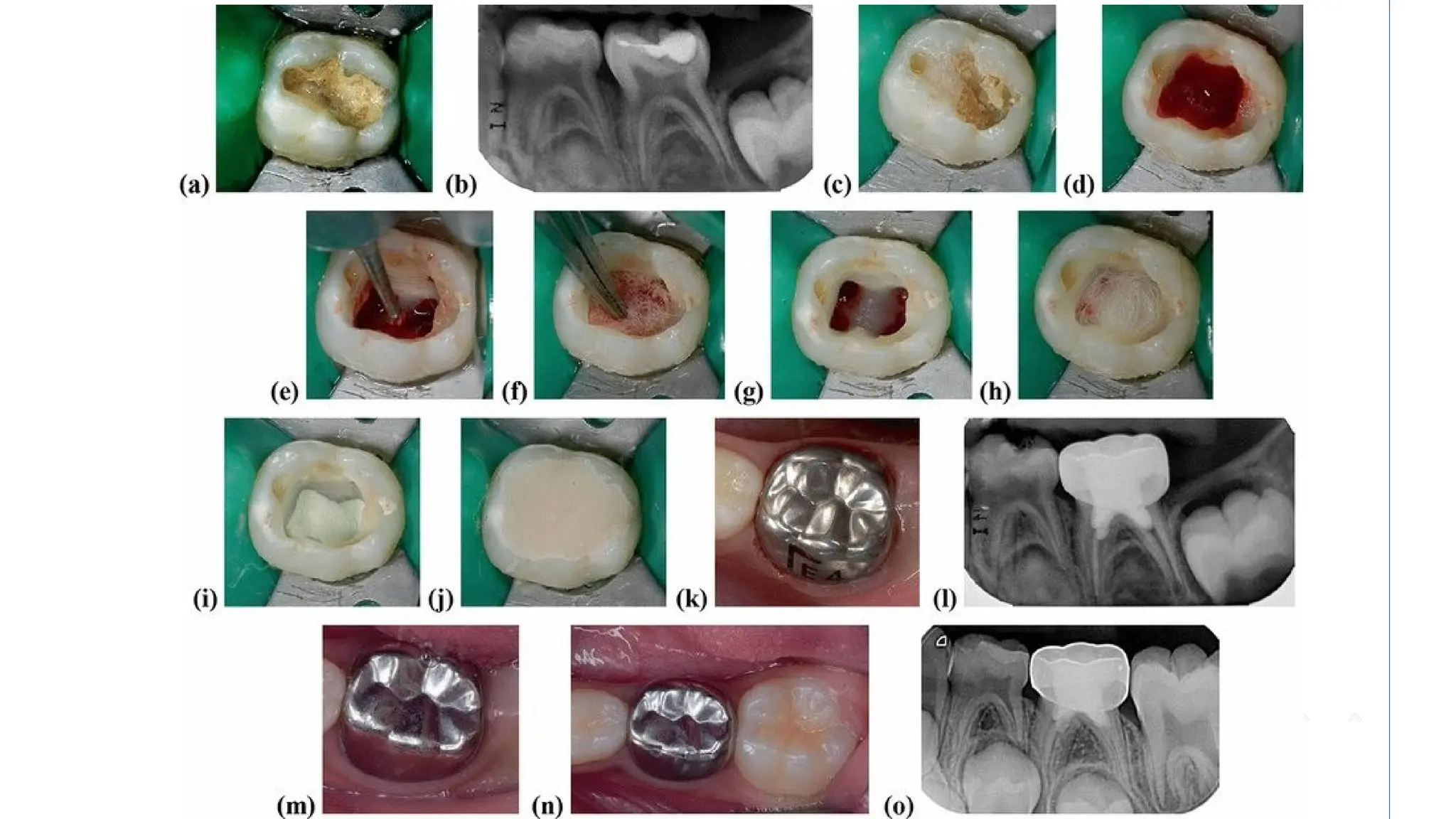
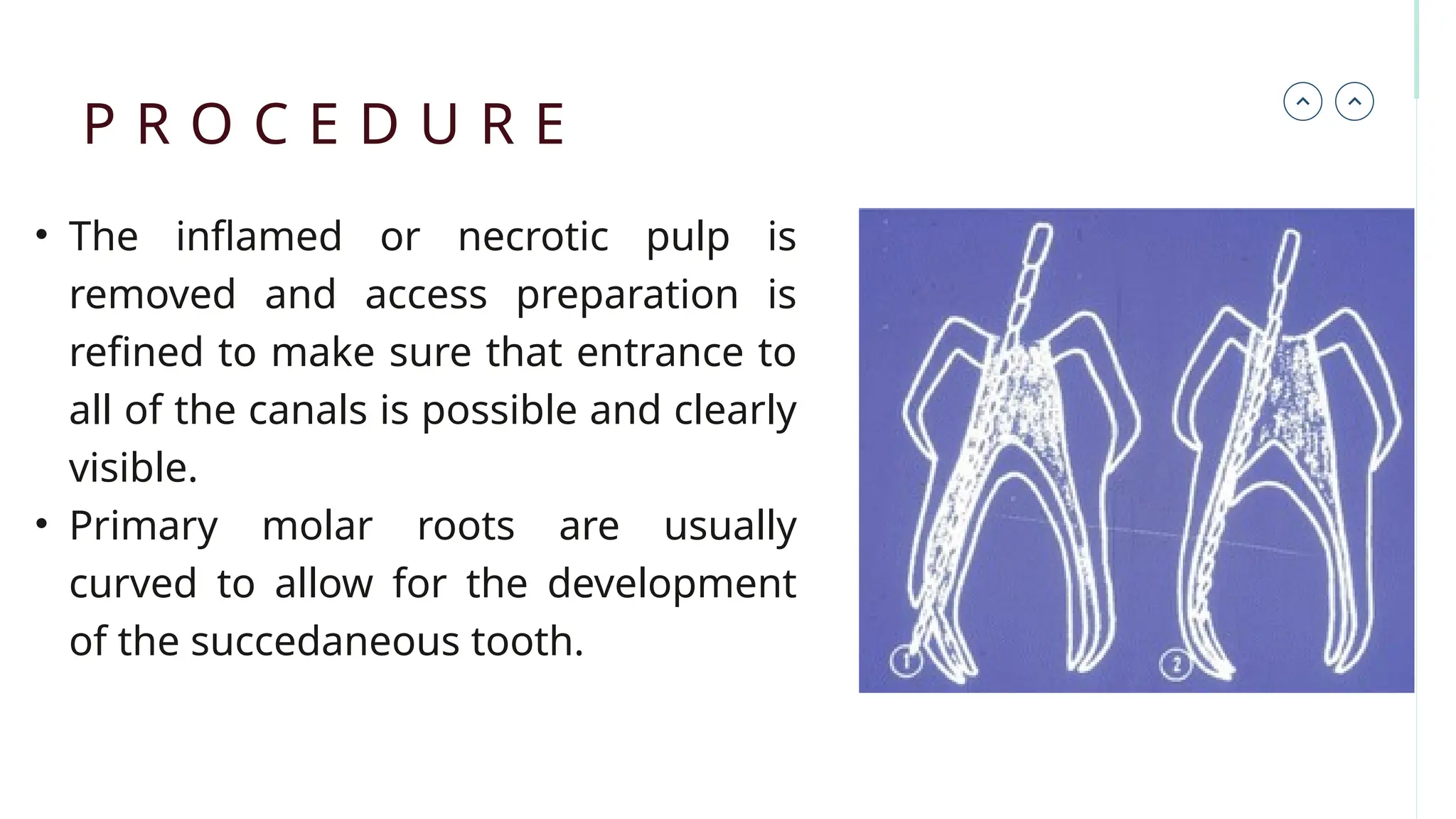
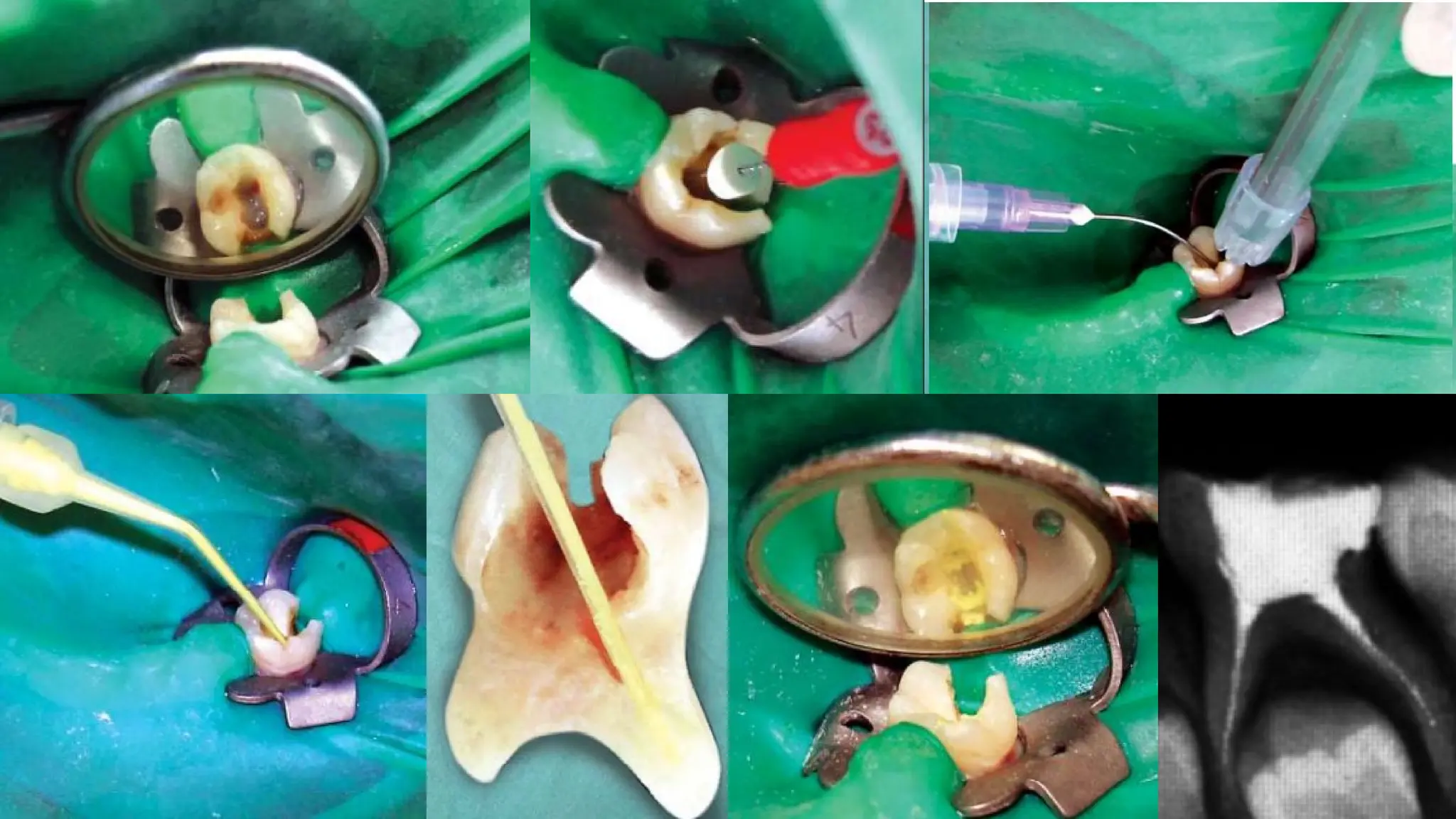
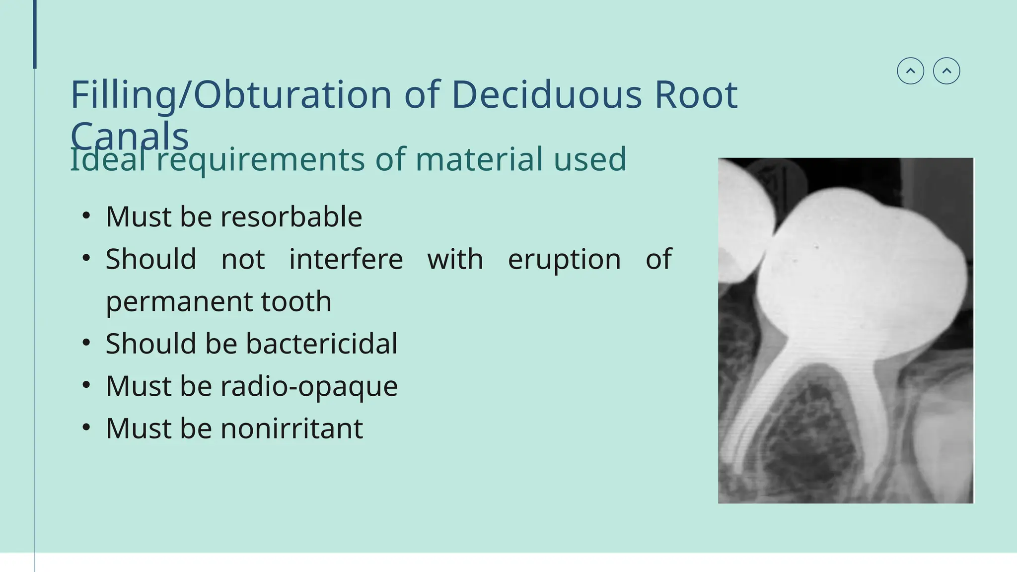
Pulp Therapy in
Primary Teeth
• Pulp treatment in primary
dentition is generally divided into
vital and non-vital pulp therapies
and assists in the preservation of
pulpally involved primary teeth in
the dental arch until the affected
tooth naturally exfoliates.
Introduction
• Pulpotomy
Types of Pulp
Therapy
• Pulpectomy
Pulpotomy
• Surgical removal of the entire coronal pulp, leaving intact the
vital tissue in the canals, followed by placement of a medicament
or dressing over the remaining pulp stump in an attempt to
promote healing and retention of this vital tissue
• Carious or mechanical
exposure of vital primary
teeth and young
permanent teeth, where
inflammation is restricted
to coronal pulp only
Indications
• History of spontaneous pain
• Swelling
• Fistula
• Tenderness to percussion
• Pathological mobility
• External/internal root resorption
• Periapical or interradicular radiolucency
• Pulp calcifications
• Pus or exudate from exposures site
• Uncontrollable bleeding from the amputated pulp stump
• half root length resorbed.
Contraindications
Procedure
• The coronal pulp is amputated, pulpal hemorrhage is controlled, and
the remaining vital radicular pulp tissue surface is treated with a long-
term clinically-successful medicament. Only MTA and formocresol are
recommended as the medicament of choice.
• Other materials or techniques such as ferric sulfate, lasers, sodium
hypochlorite, and tricalcium silicate have conditional
recommendations
• For multisurface lesions
• After the coronal pulp chamber is filled with a suitable base, the tooth
is restored with a restoration
• If there is sufficient
supporting enamel
remaining
• Some studies recommend disinfection of the pulp chamber with 3%–5%
sodium hypochlorite (NaOCl) solution prior to drying of the chamber. This is
known to dissolve and remove any remnant tissue, debris or dentinal chips
which can lead to infection of the radicular pulp.
• Many studies compared the effects of using 5% NaOCl and physiologic
saline for disinfection of the pulp chamber before placement of
pulpotomy medicament in the teeth, which showed successful treatment
outcomes with MTA.
Pulpectomy
• Removal of the entire pulp and subsequent filling of the canals of
the primary teeth with a suitable resorbable material.
Indication of Pulpectomy
• Primary teeth with pulp
inflammation extending beyond the
coronal pulp
• Roots and alveolar bone with
minimum pathologic resorption
• Primary teeth with necrotic pulp and
or periapical abscess
• Pus at the clinical pulp exposure
site.
• History of spontaneous pain
• Evidence of radicular pathologic lesion with or without caries
involvement.
• Alveolar swelling
• Continuous bleeding even after amputation of the coronal pulp
tissue during pulpotomy.
• Presence of sinus tract.
• Presence of inter-radicular or periapical radiolucency
Contraindication of Pulpectomy
• Grossly destroyed tooth that is nonrestorable clinically
• Periradicular involvement extending to the permanent tooth bud,
where the risk of damage to the permanent tooth is high.
• Root resorption—internal or external
• Extensive mobility
• Gross bone loss at the apex or at the furcation.
• Medically compromised children
• Root length remaining less than 2/3rd
• Perforation of pulpal floor
• Any dentigerous or follicular cyst present beneath
• Serious medical contraindications
1. Patient with systemic illness such as congenital ischemic heart
disease.
2. Patient with leukemia.
3. Children on long-term corticosteroids therapy
• The inflamed or necrotic pulp is
removed and access preparation is
refined to make sure that entrance to
all of the canals is possible and clearly
visible.
• Primary molar roots are usually
curved to allow for the development
of the succedaneous tooth.
P R O C E D U R E
• Endodontic files are selected and
adjusted to stop 1–2 mm short of
the radiographic apex, with the
preliminary working length
estimated according to the
preoperative radiograph.
• The instruments should be slightly bent to adjust to the curvature of
the canals, thus preventing perforations on the outer and inner
portions of the root
• The root canals are irrigated with
either 0.2 % up to 2 % chlorhexidine
solution or with 1 % up to 5 %
sodium hypochlorite (limited to one
percent sodium hypochlorite
according to the AAPD guidelines)
Irrigation
• The root canals are first irrigated with
either 0.2 % up to 2 % chlorhexidine
solution or with 1 % up to 5 % sodium
hypochlorite and then with normal
saline
• The use of sodium hypochlorite in the
primary dentition should be
performed cautiously since it is a
potent tissue irritant and must not be
extruded beyond the apex
Ideal requirements of material used
• Must be resorbable
• Should not interfere with eruption of
permanent tooth
• Should be bactericidal
• Must be radio-opaque
• Must be nonirritant
Filling/Obturation of Deciduous Root
Canals
• Zinc oxide eugenol – is used without catalyst. Lack of catalyst is
used to allow adequate working time.
• Iodoform paste – Is also being used. It consists of zinc oxide and
iodoform mixed into a paste. Its advantages over zinc oxide
eugenol are:
1.Potent bactericidal
2.Nonirritant
3.Radio-opaque
4.Chemically active until entirely resorbed
5.Good healing properties
6.Rate of resorption is faster
Different materials used
• Iodoform paste in combination with calcium hydroxide has also been
used; it is commercially available as Vitapex and Metapex. These
iodoform-containing products resorb if inadvertently pushed beyond
the apex, but the rate of resorption of the material from within the
canals is faster than the rate of physiological root resorption.
• Another root canal filling
material—a mixture of
iodoform, calcium hydroxide,
and zinc oxide—is commercially
available as Endoflas; in
addition, it has eugenol. It is
reported to resorb when
extruded beyond the apex but
resists resorption
intraradicularly. Eugenol, one of
its constituents, is known to
cause periapical irritation.
• The canals are dried thoroughly using paper points
• Lentulo spiral, pressure syringe, Jiffy’s syringe, amalgam condensor,
local anesthetic syringe, tuberculin syringe, files, etc can be used to
carry and deposit the material into the root canal.
Methods of Obturation
• Pressure syringe is one of the best devices used for obturation for the
following reasons.
1.Avoidance of air trap
2.Even amount of material is deposited
3.300 psi force produced allows the use of thick consistency
Single or two-visit
pulpectomy?
• Multiple visit pulpectomy
(MVP) involves extirpation of
the pulp tissue and
placement of intra-canal
medicament in the first visit
followed by obturation in
the subsequent visit, if the
underlying pathology still
persists it may require
additional visits.
• Single-visit pulpectomy
(SVP) involves extirpation
of pulp and filing the
canals short of the apex to
a resistance point, after
irrigation and final drying
obturating material will be
placed in same visit
• Treatment in single visit certainly has many advantages. It is less
time‑consuming, resulting in less cost for the patient. In addition,
various studies have shown that postoperative pain is equally low
when the treatment is performed in single or multiple visits.
• In fact, an argument could be made that added discomfort due to
local anesthetic or trauma from a rubber dam application experienced
after a second visit makes single‑visit endodontic treatment less
painful than multi‑visit treatment.
• However, according to
RCT study performed in
2016, single‑visit
pulpectomy can be
considered as a viable
option for the treatment
of primary teeth with
apical periodontitis.
Evaluation of Success of pulpectomy
• No purulent discharge from the gingival margin
• No abnormal mobility
• No postoperative pain
• No further resorption of root (except physiological)
• Resolution of sinus tract, by 6 month
Indirect pulp capping
• This technique shows some success in teeth with the absence of
symptomatology and with no radiographic evidence of pathosis.
Indication
• Deep caries in which the pulpal inflammation has been judged to
be minimal and complete removal of caries would probably cause
pulpal exposure.
Contraindicatio
n
• When there is wide spread inflammation or evidence of periapical
pathosis
Procedur
e
• All the caries on the cavity walls and at the DEJ are removed,
due to its closeness to the surface.
• A large round bur or spoon excavator is used to remove the
carious dentin. Round bur in slow speed is preferred, as there is
more chance of removal of large segment of dentin with
excavator
• Sedative dressing of calcium hydroxide is placed over the
remaining dentin
• Tooth is then sealed with zinc oxide eugenol and amalgam
Direct pulp capping
• Placement of a
medicament or a
nonmedicated material
on a pulp that has been
exposed, in the course
of excavating the caries,
due to fracture or due to
mechanical exposures
during routine caries
removal
Indication
• Mechanical exposures that occurs following trauma or during
cavity preparation which is <1 sq mm, surrounded by clean dentin
in an asymptomatic vital deciduous tooth
• Mechanical or carious exposures <1 sq mm, in an asymptomatic
vital young permanent tooth.
Contraindicatio
n
• Cariously exposed deciduous teeth
• Spontaneous pain
• Swelling
• Fistula
• Tenderness to percussion
• Pathologic mobility
• Root resorption—external/internal
• Periapical/interradicular radiolucency
• Pulp calcifications
• Profuse hemorrhage from the exposure site
• Pus or exudate from exposure site.
Ideal requirment for materials used for direct pulp
capping
• Stimulate reparative dentin formation
• Maintain pulpal vitality
• Bactericidal or bacteriostatic
• Adhere to dentin
• Adhere to restorative material
• Resist forces during restoration placement
• Must resist forces under restoration during lifetime of restoration
• Able to be sterilized
• Radio-opaque
• Provide bacterial seal
• Release fluoride to prevent secondary caries.
Materials used for direct pulp capping
• MTA mixing or putty
• bioceramic putty
Thank
You
Presented By: Dr. Asawer Ahmed Fayyad

PULP therapy in primary teeth..... pptx

  • 1.
  • 2.
    • Pulp treatmentin primary dentition is generally divided into vital and non-vital pulp therapies and assists in the preservation of pulpally involved primary teeth in the dental arch until the affected tooth naturally exfoliates. Introduction
  • 3.
    • Pulpotomy Types ofPulp Therapy • Pulpectomy
  • 4.
    Pulpotomy • Surgical removalof the entire coronal pulp, leaving intact the vital tissue in the canals, followed by placement of a medicament or dressing over the remaining pulp stump in an attempt to promote healing and retention of this vital tissue
  • 5.
    • Carious ormechanical exposure of vital primary teeth and young permanent teeth, where inflammation is restricted to coronal pulp only Indications
  • 6.
    • History ofspontaneous pain • Swelling • Fistula • Tenderness to percussion • Pathological mobility • External/internal root resorption • Periapical or interradicular radiolucency • Pulp calcifications • Pus or exudate from exposures site • Uncontrollable bleeding from the amputated pulp stump • half root length resorbed. Contraindications
  • 7.
    Procedure • The coronalpulp is amputated, pulpal hemorrhage is controlled, and the remaining vital radicular pulp tissue surface is treated with a long- term clinically-successful medicament. Only MTA and formocresol are recommended as the medicament of choice.
  • 8.
    • Other materialsor techniques such as ferric sulfate, lasers, sodium hypochlorite, and tricalcium silicate have conditional recommendations
  • 9.
    • For multisurfacelesions • After the coronal pulp chamber is filled with a suitable base, the tooth is restored with a restoration • If there is sufficient supporting enamel remaining
  • 10.
    • Some studiesrecommend disinfection of the pulp chamber with 3%–5% sodium hypochlorite (NaOCl) solution prior to drying of the chamber. This is known to dissolve and remove any remnant tissue, debris or dentinal chips which can lead to infection of the radicular pulp. • Many studies compared the effects of using 5% NaOCl and physiologic saline for disinfection of the pulp chamber before placement of pulpotomy medicament in the teeth, which showed successful treatment outcomes with MTA.
  • 12.
    Pulpectomy • Removal ofthe entire pulp and subsequent filling of the canals of the primary teeth with a suitable resorbable material.
  • 13.
    Indication of Pulpectomy •Primary teeth with pulp inflammation extending beyond the coronal pulp • Roots and alveolar bone with minimum pathologic resorption • Primary teeth with necrotic pulp and or periapical abscess • Pus at the clinical pulp exposure site. • History of spontaneous pain
  • 14.
    • Evidence ofradicular pathologic lesion with or without caries involvement. • Alveolar swelling • Continuous bleeding even after amputation of the coronal pulp tissue during pulpotomy. • Presence of sinus tract. • Presence of inter-radicular or periapical radiolucency
  • 15.
    Contraindication of Pulpectomy •Grossly destroyed tooth that is nonrestorable clinically • Periradicular involvement extending to the permanent tooth bud, where the risk of damage to the permanent tooth is high. • Root resorption—internal or external • Extensive mobility • Gross bone loss at the apex or at the furcation. • Medically compromised children • Root length remaining less than 2/3rd • Perforation of pulpal floor • Any dentigerous or follicular cyst present beneath
  • 16.
    • Serious medicalcontraindications 1. Patient with systemic illness such as congenital ischemic heart disease. 2. Patient with leukemia. 3. Children on long-term corticosteroids therapy
  • 17.
    • The inflamedor necrotic pulp is removed and access preparation is refined to make sure that entrance to all of the canals is possible and clearly visible. • Primary molar roots are usually curved to allow for the development of the succedaneous tooth. P R O C E D U R E
  • 18.
    • Endodontic filesare selected and adjusted to stop 1–2 mm short of the radiographic apex, with the preliminary working length estimated according to the preoperative radiograph. • The instruments should be slightly bent to adjust to the curvature of the canals, thus preventing perforations on the outer and inner portions of the root
  • 19.
    • The rootcanals are irrigated with either 0.2 % up to 2 % chlorhexidine solution or with 1 % up to 5 % sodium hypochlorite (limited to one percent sodium hypochlorite according to the AAPD guidelines) Irrigation
  • 20.
    • The rootcanals are first irrigated with either 0.2 % up to 2 % chlorhexidine solution or with 1 % up to 5 % sodium hypochlorite and then with normal saline • The use of sodium hypochlorite in the primary dentition should be performed cautiously since it is a potent tissue irritant and must not be extruded beyond the apex
  • 22.
    Ideal requirements ofmaterial used • Must be resorbable • Should not interfere with eruption of permanent tooth • Should be bactericidal • Must be radio-opaque • Must be nonirritant Filling/Obturation of Deciduous Root Canals
  • 23.
    • Zinc oxideeugenol – is used without catalyst. Lack of catalyst is used to allow adequate working time. • Iodoform paste – Is also being used. It consists of zinc oxide and iodoform mixed into a paste. Its advantages over zinc oxide eugenol are: 1.Potent bactericidal 2.Nonirritant 3.Radio-opaque 4.Chemically active until entirely resorbed 5.Good healing properties 6.Rate of resorption is faster Different materials used
  • 24.
    • Iodoform pastein combination with calcium hydroxide has also been used; it is commercially available as Vitapex and Metapex. These iodoform-containing products resorb if inadvertently pushed beyond the apex, but the rate of resorption of the material from within the canals is faster than the rate of physiological root resorption.
  • 25.
    • Another rootcanal filling material—a mixture of iodoform, calcium hydroxide, and zinc oxide—is commercially available as Endoflas; in addition, it has eugenol. It is reported to resorb when extruded beyond the apex but resists resorption intraradicularly. Eugenol, one of its constituents, is known to cause periapical irritation.
  • 26.
    • The canalsare dried thoroughly using paper points • Lentulo spiral, pressure syringe, Jiffy’s syringe, amalgam condensor, local anesthetic syringe, tuberculin syringe, files, etc can be used to carry and deposit the material into the root canal. Methods of Obturation
  • 27.
    • Pressure syringeis one of the best devices used for obturation for the following reasons. 1.Avoidance of air trap 2.Even amount of material is deposited 3.300 psi force produced allows the use of thick consistency
  • 28.
  • 29.
    • Multiple visitpulpectomy (MVP) involves extirpation of the pulp tissue and placement of intra-canal medicament in the first visit followed by obturation in the subsequent visit, if the underlying pathology still persists it may require additional visits. • Single-visit pulpectomy (SVP) involves extirpation of pulp and filing the canals short of the apex to a resistance point, after irrigation and final drying obturating material will be placed in same visit
  • 30.
    • Treatment insingle visit certainly has many advantages. It is less time‑consuming, resulting in less cost for the patient. In addition, various studies have shown that postoperative pain is equally low when the treatment is performed in single or multiple visits.
  • 31.
    • In fact,an argument could be made that added discomfort due to local anesthetic or trauma from a rubber dam application experienced after a second visit makes single‑visit endodontic treatment less painful than multi‑visit treatment.
  • 32.
    • However, accordingto RCT study performed in 2016, single‑visit pulpectomy can be considered as a viable option for the treatment of primary teeth with apical periodontitis.
  • 33.
    Evaluation of Successof pulpectomy • No purulent discharge from the gingival margin • No abnormal mobility • No postoperative pain • No further resorption of root (except physiological) • Resolution of sinus tract, by 6 month
  • 34.
    Indirect pulp capping •This technique shows some success in teeth with the absence of symptomatology and with no radiographic evidence of pathosis.
  • 35.
    Indication • Deep cariesin which the pulpal inflammation has been judged to be minimal and complete removal of caries would probably cause pulpal exposure. Contraindicatio n • When there is wide spread inflammation or evidence of periapical pathosis
  • 36.
    Procedur e • All thecaries on the cavity walls and at the DEJ are removed, due to its closeness to the surface. • A large round bur or spoon excavator is used to remove the carious dentin. Round bur in slow speed is preferred, as there is more chance of removal of large segment of dentin with excavator • Sedative dressing of calcium hydroxide is placed over the remaining dentin • Tooth is then sealed with zinc oxide eugenol and amalgam
  • 38.
    Direct pulp capping •Placement of a medicament or a nonmedicated material on a pulp that has been exposed, in the course of excavating the caries, due to fracture or due to mechanical exposures during routine caries removal
  • 39.
    Indication • Mechanical exposuresthat occurs following trauma or during cavity preparation which is <1 sq mm, surrounded by clean dentin in an asymptomatic vital deciduous tooth • Mechanical or carious exposures <1 sq mm, in an asymptomatic vital young permanent tooth.
  • 40.
    Contraindicatio n • Cariously exposeddeciduous teeth • Spontaneous pain • Swelling • Fistula • Tenderness to percussion • Pathologic mobility • Root resorption—external/internal • Periapical/interradicular radiolucency • Pulp calcifications • Profuse hemorrhage from the exposure site • Pus or exudate from exposure site.
  • 41.
    Ideal requirment formaterials used for direct pulp capping • Stimulate reparative dentin formation • Maintain pulpal vitality • Bactericidal or bacteriostatic • Adhere to dentin • Adhere to restorative material • Resist forces during restoration placement • Must resist forces under restoration during lifetime of restoration • Able to be sterilized • Radio-opaque • Provide bacterial seal • Release fluoride to prevent secondary caries.
  • 42.
    Materials used fordirect pulp capping • MTA mixing or putty • bioceramic putty
  • 43.
    Thank You Presented By: Dr.Asawer Ahmed Fayyad